

Evidence for neurogenesis in the adult mammalian substantia nigra
- PMID: 12792021
- PMCID: PMC164689
- DOI: 10.1073/pnas.1131955100
Evidence for neurogenesis in the adult mammalian substantia nigra
Abstract
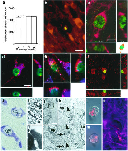
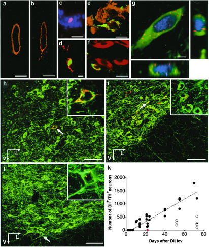
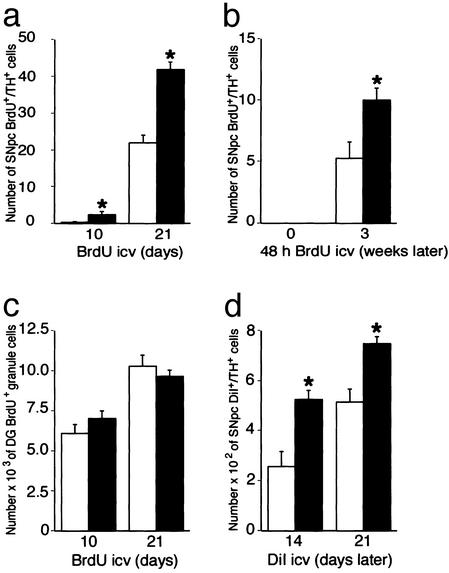
New neurons are generated from stem cells in a few regions of the adult mammalian brain. Here we provide evidence for the generation of dopaminergic projection neurons of the type that are lost in Parkinson's disease from stem cells in the adult rodent brain and show that the rate of neurogenesis is increased after a lesion. The number of new neurons generated under physiological conditions in substantia nigra pars compacta was found to be several orders of magnitude smaller than in the granular cell layer of the dentate gyrus of the hippocampus. However, if the rate of neuronal turnover is constant, the entire population of dopaminergic neurons in substantia nigra could be replaced during the lifespan of a mouse. These data indicate that neurogenesis in the adult brain is more widespread than previously thought and may have implications for our understanding of the pathogenesis and treatment of neurodegenerative disorders such as Parkinson's disease.
Figures

Comment in
-
Brain repair by cell replacement and regeneration.Proc Natl Acad Sci U S A. 2003 Jun 24;100(13):7430-1. doi: 10.1073/pnas.1332673100. Epub 2003 Jun 16. Proc Natl Acad Sci U S A. 2003. PMID: 12810949 Free PMC article. No abstract available.
-
No evidence for new dopaminergic neurons in the adult mammalian substantia nigra.Proc Natl Acad Sci U S A. 2004 Jul 6;101(27):10177-82. doi: 10.1073/pnas.0401229101. Epub 2004 Jun 21. Proc Natl Acad Sci U S A. 2004. PMID: 15210991 Free PMC article.
Similar articles
-
Early signs of neuronal apoptosis in the substantia nigra pars compacta of the progressive neurodegenerative mouse 1-methyl-4-phenyl-1,2,3,6-tetrahydropyridine/probenecid model of Parkinson's disease.Neuroscience. 2006 Jun 19;140(1):67-76. doi: 10.1016/j.neuroscience.2006.02.007. Epub 2006 Mar 14. Neuroscience. 2006. PMID: 16533572
-
Selective activation of p38 mitogen-activated protein kinase in dopaminergic neurons of substantia nigra leads to nuclear translocation of p53 in 1-methyl-4-phenyl-1,2,3,6-tetrahydropyridine-treated mice.J Neurosci. 2008 Nov 19;28(47):12500-9. doi: 10.1523/JNEUROSCI.4511-08.2008. J Neurosci. 2008. PMID: 19020042 Free PMC article.
-
The TrkB-positive dopaminergic neurons are less sensitive to MPTP insult in the substantia nigra of adult C57/BL mice.Neurochem Res. 2011 Oct;36(10):1759-66. doi: 10.1007/s11064-011-0491-5. Epub 2011 May 12. Neurochem Res. 2011. PMID: 21562748
-
Age and Parkinson's disease-related neuronal death in the substantia nigra pars compacta.J Neural Transm Suppl. 2009;(73):203-13. doi: 10.1007/978-3-211-92660-4_16. J Neural Transm Suppl. 2009. PMID: 20411779 Review.
-
Neurogenesis in substantia nigra of parkinsonian brains?J Neural Transm Suppl. 2009;(73):279-85. doi: 10.1007/978-3-211-92660-4_23. J Neural Transm Suppl. 2009. PMID: 20411786 Review.
Cited by
-
Widespread Doublecortin Expression in the Cerebral Cortex of the Octodon degus.Front Neuroanat. 2021 Apr 29;15:656882. doi: 10.3389/fnana.2021.656882. eCollection 2021. Front Neuroanat. 2021. PMID: 33994960 Free PMC article.
-
Inflammation is detrimental for neurogenesis in adult brain.Proc Natl Acad Sci U S A. 2003 Nov 11;100(23):13632-7. doi: 10.1073/pnas.2234031100. Epub 2003 Oct 27. Proc Natl Acad Sci U S A. 2003. PMID: 14581618 Free PMC article.
-
Adult human neurogenesis: from microscopy to magnetic resonance imaging.Front Neurosci. 2011 Apr 4;5:47. doi: 10.3389/fnins.2011.00047. eCollection 2011. Front Neurosci. 2011. PMID: 21519376 Free PMC article.
-
An Overview of Adult Neurogenesis.Adv Exp Med Biol. 2021;1331:77-94. doi: 10.1007/978-3-030-74046-7_7. Adv Exp Med Biol. 2021. PMID: 34453294 Review.
-
Neuroprotection by Exendin-4 Is GLP-1 Receptor Specific but DA D3 Receptor Dependent, Causing Altered BrdU Incorporation in Subventricular Zone and Substantia Nigra.J Neurodegener Dis. 2013;2013:407152. doi: 10.1155/2013/407152. Epub 2013 Nov 21. J Neurodegener Dis. 2013. PMID: 26316987 Free PMC article.
References
-
- McKay, R. D. (1997) Science 276, 66-71. - PubMed
-
- Gage, F. H. (2000) Science 287, 1433-1438. - PubMed
-
- Momma, S., Johansson, C. B. & Frisén, J. (2000) Curr. Opin. Neurobiol. 10, 45-49. - PubMed
-
- Cameron, H. A. & McKay, R. D. (2001) J. Comp. Neurol. 435, 406-417. - PubMed
-
- Gould, E., Reeves, A. J., Graziano, M. S. & Gross, C. G. (1999) Science 286, 548-552. - PubMed
MeSH terms
Substances
LinkOut - more resources
Full Text Sources
Other Literature Sources
Miscellaneous
