

Selective impairment of hippocampal neurogenesis by chronic alcoholism: protective effects of an antioxidant
- PMID: 12792022
- PMCID: PMC164688
- DOI: 10.1073/pnas.1230907100
Selective impairment of hippocampal neurogenesis by chronic alcoholism: protective effects of an antioxidant
Abstract
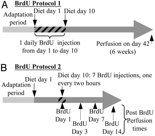
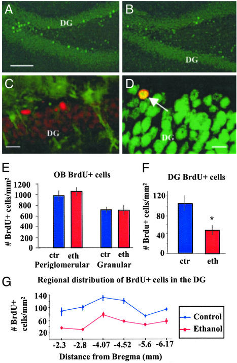
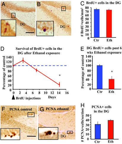
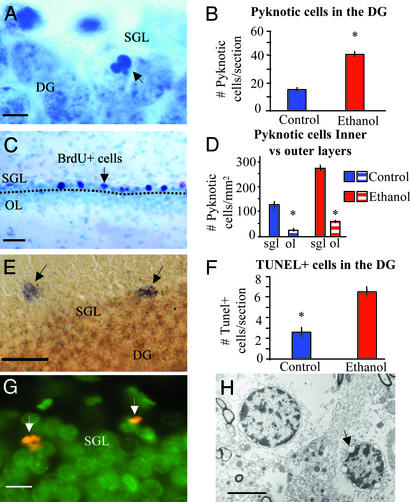
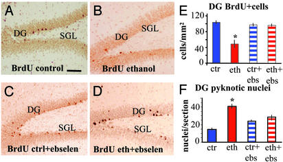
A major pathogenic mechanism of chronic alcoholism involves oxidative burden to liver and other cell types. We show that adult neurogenesis within the dentate gyrus of the hippocampus is selectively impaired in a rat model of alcoholism, and that it can be completely prevented by the antioxidant ebselen. Rats fed for 6 weeks with a liquid diet containing moderate doses of ethanol had a 66.3% decrease in the number of new neurons and a 227-279% increase in cell death in the dentate gyrus as compared with paired controls. Neurogenesis within the olfactory bulb was not affected by alcohol. Our studies indicate that alcohol abuse, even for a short duration, results in the death of newly formed neurons within the adult brain and that the underlying mechanism is related to oxidative or nitrosative stress. Moreover, these findings suggest that the impaired neurogenesis may be a mechanism mediating cognitive deficits observed in alcoholism.
Figures
Similar articles
-
Ebselen prevents chronic alcohol-induced rat hippocampal stress and functional impairment.Alcohol Clin Exp Res. 2007 Mar;31(3):486-92. doi: 10.1111/j.1530-0277.2006.00329.x. Alcohol Clin Exp Res. 2007. PMID: 17295734
-
Chronic alcohol exposure reduces hippocampal neurogenesis and dendritic growth of newborn neurons.Eur J Neurosci. 2005 May;21(10):2711-20. doi: 10.1111/j.1460-9568.2005.04120.x. Eur J Neurosci. 2005. PMID: 15926919
-
Chronic systemic D-galactose exposure induces memory loss, neurodegeneration, and oxidative damage in mice: protective effects of R-alpha-lipoic acid.J Neurosci Res. 2006 Jun;83(8):1584-90. doi: 10.1002/jnr.20845. J Neurosci Res. 2006. PMID: 16555301
-
Melatonin maintains adult hippocampal neurogenesis and cognitive functions after irradiation.Prog Neurobiol. 2010 Jan 11;90(1):60-8. doi: 10.1016/j.pneurobio.2009.10.019. Epub 2009 Oct 24. Prog Neurobiol. 2010. PMID: 19857546 Review.
-
Alcohol, neural stem cells, and adult neurogenesis.Alcohol Res Health. 2003;27(2):197-204. Alcohol Res Health. 2003. PMID: 15303631 Free PMC article. Review.
Cited by
-
The effects of chronic alcoholism on cell proliferation in the human brain.Exp Neurol. 2013 Sep;247:9-18. doi: 10.1016/j.expneurol.2013.03.020. Epub 2013 Mar 28. Exp Neurol. 2013. PMID: 23541433 Free PMC article.
-
Alterations of theta power and synchrony during encoding in young adult binge drinkers: Subsequent memory effects associated with retrieval after 48 h and 6 months.Front Psychol. 2022 Dec 15;13:1061016. doi: 10.3389/fpsyg.2022.1061016. eCollection 2022. Front Psychol. 2022. PMID: 36591031 Free PMC article.
-
The Effect of Dehydroepiandrosterone Treatment on Neurogenesis, Astrogliosis and Long-Term Cocaine-Seeking Behavior in a Cocaine Self-Administration Model in Rats.Front Neurosci. 2021 Nov 26;15:773197. doi: 10.3389/fnins.2021.773197. eCollection 2021. Front Neurosci. 2021. PMID: 34899172 Free PMC article.
-
Effects of Chronic Voluntary Alcohol Drinking on Thiamine Concentrations, Endoplasmic Reticulum Stress, and Oxidative Stress in the Brain of Crossed High Alcohol Preferring Mice.Neurotox Res. 2019 Nov;36(4):777-787. doi: 10.1007/s12640-019-00032-y. Epub 2019 Apr 10. Neurotox Res. 2019. PMID: 30972556 Free PMC article.
-
Type 2 Neural Progenitor Cell Activation Drives Reactive Neurogenesis after Binge-Like Alcohol Exposure in Adolescent Male Rats.Front Psychiatry. 2017 Dec 15;8:283. doi: 10.3389/fpsyt.2017.00283. eCollection 2017. Front Psychiatry. 2017. PMID: 29326611 Free PMC article.
References
-
- American Psychiatric Association (1994) Diagnostic and Statistical Manual of Mental Disorders (Am. Psychiatr. Assoc., Washington, DC), 4th Ed.
-
- Harper, C. (1998) J. Neuropathol. Exp. Neurol. 57, 101-110. - PubMed
-
- Albert, M. S., Butters, N. & Brandt, J. (1980) J. Stud. Alcohol 41, 1071-1081. - PubMed
-
- Korbo, L. (1999) Alcohol Clin. Exp. Res. 23, 164-168. - PubMed
-
- Walker, D. W., Barnes, D. E., Zornetzer, S. F., Hunter, B. E. & Kubanis, P. (1980) Science 209, 711-713. - PubMed
Publication types
MeSH terms
Substances
LinkOut - more resources
Full Text Sources
Other Literature Sources
Medical
