. 2003 Jul 15;550(Pt 2):365-72.
doi: 10.1113/jphysiol.2002.036400. Epub 2003 Jun 6.

Molecular dissection of the inward rectifier potassium current (IK1) in rabbit cardiomyocytes: evidence for heteromeric co-assembly of Kir2.1 and Kir2.2

Affiliations

Molecular dissection of the inward rectifier potassium current (IK1) in rabbit cardiomyocytes: evidence for heteromeric co-assembly of Kir2.1 and Kir2.2

Carsten Zobel et al. J Physiol. .

Abstract

Cardiac inward rectifier K+ currents (IK1) play an important role in maintaining resting membrane potential and contribute to late phase repolarization. Members of the Kir2.x channel family appear to encode for IK1. The purpose of this study was to determine the molecular composition of cardiac IK1 in rabbit ventricle. Western blots revealed that Kir2.1 and Kir2.2, but not Kir2.3, are expressed in rabbit ventricle. Culturing rabbit myocytes resulted in an approximately 50% reduction of IK1 density after 48 or 72 h in culture which was associated with an 80% reduction in Kir2.1, but no change in Kir2.2, protein expression. Dominant-negative (DN) constructs of Kir2.1, Kir2.2 and Kir2.3 were generated and tested in tsA201 cells. Adenovirus-mediated over-expression of Kir2.1dn, Kir2.2dn or Kir2.1dn plus Kir2.2dn in cultured rabbit ventricular myocytes reduced IK1 density equally by 70% 72 h post-infection, while AdKir2.3dn had no effect, compared to green fluorescent protein (GFP)-infected myocytes. Previous studies indicate that the [Ba2+] required for half-maximum block (IC50) differs significantly between Kir2.1, Kir2.2 and Kir2.3 channels. The dependence of IK1 on [Ba2+] revealed a single binding isotherm which did not change with time in culture. The IC50 for block of IK1 was also unaffected by expression of the different DN genes after 72 h in culture. Taken together, these results demonstrate functional expression of Kir2.1 and Kir2.2 in rabbit ventricular myocytes and suggest that macroscopic IK1 is predominantly composed of Kir2.1 and Kir2.2 heterotetramers.


Figures

Figure 1
Figure 1. Effect of culture on IK1 in rabbit ventricular myocytes
A, typical Ba2+-subtracted membrane currents elicited by 500 ms voltage steps ranging from −130 mV to −10 mV (by 10 mV) from a holding potential of −40 mV. B, peak IK1 densities from myocytes cultured for 2 h (□) were significantly decreased (P < 0.05) after culturing for 48 h (▵) but stayed unchanged upon further culturing (72h, •) for membrane potentials between −130 and −80 mV.
Figure 2
Figure 2. Western blots for native Kir2.1, Kir2.2 and Kir2.3 proteins in rabbit ventricular myocytes
Western blots showing typical signals obtained by probing rabbit ventricular myocyte preparations harvested after 2 or 48 h in culture along with rabbit brain controls using specific antibodies for Kir2.1 (A), Kir2.2 (B) or Kir2.3 (C). The bar graphs show summarized results of densitometric measurements for Kir2.1 (n = 3) and Kir2.2 (n = 7) after 48 h in culture normalized to expression levels after 2 h in culture.
Figure 3
Figure 3. Validation of dominant-negative (DN) genes in tsA201 cells
Bar graphs showing the membrane conductance measured in tsA201 cells transfected with either wild-type Kir2.1 (A) or Kir2.2 (B) genes. Equimolar co-transfection with either Kir2.1dn or Kir2.2dn constructs significantly diminished the slope conductance of the wild-type channels. Doubling the amount of wild-type construct (‘Kir2.1 double’ or ‘Kir2.2 double’) increased the slope conductance correspondingly. Membrane potentials were held at −40 mV and voltage steps from −130 to −10 mV in 10 mV increments were applied for 500 ms with an interval of 1 s. Slope conductance was calculated from the linear range of the current–voltage relationship between −130 and −80 mV). * Significantly different at P < 0.05 relative to Kir2.1 (A) or Kir2.2 (B).
Figure 4
Figure 4. Effect of infection of different AdKir2.xdn constructs on IK1 in cultured rabbit ventricular myocytes
A, original traces showing typical Ba2+-substracted IK1 recorded from myocytes 72 h after infection with the indicated constructs. B, Ba2+-subtracted peak currents as a function of voltage in cultured rabbit myocytes 72 h after infection. IK1 density (from −130 mV to −90 mV) decreased significantly in myoctyes infected with AdKir2.1dn, AdKir2.1dn or AdKir2.1dn plus AdKir2.1dn compared to the control myocytes (AdGFP, P < 0.05). No significant difference was detected between IK1 density recorded after infection with AdKir2.1dn, AdKir2.2dn or AdKir2.1dn plus AdKir2.2dn.
Figure 5
Figure 5. Effects of a dominant-negative Kir2.3 construct
A, slope conductance measured in tsA201 cells expressing Kir2.3 or Kir2.3 plus Kir2.3dn. Cells were held at a membrane potential of −40 mV and voltage steps from −130 to −10 mV in 10 mV increments were applied for 500 ms with a 1 s interval. Slope conductance was calculated from the linear range of the current–voltage relationship (−130 to −80 mV). B, Ba2+-subtracted peak IK1 as a function of voltage measured in rabbit ventricular myocytes expressing either AdGFP or AdKir2.3dn. * Significant difference at P < 0.05 between Kir2.3 alone and Kir2.3 plus Kir2.3dn.
Figure 6
Figure 6. Effects of infection with AdKir2.X genes on transient outward K+ currents in cultured rabbit ventricular myocytes
A, typical recordings of Ito measured at +60 mV with a prepulse at +40 mV to inactivate sodium currents in myocytes infected with AdGFP or AdKir2.1dn plus AdKir2.2dn 72 h post-infection. B, summary of the effects of infection on Ito in cultured myocytes following infection with the indicated AdKir2.X genes.
Figure 7
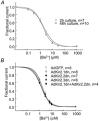
Figure 7. Blockade of cardiac IK1 by Ba2+ in cultured rabbit ventricular myocytes
A, Ba2+ dose–response curves for IK1 in non-infected myocytes cultured for 2 h or 48 h at −100 mV. The data were fitted with a single binding isotherm IBa/ICon = 1/(1 + [Ba2+]n/IC50n) where IBa is the IK1 in the presence of Ba2+ while ICon is the IK1 before application of Ba2+. B, Ba2+ dose–response curve for IK1 measured at −100 mV in myocytes cultured for 48 h following over-expression of various AdKir2.x genes as indicated. Despite marked differences in the level of IK1 in the various groups, there was no measurable difference in the IC50 values for Ba2+.

References

    1. Barry DM, Trimmer JS, Merlie JP, Nerbonne JM. Differential expression of voltage-gated K+ channel subunits in adult rat heart. Relation to functional K+ channels. Circ Res. 1995;77:361–369. - PubMed
    1. Brahmajothi MV, Morales MJ, Liu S, Rasmusson RL, Campbell DL, Strauss HC. In situ hybridization reveals extensive diversity of K+ channel mRNA in isolated ferret cardiac myocytes. Circ Res. 1996;78:1083–1089. - PubMed
    1. Burnashev NA, Zilberter YI. Two types of single inward rectifying potassium channels in rat myocardial cells. Gen Physiol Biophys. 1986;5:495–504. - PubMed
    1. Cho HC, Tsushima RG, Nguyen TT, Guy HR, Backx PH. Two critical cysteine residues implicated in disulfide bond formation and proper folding of Kir2. 1. Biochemistry. 2000;39:4649–4657. - PubMed
    1. Christe G. Localization of K(+) channels in the tubules of cardiomyocytes as suggested by the parallel decay of membrane capacitance, IK(1) and IK(ATP) during culture and by delayed IK(1) response to barium. J Mol Cell Cardiol. 1999;31:2207–2213. - PubMed

Publication types

Substances

LinkOut - more resources