Review
. 2003 Jul 15;550(Pt 2):337-46.
doi: 10.1113/jphysiol.2003.043299. Epub 2003 Jun 6.

Interstitial cells: involvement in rhythmicity and neural control of gut smooth muscle

Affiliations
Review

Interstitial cells: involvement in rhythmicity and neural control of gut smooth muscle

G D S Hirst et al. J Physiol. .

Abstract

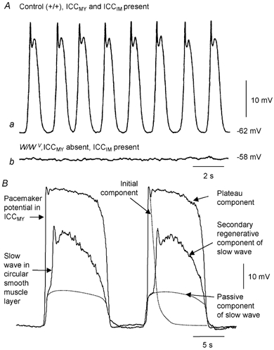
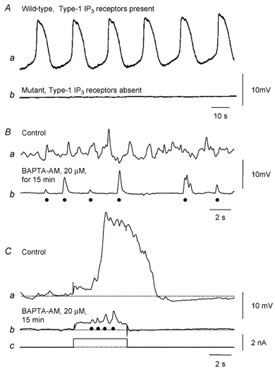
Many smooth muscles display spontaneous electrical and mechanical activity, which persists in the absence of any stimulation. In the past this has been attributed largely to the properties of the smooth muscle cells. Now it appears that in several organs, particularly in the gastrointestinal tract, activity in smooth muscles arises from a separate group of cells, known as interstitial cells of Cajal (ICC), which are distributed amongst the smooth muscle cells. Thus in the gastrointestinal tract, a network of interstitial cells, usually located near the myenteric plexus, generates pacemaker potentials that are conducted passively into the adjacent muscle layers where they produce rhythmical membrane potential changes. The mechanical activity of most smooth muscle cells, can be altered by autonomic, or enteric, nerves innervating them. Previously it was thought that neuroeffector transmission occurred simply because neurally released transmitters acted on smooth muscle cells. However, in several, but not all, regions of the gastrointestinal tract, it appears that nerve terminals, rather than communicating directly with smooth muscle cells, preferentially form synapses with ICC and these relay information to neighbouring smooth muscle cells. Thus a set of ICC, which are distributed amongst the smooth muscle cells of the gut, are the targets of transmitters released by intrinsic enteric excitatory and inhibitory nerve terminals: in some regions of the gastrointestinal tract, the same set of ICC also augment the waves of depolarisation generated by pacemaker ICC. Similarly in the urethra, ICC, distributed amongst the smooth muscle cells, generate rhythmic activity and also appear to be the targets of autonomic nerve terminals.


Figures

Figure 1
Figure 1. Pacemaker role of ICCMY in small intestine and gastric antrum
A shows intracellular recordings of electrical activity in the circular muscle of the small intestine of mice; both recordings are in the presence of nifedipine. Aa shows the regular discharge of slow waves recorded from a control tissue. Ab shows that slow waves are absent in the small intestine of W/WV mice: these tissues contain ICCDMP and normal smooth muscle cells but lack ICCMY. (Modified from Ward et al. 1994.) B shows simultaneous intracellular recordings from ICCMY and the circular muscle layer of guinea-pig gastric antrum. The onset of pacemaker depolarisation is seen to precede the onset of depolarisation in the circular muscle layer. Each pacemaker potential consist of two components, an initial component, followed by a plateau component. Furthermore each slow wave consists of two components, an initial passive component, whose time course is shown by a dotted line, and a secondary regenerative component, superimposed. (Modified from Hirst et al. 2002c.)
Figure 2
Figure 2. Generation of secondary regenerative component of antral slow waves by ICCIM located in the circular smooth muscle layer
A schematic diagram showing a transverse section of the antral wall with an outer longitudinal layer (open circles at top), separated from the circular muscle layer (ellipsoids at bottom) by ICCMY. The circular layer consists of smooth muscle cells (dark shading) and ICCIM (light shading). Aa and b show intracellular recordings of electrical activity in the circular layer of the gastric antrum, from control and W/WV mice, respectively; both recordings are in the presence of nifedipine. Aa shows the regular discharge of slow waves recorded from a control tissue; each slow wave consists of a passive component, generated by ICCMY, and a secondary regenerative component (see Fig. 1). Ab shows that slow waves in the antrum of W/WV mice, which contain ICCMY but lack ICCIM, consist only of the passive component generated by ICCMY. (Modified from Dickens et al. 2001.) B shows traces which compare the time course of a regenerative potential (Ba), initiated by injecting depolarising current into a bundle of circular muscle (Bc), with the time course of the associated change in [Ca2+]i obtained by averaging 30 successive responses (Bb). Note that the onset of the increase in [Ca2+]i lags the onset of applied depolarisation by some 900 ms but corresponds with the start of the regenerative response. (Modified from Hirst et al. 2002b.)
Figure 3
Figure 3. Generation of electrical activity by antral ICC depends upon IP3 receptors and discharge of unitary potentials
Aa and b show intracellular recordings of electrical activity in the circular layer of the gastric antrum, from control and mutant mice which lack type-1 IP3 receptors; both recordings were made in the presence of nifedipine. Aa shows the regular discharge of slow waves recorded from a control tissue. In contrast when recordings were made from tissues lacking type-1 IP3 receptors, slow waves were not detected (Ab). (Modified from Suzuki et al. 2001.) B shows membrane potential recordings from an isolated bundle of antral circular muscle from the guinea-pig. In control solution the resting potential displays an ongoing discharge of membrane noise (Ba); after buffering [Ca2+]i to low levels, individual unitary potentials (•) are detected (Bb). Ca shows a regenerative potential initiated in control solution. After buffering [Ca2+]i to low levels, depolarising currents (Cc) cause an increase in the frequency of occurrence of unitary potentials (•; Cb). (Modified from Edwards et al. 1999.)
Figure 4
Figure 4. Involvement of ICCIM in responses to inhibitory and excitatory nerve stimulation in the stomach
Nerve stimulation evoked a biphasic response consisting of an EJP, followed by an IJP in the fundus of control mice (Aa). After abolishing the IJP with Nω-nitro-L-arginine (L-NA), nerve stimulation evoked an EJP (4Ab) whose amplitude was little changed by inhibiting cholinesterases with neostigmine (Ac); in neostigmine, the cholinergic EJP was followed by a slower depolarisation (Ac). Both excitatory responses were abolished by atropine (Ad). When similar recordings were made from Sl/Sld mutants, which lack ICCIM, nerve stimulation failed to evoke either an EJP or an IJP (Ba and b) but in the presence of neostigmine an atropine-sensitive slow depolarisation was detected (Bc and d; modified from Beckett et al. 2003). In an isolated bundle of circular muscle, dissected from guinea-pig antrum, after abolishing the effects of inhibitory nerve stimulation with apamin and L-NA, excitatory nerve stimulation evoked a regenerative potential (Ca). The regenerative potential, but not the response of smooth muscle cells to neurally released ACh, was abolished by hyperpolarisation (Ca). The excitatory response involving ICCIM was abolished by anthracene-9-carboxylic acid (9-AC) but the response of smooth muscle cells to neurally released ACh persisted (Cb). (Modified from Hirst et al. 2002c.)

References

    1. Beckett EAH, Horiguchi K, Khoyi M, Sanders KM, Ward SM. Loss of enteric motor neurotransmission in the gastric fundus of Sl/Sld mice. J Physiol. 2002;543:871–887. - PMC - PubMed
    1. Burns AJ, Lomax AE, Torihashi S, Sanders KM, Ward SM. Interstitial cells of Cajal mediate inhibitory neurotransmission in the stomach. Proc Natl Acad Sci U S A. 1996;93:12008–12013. - PMC - PubMed
    1. Chowdhury JU, Pang YW, Huang SM, Tsugeno M, Tomita T. Sustained contraction produced by caffeine after ryanodine treatment in the circular muscle of the guinea-pig gastric antrum and rabbit portal vein. Br J Pharmacol. 1995;114:1414–1418. - PMC - PubMed
    1. Connor JA, Prosser CL, Weems WA. A study of pace-maker activity in intestinal smooth muscle. J Physiol. 1974;240:671–701. - PMC - PubMed
    1. Cousins HM, Edwards FR, Hirst GD. Neuronally released and applied acetylcholine on the longitudinal muscle of the guinea-pig ileum. Neuroscience. 1995;65:193–207. - PubMed

Publication types

LinkOut - more resources