. 2003 Jun 24;100(13):7638-43.
doi: 10.1073/pnas.1230693100. Epub 2003 Jun 6.

The Notch ligand Delta1 is sequentially cleaved by an ADAM protease and gamma-secretase

Affiliations

The Notch ligand Delta1 is sequentially cleaved by an ADAM protease and gamma-secretase

Emmanuelle Six et al. Proc Natl Acad Sci U S A. .

Abstract

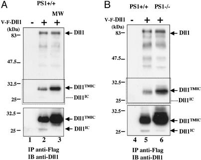
Notch signaling is involved in numerous cell fate decisions in invertebrates and vertebrates. The Notch receptor is a type I transmembrane (TM) protein that undergoes two proteolytic steps after ligand binding, first by an ADAM (a distintegrin and metalloprotease) in the extracellular region, followed by gamma-secretase-mediated cleavage inside the TM domain. We demonstrate here that the murine ligand Delta1 (Dll1) undergoes the same sequence of cleavages, in an apparently signal-independent manner. Identification of the ADAM-mediated shedding site localized 10 aa N-terminal to the TM domain has enabled us to generate a noncleavable mutant. Kuzbanian/ADAM10 is involved in this processing event, but other proteases can probably substitute for it. We then show that Dll1 is part of a high-molecular-weight complex containing presenilin1 and undergoes further cleavage by a gamma-secretase-like activity, therefore releasing the intracellular domain that localizes in part to the nucleus. Using the shedding-resistant mutant, we demonstrate that this gamma-secretase cleavage depends on prior ectodomain shedding. Therefore Dll1 is a substrate for regulated intramembrane proteolysis, and its intracellular region possibly fulfills a specific function in the nucleus.


Figures

Fig. 1.
Fig. 1.
Murine Dll1 undergoes a constitutive ectodomain shedding. (A) Schematic map of the tagged Dll1 molecule used in B and C. (B and C) Pulse– chase analysis of Dll1. HEK293T cells were transfected with V-F-Dll1. After 24 h the cells were pulsed with [35S]Met (t0) for 20 min and chased for 0.5, 1, 2, 4, and 6 h. Cell extracts were immunoprecipitated with anti-VSV (B) or anti-Flag (C) antibodies. Culture media were immunoprecipitated with anti-VSV antibody (B Lower). Dll1 indicates full-length Dll1, Dll1TMIC is the membrane-associated processing product, and Dll1EC is the shedding product. Molecular mass markers are indicated on the left.
Fig. 2.
Fig. 2.
Identification of the EC cleavage site, generation of a Dll1 mutant for this cleavage, and processing of Dll1 in Kuz–/– cells. (A) Alignment of mDll1 with Dll1-D8 and Dll1-Apa mutants and Delta orthologs, in the juxtamembrane region (amino acids 516–545 of mDll1). The two cleavage sites identified by Mishra-Gorur et al. (19) in Drosophila Delta and the identified cleavage site of Dll1 are indicated. (B) Identification of the cleavage site. HEK293T cells were transfected with Delta-Myc6. After 24 h, cells were labeled with [3H]Leu or [35S]Met. Whole-cell extracts were immunoprecipitated with anti-Myc antibody. By radio sequencing of formula image a 35S peak was detected at cycle 1 and a 3H peak at cycle 19. The corresponding amino acid sequence is shown (Lower). (C) Analysis of the noncleavable Dll1-D8 and Dll1–Apa mutants. HEK293T cells were transfected with WT Dll1 (lane 2), Dll1-D8 (lane 3), or Dll1-Apa (lane 4), and whole-cell extracts were blotted with Dll1 antiserum. (D) Proteolytic cleavage of Dll1 is reduced in Kuz–/– cells. Kuz +/+ (lanes 1 and 2) and –/– (lanes 3 and 4) MEFs were infected with a retrovirus encoding GFP alone (MIG) or V-F-Dll1-internal ribosomal entry site-GFP (V-F-Dll1). GFP-enriched pools of cells were analyzed by immunoblotting of whole-cell extracts by using anti-Flag antibody. Equal protein loading was controlled by using an anti-GFP antibody (the level of GFP being correlated with the level of Dll1 expression).
Fig. 3.
Fig. 3.
Full-length Dll1 and its proteolytic fragment Dll1TMIC are present in distinct complexes. (A) Gel filtration analysis of extracts from 293T cells transfected with Dll1. Whole-cell extracts were fractionated through a Superose 6 column. Fractions were analyzed by immunoblotting, using Dll1 antiserum (Upper), followed by PS1 antiserum (Lower) after membrane stripping. Positions of full-length Dll1, Dll1TMIC, and the N-terminal processing product of PS1 (PS1 NTF) are indicated on the right. (B) Dll1 interacts with PS1. HEK293T cells were transfected with plasmids encoding V-F-Dll1 and/or PS1 as indicated. Whole-cell extracts (WE; lane 1) and anti-PS1 immunoprecipitates (IP anti-PS1; lanes 2–4) were analyzed on SDS/PAGE and immunoblotted with an anti-Flag antibody. The position of Igs is indicated on the right.
Fig. 4.
Fig. 4.
Dll1 is cleaved by a presenilin-dependent γ-secretase activity. PS1 +/+ and –/– MEFs were infected by a retrovirus encoding GFP alone (–) or V-F-Dll1-internal ribosomal entry site-GFP (+). (A) Dll1IC release is inhibited by MW167. GFP-enriched pools of PS1+/+ cells were analyzed by immunoprecipitation using the anti-Flag antibody followed by immunoblotting with the Dll1 antiserum. (Upper) Short exposure. (Lower, corresponding to the boxed region) Long exposure. Dll1IC indicates the position of the soluble IC fragment. In lane 3, PS1+/+ cells were treated for 15 h in the presence of the γ-secretase inhibitor MW167 (MW) before lysis. (B) Absence of Dll1IC release in PS1–/– cells. A similar experiment to lanes 1 and 2 of A was performed in parallel in PS1+/+ (lanes 4 and 5) and PS1–/– (lane 6) cells.
Fig. 5.
Fig. 5.
Generation of Dll1TMIC is a preliminary requirement for γ-secretase cleavage to occur. (A) Dll1-Apa and Dll1-D8 do not undergo γ-secretase cleavage. HEK293T cells were transfected with V-F-Dll1 (lanes 1, 4, and 7), V-F-Dll1-Apa (lanes 2, 5, and 8), and V-F-Dll1-D8 (lanes 3, 6, and 9) and treated with lactacystin for 3 h. Membrane, cytosolic, and nuclear extracts were prepared and analyzed by immunoblotting using the anti-Flag antibody, followed by immunoblotting with anti-β-tubulin (a cytosolic marker) and anti-CSL (a nuclear marker) antibodies as a control for the purity of the fractions and equal protein loading. (B) Dll1IC localized to the nucleus. HeLa cells were transfected with Dll1IC-V5 and treated with lactacystin for 3 h. Cells were stained with the anti-V5 antibody (Left) and Hoechst for nuclear staining (Center). Light blue in the merged image (Right) indicates colocalization.

Similar articles

Cited by

References

    1. Artavanis-Tsakonas, S., Rand, M. D. & Lake, R. J. (1999) Science 284, 770–776. - PubMed
    1. Logeat, F., Bessia, C., Brou, C., Lebail, O., Jarriault, S., Seidah, N. G. & Israël, A. (1998) Proc. Natl. Acad. Sci. USA 95, 8108–8112. - PMC - PubMed
    1. Blaumueller, C. M., Qi, H. L., Zagouras, P. & Artavanis-Tsakonas, S. (1997) Cell 90, 281–291. - PubMed
    1. Fleming, R. J., Purcell, K. & Artavanis-Tsakonas, S. (1997) Trends Cell Biol. 7, 437–441. - PubMed
    1. Lendahl, U. (1998) BioEssays 20, 103–107. - PubMed

Publication types

MeSH terms