Review
. 2003 Jun;67(2):238-76, table of contents.
doi: 10.1128/MMBR.67.2.238-276.2003.

Prophage genomics

Affiliations
Review

Prophage genomics

Carlos Canchaya et al. Microbiol Mol Biol Rev. 2003 Jun.

Erratum in

  • Microbiol Mol Biol Rev. 2003 Sep;67(3):473

Abstract

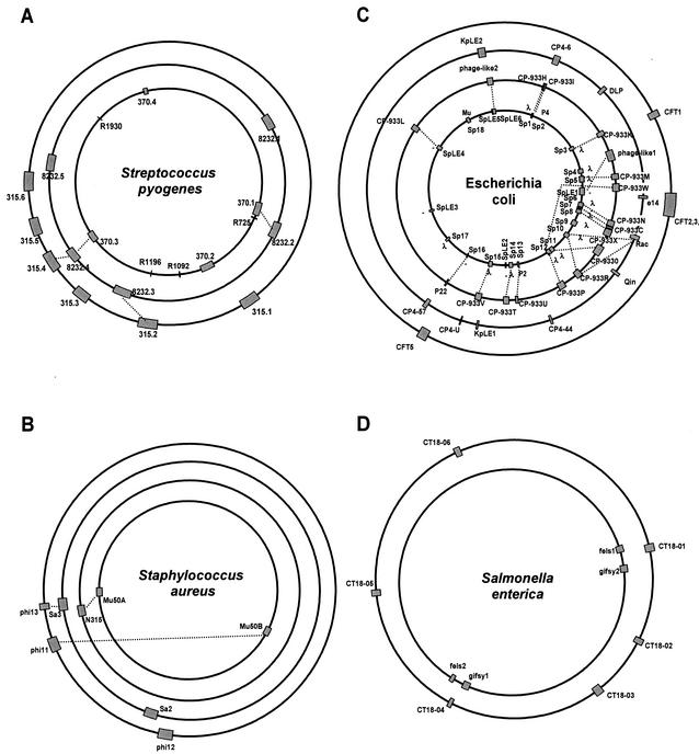
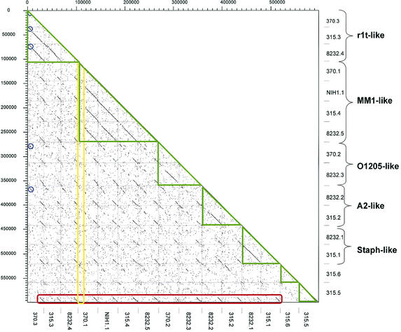
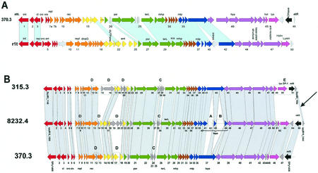
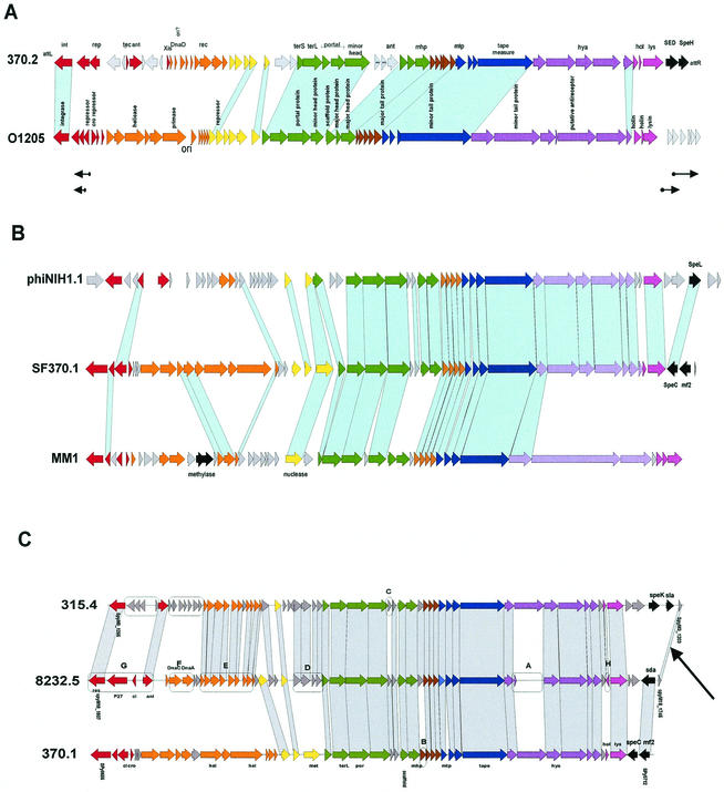
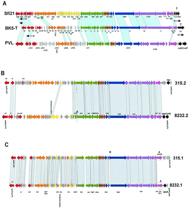
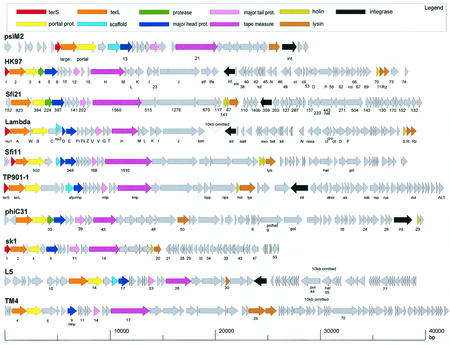
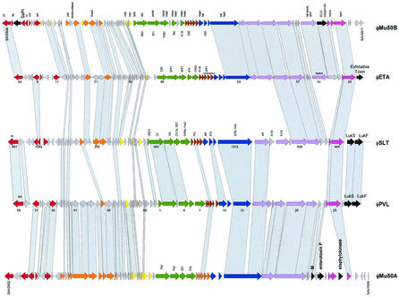
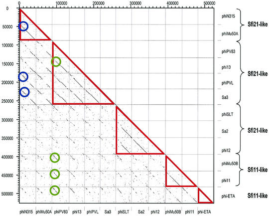
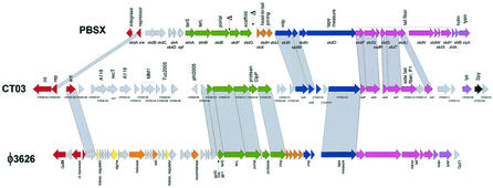
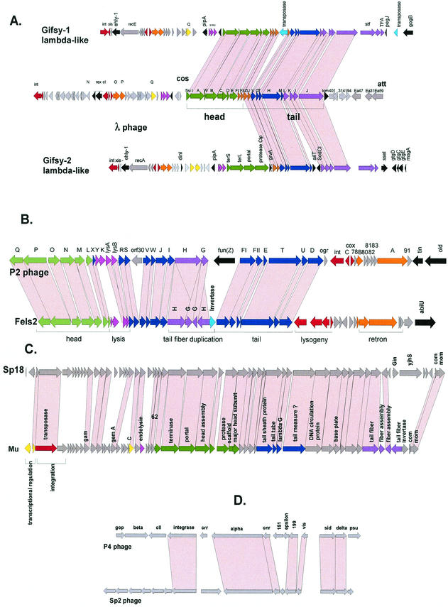
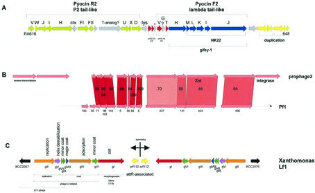
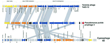
The majority of the bacterial genome sequences deposited in the National Center for Biotechnology Information database contain prophage sequences. Analysis of the prophages suggested that after being integrated into bacterial genomes, they undergo a complex decay process consisting of inactivating point mutations, genome rearrangements, modular exchanges, invasion by further mobile DNA elements, and massive DNA deletion. We review the technical difficulties in defining such altered prophage sequences in bacterial genomes and discuss theoretical frameworks for the phage-bacterium interaction at the genomic level. The published genome sequences from three groups of eubacteria (low- and high-G+C gram-positive bacteria and gamma-proteobacteria) were screened for prophage sequences. The prophages from Streptococcus pyogenes served as test case for theoretical predictions of the role of prophages in the evolution of pathogenic bacteria. The genomes from further human, animal, and plant pathogens, as well as commensal and free-living bacteria, were included in the analysis to see whether the same principles of prophage genomics apply for bacteria living in different ecological niches and coming from distinct phylogenetical affinities. The effect of selection pressure on the host bacterium is apparently an important force shaping the prophage genomes in low-G+C gram-positive bacteria and gamma-proteobacteria.


Figures

FIG. 1.
FIG. 1.
Prophage content of four human bacterial pathogens. The prophages are indicated as shaded boxes on the bacterial genome maps. The lengths of the boxes correspond to the relative sizes of the prophage DNA with respect to the bacterial chromosome. Note that the circumference of the bacterial genomes does not correspond to their relative length. Prophages with extensive DNA sequence identity are linked by dotted lines. (A) S. pyogenes genomes of the sequenced M1, M18, and M3 strains (from center to periphery). (B) S. aureus genomes of the sequenced Mu50 (center), N315, MW2, and 8325 strains. (C) E. coli genomes from the O157:H7 Sakai (center) and O157:H7 EDL933 strains, the laboratory strain K-12, and the uropathogenic strain CFT073. (D) S. enterica serovar Typhimurium LT2 (center) and serovar Typhi CT18.
FIG. 2.
FIG. 2.
Dot plot matrix for the currently available 15 S. pyogenes prophages identified by their prophage names on the x and y axes. According to their structural genes, the prophages were classified into distinct groups (green triangles) and annotated on the left ordinate. The prophage genomes were aligned with their integrase gene to the left (top). The extent of the conservation of the tail fiber and lysis genes is highlighted by the red box. The lack of conservation of the lysogeny and DNA replication genes is demonstrated by the yellow box. The largest group of prophages sharing early genes is indicated by the blue circles.
FIG. 3.
FIG. 3.
r1t-like S. pyogenes prophages. (A) Alignment of the S. pyogenes prophage 370.3 with the L. lactis prophage r1t. (B) Alignment of r1t-like prophages from the three sequenced S. pyogenes genomes. Genes sharing sequence relationships are linked by shading. Boxes A to E mark features discussed in the text. The prophage modules, as identified by bioinformatic analysis, are color coded. Lysogeny, red; DNA replication, orange; transcriptional regulation?, yellow; DNA packaging and head, green; head-to-tail joining, brown; tail, blue; tail fiber, mauve; lysis, violet; lysogenic conversion, black; unattributed genes, grey. Selected genes were annotated: int, integrase; cI/cro, repressors; xis, excisionase; repl, replication; rec, recombination; ant, antirepressor; por, portal; terL, large subunit terminase; mhp/mtp, major head/tail protein; hya, hyaluronidase; hol, holin; lys, lysin.
FIG. 4.
FIG. 4.
Sfi11-like S. pyogenes prophages. (A) Alignment of S. pyogenes prophage 370.2 with S. thermophilus prophage O1205. The arrows under the map indicate phage O1205 transcripts detected in the lysogen. (B) Alignment of S. pyogenes prophage 370.1 with S. pyogenes prophage NIH1.1 and S. pneumoniae prophage MM1. (C) Alignment of Sfi11-like prophages from the three sequenced S. pyogenes genomes. For annotations, see Fig. 3.
FIG. 5.
FIG. 5.
Sfi21-like prophages. (A) Alignment of S. thermophilus prophage Sfi21, L. lactis prophage BK5-T, and S. aureus prophage PVL. Sfi21 and BK5-T genes transcribed in the lysogenic host are indicated by arrows under the gene map. Genes are annotated as in the original publications (103, 129, 134). (B and C) Alignment of the Sfi21-like S. pyogenes prophages of the A2-like (B) and Staphylococcus-like (C) subgroups.
FIG. 6.
FIG. 6.
Alignment of the gene maps from bacteriophages belonging to a postulated lambda-supergroup of Siphoviridae. The viruses represent prophages from Archaea (Methanobacterium virus psiM2), γ-proteobacteria (E. coli phages HK97 and lambda), low-G+C gram-positive bacteria (S. thermophilus phages Sfi21 and Sfi11; L. lactis phages TP901-1 and sk1) and high-G+C gram-positive bacteria (Streptomyces phage phiC31; Mycobacterium phages L5 and TM4). Virulent phages are underlined. To better visualize the similarity between the structural gene clusters from this diverse group of phages, the phage genome maps are aligned starting with the terminase genes at the left. Structural genes are identified by a color code, as indicated at the top. Selected ORFs are numbered to facilitate the orientation with the GenBank entry.
FIG.7.
FIG.7.
Alignment of the five major types of S. aureus prophages. phiMu50A and phiMu50B are prophages extracted from the sequenced Mu50 S. aureus strain, while phages ETA, SLT, and PVL were induced from unsequenced S. aureus strains. The phage genes are color coded as in Fig. 3. Sequence-related genes between the prophages are linked by shading. Sequence matches of structural genes to those in other than staphylococcal phages are indicated (Bacillus, 105, SPP1, SPBc; Listeria, PSA, A118; Lactobacillus, adh, g1e; Lactococcus, TP901-1; Streptococcus, Sfi21, Sfi11, 1205, MM1 Spy; E. coli, N15; Pseudomonas, D3). Selected ORFs are numbered to facilitate the orientation with the GenBank entry.
FIG. 8.
FIG. 8.
Dot plot matrix for the currently available 12 S. aureus prophages. The prophages are identified by their names on the x and y- axes. According to the structural genes, the prophages were classified into five distinct groups (red triangles) and annotated on the left ordinate. Two groups of phages sharing relatively highly conserved early genes are marked by blue and green circles.
FIG. 9.
FIG. 9.
Clostridium prophages. The genome of C. tetani prophage 3 (center) is aligned with B. subtilis prophage PBSX (top) and C. perfringens prophage phi3626 (bottom). Genes related at the protein sequence level are connected by shading. Putative gene functions are indicated, and the modular structure of the prophage genomes is indicated by a color code, as in Fig. 3. Two likely deletions in the head gene cluster of PBSX are marked by a delta symbol.
FIG.10.
FIG.10.
Typical prophages found in the genomes of E. coli and Salmonella. (A) Alignment of the Salmonella prophages Gifsy-1 and Gifsy-2 with E. coli phage lambda. Genes with sequence similarity at the protein level are linked by shading. For the color coding of the genes, see Fig. 3. (B) Alignment of the P2-like Salmonella prophage Fels-2 with E. coli phage P2. P2 genes were annotated and color coded according to their attribution to phage modules. Genes linked by sequence similarity are connected by shading. (C) Alignment of the O157 prophage Sp18 with E. coli phage Mu. (D) Alignment of the O157 prophage Sp2 with E. coli satellite phage P4.
FIG. 11.
FIG. 11.
Pseudomomas phage-derived bacteriocin and filamentous phages from Pseudomonas and Xanthomonas. (A) The gene map of a prophage-like DNA element in P. aeruginosa encoding the bacteriocins pyocin R2 and F2 resembling phage tail modules. Genes with sequence identity to phage tail genes from phage P2 are colored in green, and those with sequence identity to genes from lambdoid phages are in red (Pseudomonas phage D3) or blue (Salmonella phage Gifsy-1 and E. coli phage HK22). The extent of lambda-like and P2-like tail genes is marked by brackets. Further unattributed likely phage tail genes are shown in yellow. For easier orientation, the gene numbers of the first and last genes of this likely recombined prophage remnant are provided. (B) P. aeruginosa PAO1 prophage 2 aligned with filamentous Pseudomonas phage Pf1. The numbers between the maps indicate percent protein sequence identity. The numbers below the Pf1 gene map identify the phage genes from the GenBank entry. (C) Lf-like filamentous prophage from the X. campestris genome. The symmetry axis of the gene map is indicated, as well as the tentative gene functions and sequence similarities to other filamentous phages.
FIG. 12.
FIG. 12.
A new class of prophages in P. putida. P. putida prophage 3 (center) is aligned with Y. enterocolitica T7-like virulent phage phiYe03-12 (top) and cyanophage P60. Genes with protein sequence similarities are linked by shading. Putative gene functions are indicated, as well as their attribution to T7 class I early (yellow), class II DNA metabolism (orange), and class III morphogenesis modules (blue).
FIG. 13.
FIG. 13.
Prophages from bacterial plant pathogens. (A) Prophage content of X. fastidiosa strain 9a5c. Prophages linked by high DNA sequence identity are connected by double-arrowed lines. (B) Alignment of the X. fastidiosa prophages XfP3 and XfP4. The degree of protein sequence identity is reflected by different intensities of red shading. Suspected gene functions are indicated. (C) Alignment of the X. fastidiosa prophages XfP1 and XfP2. Selected genes are annotated. Genes with protein sequence similarity to phage APSE-1 from an endosymbiont of a pea aphid are marked in violet, and those with similarity to phage WO from the Drosophila endosymbiont Wolbachia are marked in yellow. Genes corresponding to lambda-like DNA-packaging genes are in dark green, while tail genes resembling P2 phage proteins are in light green. The gene numbers for the first and last genes of the prophages are indicated to facilitate orientation on the Xylella genome. Candidates for virulence-associated genes are marked with vap. Prophage genes with protein sequence identity are linked by shading. The different shades of red indicate >90%, >80%, >70%, and any protein sequence identity. (D) Alignment of the X. campestris prophage XCCP1 (center) with X. axonopodis prophage XACP1 (top) and Salmonella prophage Fels-2 (bottom). Note that the leftmost XACP1 genes are virtually translocated and inverted to allow an optimal alignment with the XCCP1 gene map.

Similar articles

Cited by

References

    1. Ackermann, H. W. 1998. Tailed bacteriophages: the order caudovirales. Adv. Virus Res. 51:135-201. - PMC - PubMed
    1. Akman, L., A. Yamashita, H. Watanabe, K. Oshima, T. Shiba, M. Hattori, and S. Aksoy. 2002. Genome sequence of the endocellular obligate symbiont of tsetse flies, Wigglesworthia glossinidia. Nat. Genet. 32:402-407. - PubMed
    1. Allison, G. E., D. Angeles, N. Tran-Dinh, and N. K. Verma. 2002. Complete genomic sequence of SfV, a serotype-converting temperate bacteriophage of Shigella flexneri. J. Bacteriol. 184:1974-1987. - PMC - PubMed
    1. Auvray, F., M. Coddeville, R. C. Ordonez, and P. Ritzenthaler. 1999. Unusual structure of the attB site of the site-specific recombination system of Lactobacillus delbrueckii bacteriophage mv4. J. Bacteriol. 181:7385-7389. - PMC - PubMed
    1. Auvray, F., M. Coddeville, P. Ritzenthaler, and L. Dupont. 1997. Plasmid integration in a wide range of bacteria mediated by the integrase of Lactobacillus delbrueckii bacteriophage mv4. J. Bacteriol. 179:1837-1845. - PMC - PubMed

Publication types

LinkOut - more resources