Review
. 2003 Jun;67(2):277-301, table of contents.
doi: 10.1128/MMBR.67.2.277-301.2003.

Conjugative plasmid transfer in gram-positive bacteria

Affiliations
Review

Conjugative plasmid transfer in gram-positive bacteria

Elisabeth Grohmann et al. Microbiol Mol Biol Rev. 2003 Jun.

Abstract

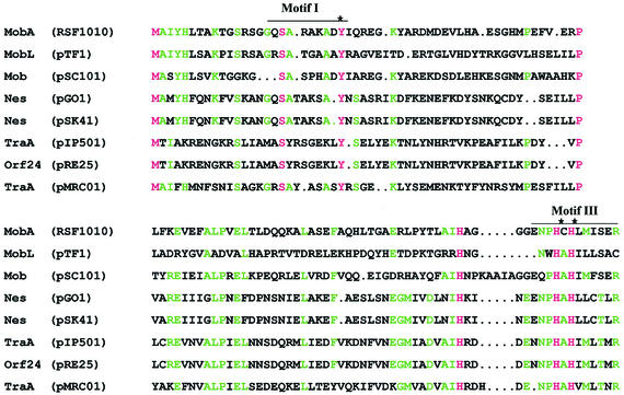
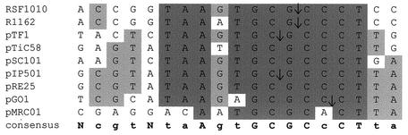
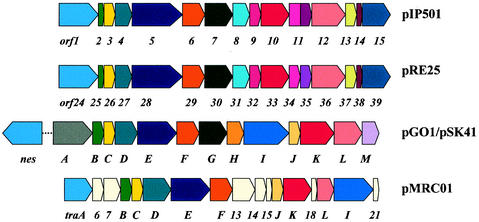
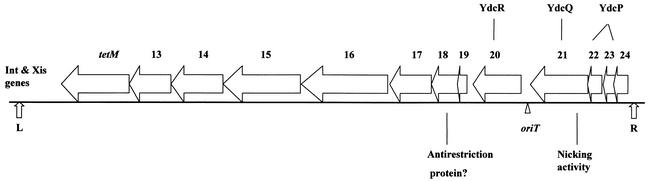
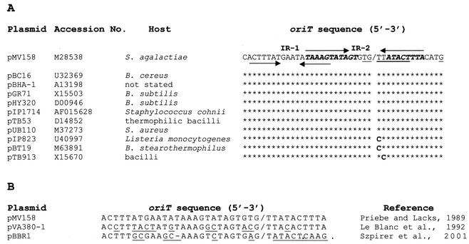
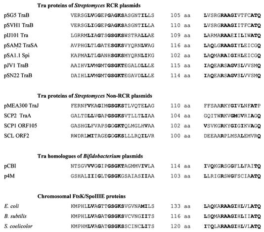
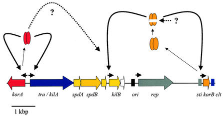
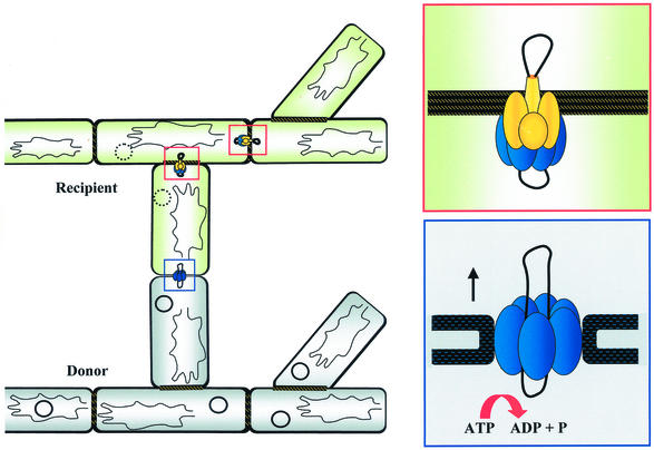
Conjugative transfer of bacterial plasmids is the most efficient way of horizontal gene spread, and it is therefore considered one of the major reasons for the increase in the number of bacteria exhibiting multiple-antibiotic resistance. Thus, conjugation and spread of antibiotic resistance represents a severe problem in antibiotic treatment, especially of immunosuppressed patients and in intensive care units. While conjugation in gram-negative bacteria has been studied in great detail over the last decades, the transfer mechanisms of antibiotic resistance plasmids in gram-positive bacteria remained obscure. In the last few years, the entire nucleotide sequences of several large conjugative plasmids from gram-positive bacteria have been determined. Sequence analyses and data bank comparisons of their putative transfer (tra) regions have revealed significant similarities to tra regions of plasmids from gram-negative bacteria with regard to the respective DNA relaxases and their targets, the origins of transfer (oriT), and putative nucleoside triphosphatases NTP-ases with homologies to type IV secretion systems. In contrast, a single gene encoding a septal DNA translocator protein is involved in plasmid transfer between micelle-forming streptomycetes. Based on these clues, we propose the existence of two fundamentally different plasmid-mediated conjugative mechanisms in gram-positive microorganisms, namely, the mechanism taking place in unicellular gram-positive bacteria, which is functionally similar to that in gram-negative bacteria, and a second type that occurs in multicellular gram-positive bacteria, which seems to be characterized by double-stranded DNA transfer.


Figures

FIG. 1.
FIG. 1.
Alignment of conjugative DNA relaxases of the IncQ-type family. Amino acid positions that are conserved throughout are shown in pink. Green letters mark positions that are conserved in at least five of the eight proteins. The delimitations of two conserved motifs that were identified first in IncP-like relaxases are indicated by lines above the sequence block. The active tyrosine in motif I and the two histidines in motif III that are conserved in all conjugative DNA relaxases are marked with asterisks. GenBank/EMBL accession numbers: MobA (RSF1010), X04830; MobL (pTF1), S12190; Mob (pSC101), P14492; Nes (pGO1), U50629; Nes (pSK41), AF051917; TraA (pIP501), L39769; Orf24 (pRE25), X92945; TraA (pMRC01), NC_001949.
FIG. 2.
FIG. 2.
Alignment of oriT nick regions. Nucleotides conserved in the nick regions of at least eight of nine plasmids of the RSF1010 oriT family are indicated by dark grey shading. Nucleotides that are conserved in at least five of the nine plasmids are indicated by light grey shading. A consensus sequence is also shown. Nucleotides conserved in all oriT regions are shown in capital letters; positions conserved in at least five of the nine sequences are indicated in lowercase letters. The cleavage sites determined experimentally are indicated by arrows. GenBank/EMBL accession numbers: RSF1010, M28829; R1162, M13380; pTF1, X52699; pTiC58, M95646; pSC101, X01654; pIP501, L39769; pRE25, X92945; pGO1, U50629; pMRC01, NC_001949. Modified from reference .
FIG. 3.
FIG. 3.
Comparison of the transfer regions of pIP501, pRE25, pGO1, pSK41, and pMRC01. Similar gene products are shown in the same color. Cream-colored boxes represent tra genes unique to pMRC01. The putative transfer proteins of pRE25 and pIP501 show a high degree of identity (between 80 and 100%). Orf1 to Orf6, Orf8 to Orf9, and Orf14 are 100% identical to the corresponding pRE25 gene products. Orf13 (262 amino acids) is significantly larger than the corresponding Orf37 (231 amino acids) encoded by pRE25. In pIP501, one big Orf (Orf11, 306 amino acids) comprises the regions of the corresponding Orf34 and Orf35 in pRE25. The gene products of the transfer region of pGO1 (trsA to trsM) and pSK41 (traA to traM) also exhibit a very high degree of similarity (between 97 and 98% identity). Tra proteins of pMRC01 show 25 to 42% identity to the corresponding proteins of pGO1 (59). pGO1 and pSK41 encode at least one additional tra gene, nes, located outside the transfer region. In pSK41, the distance between nes and the tra-region is approximately 11 kb. Specific single-strand nicking mediated by Nes at the respective oriT site was demonstrated for pGO1 (48).
FIG. 4.
FIG. 4.
Map of the conjugative transposon Tn916 with open reading frames thought to be related to conjugation. The genes related to excision of Tn916, int and xis, are shown near the left end of the transposon. tetM is the inducible tetracycline resistance determinant of the transposon. Orf13 and Orf14 have homologues (near identity) within Tn5397, a conjugative transposon in Clostridium difficile. Orf15 and Orf16 each have homologues on both pAD1 and pAM373. Orf18 has similarity to the bacterial antirestriction proteins, Ard of plasmid Coll-b-P9 and ArdA of pKM101 (72). Orf20 is a homologue of the hypothetical YdcR in B. subtilis; Orf22 and Orf23, which have some similarity to each other, are both homologues of the hypothetical YdcP. Orf21 has nonspecific nicking activity that is likely to be related to an origin-involved transfer event (44). Orf21 has strong homology to a B. subtilis hypothetical protein (YdcQ), and an internal segment resembles the FtsK/SpoIIIE family (14). Modified from reference 44.
FIG. 5.
FIG. 5.
The oriT region of the pMV158 family of plasmids. (A) Features of the pMV158 oriT and its conservation among RCR-plasmids. The inverted repeats (IR) are indicated by arrows above and below the sequence. Nucleotides protected from DNase I cleavage by MobM are indicated in boldface italics, whereas the extended −10 promoter region of the mobM gene is underlined. The last nucleotide depicted (G, underlined) is the mobM transcription initiation site in L. lactis. The MobM-mediated nick (l) and the changes in the nucleotide sequence (boldface) are indicated. (B) Characterized oriT regions of three plasmids with similar mobilization cassette and the same nick site (l). Differences are underlined. DNA from plasmid pVA380-1 is a substrate of pMV158 MobM.
FIG. 6.
FIG. 6.
Pock formation, indicating the intramycelial plasmid spreading. (A) If spores of a donor strain carrying a self-transmissible plasmid are mixed with an excess of plasmid-free recipient spores, characteristic growth retardation zones (pocks) are formed, indicating the area where the recipient mycelium has acquired a plasmid. The size of the pocks depends on the action of the spd genes. (B) In S. lividans pock formation is also associated with the induction of the red-pigmented antibiotic actinorhodin. (C) A pIJ101-carrying donor was streaked on a lawn of a recipient expressing gfp from Aequorea victoria. Since the aerial mycelium and spores of S. lividans show red autofluorescence, only the pock regions, where morphological differentiation is retarded, light up green.
FIG. 7.
FIG. 7.
Gene organization of actinomycete plasmids. All plasmids are drawn starting from the GntR-type regulatory gene. Identical colors indicate similar function. Red, GntR-type regulator; yellow, spread genes; orange, spdB2; blue, tra; white, Orf; grey, replication gene; light red, regulatory gene; pink, mutT homologue; green, integration/excision/recombination gene; brown, transposon. Arrows indicate regions with promoter activity. (A) Streptomyces RCR plasmids. (B) Non-RCR-type actinomycete plasmids. Only the putative transfer region of the 356-kb linear SCP1 plasmid is shown. GenBank/EMBL accession numbers: pSG5, X80774; pSVH1, Muth (unpublished); pIJ101, M21778; pSB24.2, M32513; pJV1, U23762; pSN22, D14281; pSNA1, AJ243257; pSAM2, AJ005260; pSA1.1, AB010724; pMEA300, L36679; SCP2 AL645771; and SCP1, AL590463.
FIG. 8.
FIG. 8.
Conserved sequence motifs of the septal DNA translocator proteins. The proteins of the septal DNA translocator family share a nucleotide-binding site (Walker box A) and a conserved RAAGI motif located 100 to 133 amino acids (aa) downstream of the NTP-binding site. These motifs are also present (with wider spacing) in the hexameric ring helicase TrwB of plasmid R388 (86).
FIG. 9.
FIG. 9.
Transcriptional regulation of the transfer functions of the S. lividans plasmid pIJ101. The GntR-type repressor KorA represses the transcription of tra/kilA and korA by binding to the overlapping promoter regions (204). The KilB repressor KorB is synthesized as a 10-kDa protein and subsequently processed to a 6-kDa molecule which binds to the kilB promoter 50-fold more efficiently than to the korB promoter (206). By an unknown mechanism, KorA also overrides the lethal action of unregulated kilB expression.
FIG. 10.
FIG. 10.
Model for conjugative plasmid transfer in Streptomyces. The hyphal tips of a plasmid-carrying donor and a recipient mycelium grow together without the need for a plasmid-encoded aggregation system. In the presence of a Tra protein, the hyphae fuse. Tra multimers form a ring-shaped structure around the double-stranded plasmid (lower inset). Depending on ATP hydrolysis, the unprocessed plasmid molecule is translocated through the Tra pore into the recipient. The newly incoming plasmid is subsequently distributed to the neighboring mycelial compartments via the hydrophobic Spd proteins, which form a pore-like structure in the septal crosswalls (upper inset). This plasmid spreading is manifested by the formation of pock-like inhibition zones (Fig. 6).

References

    1. An, F. Y., and D. B. Clewell. 1997. The origin of transfer (oriT) of the enterococcal pheromone-responding, cytolysin plasmid pAD1 is located within the repA determinant. Plasmid 37:87-94. - PubMed
    1. An, F. Y., and D. B. Clewell. 2002. Identification of the cAD1 sex pheromone precursor in Enterococcus faecalis. J. Bacteriol. 184:1880-1887. - PMC - PubMed
    1. Anderson, D. J., and L. L. McKay. 1984. Genetic and physical characterization of recombinant plasmids associated with cell aggregation and high-frequency conjugal transfer in Streptococcus lactis M3. J. Bacteriol. 158:954-962. - PMC - PubMed
    1. Andrup, L., J. Damgaard, and K. Wassermann. 1993. Mobilization of small plasmids in Bacillus thuringiensis subsp. israelensis is accompanied by specific aggregation. J. Bacteriol. 175:6530-6536. - PMC - PubMed
    1. Andrup, L., O. Jorgensen, A. Wilcks, L. Smidt, and G. B. Jensen. 1996. Mobilization of “nonmobilizable” plasmids by the aggregation-mediated conjugation system of Bacillus thuringiensis. Plasmid 36:75-85. - PubMed

Publication types

LinkOut - more resources