

Review
doi: 10.1128/EC.2.3.390-397.2003.
Relationship between switching and mating in Candida albicans
Affiliations
- PMID: 12796284
- PMCID: PMC161441
- DOI: 10.1128/EC.2.3.390-397.2003
Item in Clipboard
Review
Relationship between switching and mating in Candida albicans
Eukaryot Cell.
2003 Jun.
No abstract available
Figures
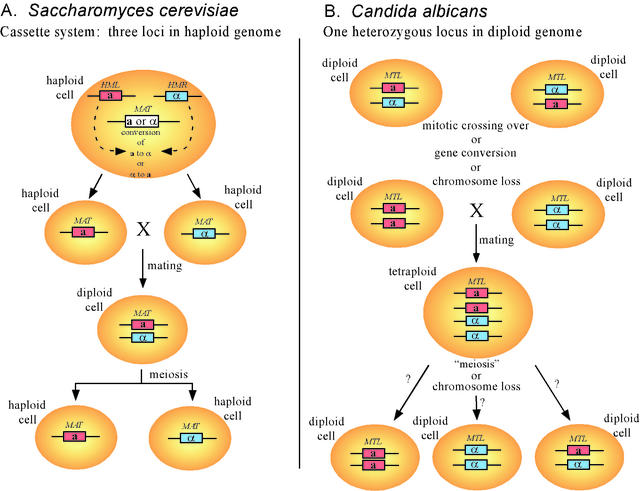
The configuration of mating type loci and the mechanisms for generating cells of the opposite mating type are different for S. cerevisiae and C. albicans. Note that while S. cerevisiae contains a cassette system that includes two silent loci and one expressed locus, C. albicans is normally heterozygous for mating type at one locus. While S. cerevisiae changes mating type at the expression locus with no loss of the alternate mating type information, C. albicans loses the information of one of the two mating types when it expresses a mating type. HML, homothallic mating locus left; HMR, homothetic mating locus right; MAT, mating type locus; MTL, mating type-like locus.
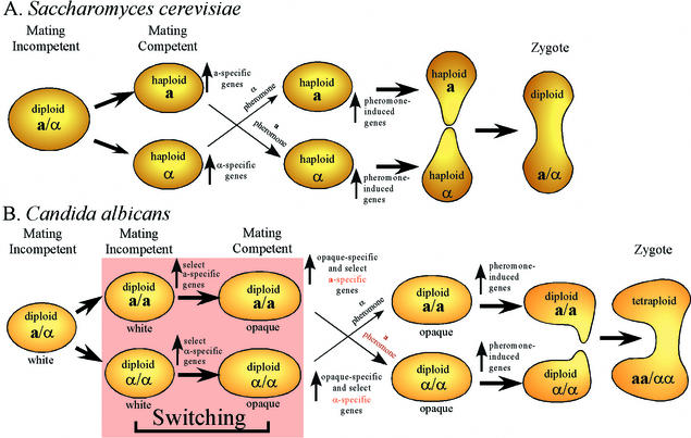
C. albicans has inserted an extra developmental step, the switch from white to opaque, into the mating process. In S. cerevisiae, a and α cells are immediately mating competent, and all a-specific and α-specific genes are upregulated. In C. albicans, a homozygous a or α cell is not mating competent unless it switches to the opaque phenotype. In C. albicans, upregulation of a-specific and α-specific gene expression is divided between the transition to a homozygous state and the transition from white to opaque.
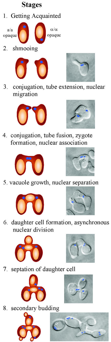
Cell biology of mating in C. albicans. Models are drawn of the stages in the left-hand vertical column, and combined-phase images of the cell bodies and zygotes and fluorescent images of the nuclei are presented in the right-hand vertical column.
References
-
- Calderone, R. A. 2002. Candida and candidiasis. ASM Press, Washington, D.C.
Publication types
MeSH terms
Grants and funding
LinkOut - more resources
Full Text Sources
