. 2003 Jul;206(Pt 14):2441-57.
doi: 10.1242/jeb.00433.

Food transport in the C. elegans pharynx

Affiliations

Food transport in the C. elegans pharynx

Leon Avery et al. J Exp Biol. 2003 Jul.

Abstract

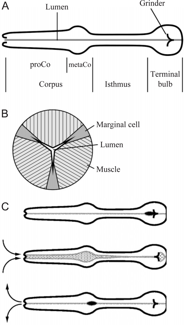
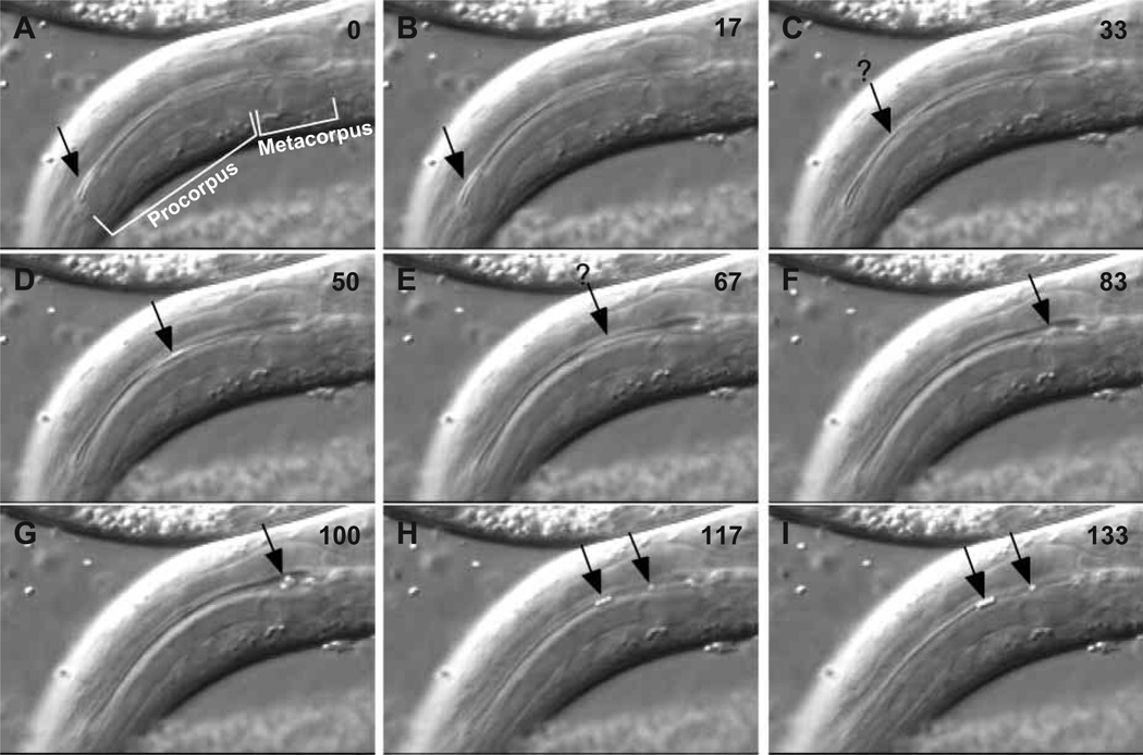
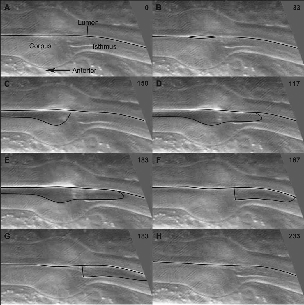
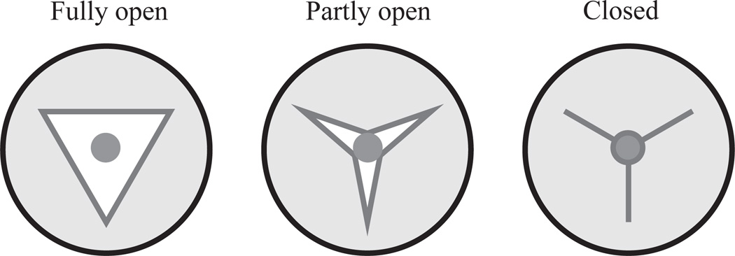
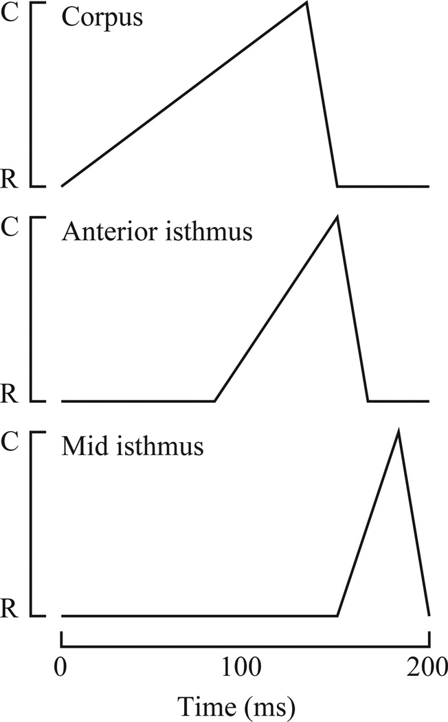
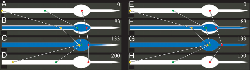
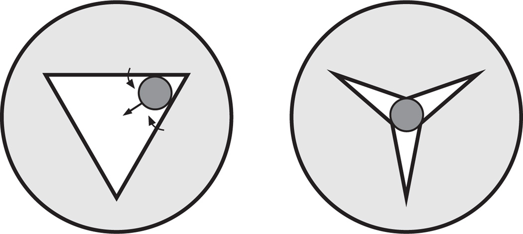
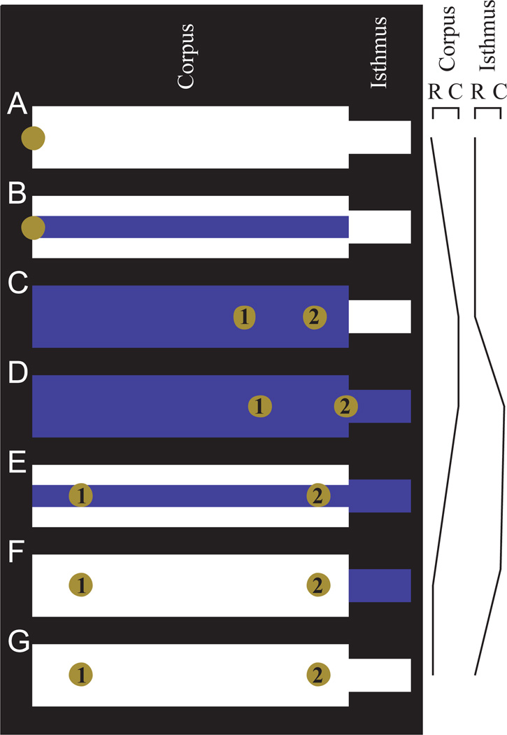
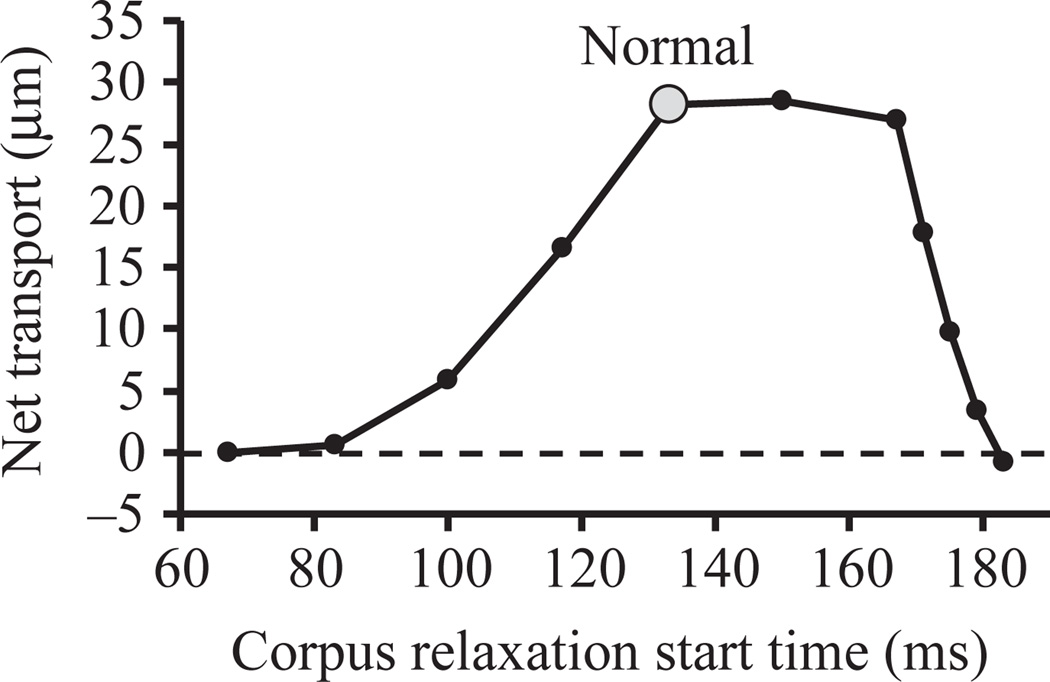
Pumping of the C. elegans pharynx transports food particles (bacteria) posteriorly. We examined muscle motions to determine how this posterior transport is effected. We find that the motions of the middle section of the pharynx, the anterior isthmus, are delayed relative to the anterior section, the corpus. Simulations in which particles are assumed to move at mean fluid velocity when not captured by the walls of the pharyngeal lumen show that delayed isthmus motions do indeed cause net particle transport; however, the amount is much less than in the real pharynx. We propose that the geometry of the pharyngeal lumen forces particles to the center, where they move faster than mean fluid velocity. When this acceleration is incorporated into the simulation, particles are transported efficiently. The transport mechanism we propose explains past observations that the timing of muscle relaxation is important for effective transport. Our model also makes a prediction, which we confirm, that smaller bacteria are better food sources for C. elegans than large ones.


Figures

Fig. 1
Fig. 1
Pharyngeal anatomy and pharyngeal pumping. (A) The pharynx is a tubular muscle divided into three regions: the corpus, connected to the mouth at the anterior end, the isthmus, and the terminal bulb, connected to the intestine at the posterior end. The corpus is further subdivided into a cylindrical or tapered anterior section, the procorpus (proCo), and a posterior bulb, the metacorpus (metaCo). (B) Simplified schematic cross-section through the pharynx. The lumen is shown almost closed, and is approximately to scale. The hatching in the muscle cells shows the orientation of their acto-myosin filaments. When they contract the muscle cells become thinner in the radial dimension and longer circumferentially, pulling the lumen open. Three intermediate filament-containing marginal cells anchor the apices of the lumen. (C) Pharyngeal pumping is a contraction–relaxation cycle involving the corpus, anterior half of the isthmus and terminal bulb. The cycle begins with the nearsimultaneous contraction of these muscles. Contraction of the radially oriented muscles of the corpus and anterior isthmus opens the lumen. Because the posterior isthmus remains closed during this time, the lumen is filled by liquid sucked in through the mouth, carrying with it suspended food particles. Contraction of the terminal bulb rotates the plates of the grinder, breaking bacteria in the terminal bulb and passing the debris back to the intestine. After contraction there is a near-simultaneous relaxation, returning the grinder to its resting position and closing the lumen of the corpus and anterior isthmus. Liquid is expelled from the corpus and anterior isthmus, but food particles are trapped and transported posteriorly. A second motion of the pharyngeal muscles, posterior isthmus peristalsis (not shown here) carries food from the anterior isthmus back to the terminal bulb.
Fig. 2
Fig. 2
Particle motions. This sequence of consecutive video fields shows the transport of 3 or 4 latex beads from the anterior end of the procorpus to the posterior procorpus or metacorpus. Time in ms is shown in the upper right corner of each image. (A) At time 0 the pharynx is relaxed (lumen closed) and the particles (black arrow), slightly out of focus but detectable by their refractility, are lined up in the anterior procorpus. (B) The contraction has begun (visible as a slight expansion of the procorpus lumen), but the particles have not moved. (C–G) Contraction progresses and the particles are drawn back to the metacorpus. The black arrow indicates the particles. They are not clearly visible in C and E, probably because they are out of focus or moving too rapidly or both; here the black arrow is a guess at their location. Between G and H the corpus muscles partly relaxed and the particles moved forward, three to the posterior procorpus and one to anterior metacorpus. (We do not know whether this fourth particle was at the anterior procorpus at time 0, or out of focus at a more posterior position.) (I) Relaxation is complete. The beads do not move between H and I.
Fig. 3
Fig. 3
Anterior isthmus motions. Non-consecutive series of video fields shows the motions of the metacorpus and anterior isthmus during a pump. Black lines show the lumen; time in ms is in the upper right corner of each image.(A) The pharynx is fully relaxed, the lumen closed. At 33 ms (B) contraction (opening) of the metacorpus is barely visible, and at 100 ms (C) contraction is complete and the corpus lumen fully open. Only at 117 ms (D), after the corpus has fully contracted, does the anteriormost part of the isthmus open. The metacorpus remains fully contracted until 150 ms (E), then goes from fully open to fully closed between two consecutive video fields at 150 and 167 ms (F). In the meantime the isthmus contraction proceeds, gradually opening more posteriorly until at 183 ms (G) the entire anterior half of the isthmus is open. At 233 ms (H) the isthmus is fully relaxed.
Fig. 4
Fig. 4
Particle trapping by the walls of the pharyngeal lumen. The pharyngeal lumen has a triradiate shape, with apices anchored by intermediate-filament-containing marginal cells and muscle cells on each of the three sides. Contraction of the muscles pulls the lumen open. We do not know the shape of the open lumen, but have assumed an equilateral triangle for simplicity (Fully open). (Note that the size of the lumen is exaggerated for clarity.) In this configuration particles are free to move with the fluid. As the muscles relax the lumen closes. At the point where the diameter of the lumen (defined as the diameter of an inscribed circle) equals the diameter of a particle contained within it, we assume that the particle is held by the walls and no longer moves (Partly open). Liquid can still flow through the three radii. As relaxation continues the particle remains immobile and the walls deform around it (Closed).
Fig. 5
Fig. 5
Summary of corpus and isthmus motions. For simulation, muscle motions were assumed to be piecewise linear as diagrammed here (i.e. the extent of contraction varies linearly with time, except at discrete breakpoints where its rate of change with time abruptly changes.) The corpus contracts from 0 to 133 ms and relaxes from 133 to 150 ms. C indicates the fully contracted position and R the fully relaxed position. The entire corpus contracts and relaxes as a unit. The anterior isthmus contracts from 83 to 150 ms and relaxes from 150 to 167 ms; the middle of the isthmus (the most posterior region included in the simulation) contracts from 150 to 183 ms and relaxes from 183 to 200 ms. Between the anterior and middle isthmus the times at which contraction starts, relaxation starts, and relaxation ends vary linearly. These times are based on the video sequence from which Fig. 3 was abstracted, but the precise times of pharyngeal motions are quite variable. Important features consistently seen are the delay of isthmus motions with respect to the corpus, and the progression of motion from the anterior to the middle isthmus.
Fig. 6
Fig. 6
Simulated particle motions in the pharyngeal lumen. Simulated motions of three particles in the corpus and anterior isthmus. Anterior is to the left; the posterior end of each diagram (the pointed end) represents the middle of the isthmus. (The posterior half of the isthmus and the terminal bulb were not included in the simulation and are not pictured here.) The white region is the maximum possible opening of the lumen, and blue represents the opening at any particular point in time. Time in ms is given in the upper right corner of each panel. (A–D) Motions were simulated as shown in Fig. 5. (E–H) The isthmus was assumed to move in synchrony with the corpus, so that muscle motions are precisely reciprocal. (B,F) (83 ms) The beginning of anterior isthmus contraction, when the corpus is partly contracted; (C,G) (133 ms) maximum corpus contraction, and (D,H) the end of a full cycle. (Because the isthmus motions are not delayed in E–H, the cycle is only 150 ms long.) In A–D, each particle has a slightly more posterior position at the end of the cycle than at the beginning, i.e. there is net transport. In E–H, particles end up precisely where they began, showing that net transport requires the delayed isthmus motions shown in A–D.
Fig. 7
Fig. 7
Successive positions of a particle during repetitive pumping, showing its position at the beginning of each of 15 successive cycles. The particle was placed at the mouth (left) at the beginning of the first cycle. The 14th cycle carried it into the isthmus. In the anterior metacorpus some of the pictures of the particle are displaced vertically so that each successive position can be clearly seen, but this is not meant to imply an off-center position.
Fig. 8
Fig. 8
Particles are pushed to the center of the closing pharyngeal lumen. This figure diagrams how we believe the shape and motions of the pharyngeal lumen during relaxation would push particles to the center. See text for further explanation.
Fig. 9
Fig. 9
Centered particles are transported more efficiently. Four simulations were run in which we measured the final position after a single cycle of a single particle placed at the mouth at time zero. The brown particle was assumed to move at mean fluid velocity as in previous simulations. The green and red particles move at 1.4 and 2.0 times mean fluid velocity, respectively, and the blue particle moves at the calculated velocity of the fluid at the center of the lumen, which varies from 2.2 to 3.5 times mean velocity depending on how open the lumen is at a particular moment. See text for further explanation.
Fig. 10
Fig. 10
The four-stage pharynx: a simple model, which captures the essence of our proposed mechanism for particle transport in the corpus. It is so named because the motions occur in four non-overlapping stages: corpus contraction, anterior isthmus contraction, corpus relaxation and anterior isthmus relaxation. (The contractions of the isthmus and corpus overlap, in the sense that both are often contracted at the same time, but the motions do not overlap: when one is moving, the other holds its position.) Additionally, the corpus and isthmus are each uniform in diameter and motion along their entire lengths. In this figure the movement of a particle placed at the mouth at the beginning of a cycle is followed, and the results are shown for the particle moving at mean fluid velocity (particle 1) or at twice mean fluid velocity (particle 2). The particle has a diameter 1/3 that of the maximum corpus diameter, and the volume of the anterior isthmus is 5% that of the corpus. (B,E) Snapshots taken when the corpus is 1/3 open; it is at these points that the particles become free to move (B) or stop moving (E), because they are held by the walls of the corpus lumen.
Fig. 11
Fig. 11
Relaxation timing affects transport. Summary of a series of simulations in which the timing of corpus relaxation was varied from the base values in Fig. 5 while isthmus motions were held constant, and the transport of a particle placed at the mouth at time zero moving at center fluid velocity was measured. (Negative net transport means that a particle placed within the corpus at time zero was transported anteriorly that distance toward the mouth.) The point labeled ‘normal’ corresponds to the movement in Fig. 5 and to the blue particle in Fig. 9. For earlier than normal relaxation time, the corpus contracted linearly from time zero to the time plotted on the x axis. For later than normal relaxation time, the corpus contracted linearly from 0 to 133 ms, remained contracted until the time plotted, then relaxed. (We also did a series of simulations in which the corpus contracted linearly from time zero to the beginning of relaxation with no pause in the fully contracted state. The results are similar except that the steep fall-off in transport begins at 167 ms instead of 171 ms and is more severe.) Relaxation always took 1/60 s. In this series, anterior isthmus contraction always began at 150 ms. This graph can however be used to predict the results of simulations in which isthmus contraction began at other times by scaling the x-axis. For instance, if corpus relaxation began at 120 ms and isthmus relaxation began at 180 ms, net transport would be the same as if corpus relaxation began at 100 ms and isthmus relaxation at 150 ms (5.9 µm from the graph), since (120, 180) can be obtained by multiplying (100, 150) by 1.2. For this statement to be exactly true, all other times, e.g. the time at which mid-isthmus relaxation begins, would also have to be multiplied by 1.2.
Fig. 12
Fig. 12
Effect of bacterial size on edibility. The growth rates (days–1) of wild-type worms and three different feeding-defective mutants on 14 different bacterial strains. The bacteria are listed in order of decreasing eat-2 growth rate. Above are photographs of each of the bacterial strains as they appear when isolated from lawns on nematode growth medium. The scale bar applies to all pictures. In addition, 0.8 µm blue-dyed latex beads were mixed in with the bacteria as an internal size standard. These are identified by black dots. A strong although not perfect inverse correlation between growth rate and bacterial size is obvious. B7, Pseudomonas sp.; H39, Comamonas sp.; W11, Pseudomonas sp.; H10, Unidentified; H-12, Acinetobacter junii; H26, Pantoea sp.; HB101, Escherichia coli; H25, Acinetobacter sp.; H28, Bacillus simplex; S4, Panteoa dispersa; S3, Bacillus licheniformis; DA837, Escherichia coli; S9, Bacillus sp.; S13, Bacillus cereus; L10, Bacillus megaterium.
Fig. 13
Fig. 13
Parametrization of pharyngeal lumen opening, showing how the parameters r and rmax were defined. The white area represents a cross-section of the lumen open to radius r, and the tinted area the maximum possible opening rmax for this cross-section.
Fig. 14
Fig. 14
Calculated flow field in the 30% open pharyngeal lumen. (A) The shape of the pharyngeal lumen at 30% of its maximum opening as a 289×250 pixel image. This image was used directly as input for numerical solution of Poisson’s equation (B).

Similar articles

Cited by

References

    1. Albertson DG, Thomson JN. The pharynx of Caenorhabditis elegans. Phil. Trans. R. Soc. Lond. B. 1976;275:299–325. - PubMed
    1. Avery L. The genetics of feeding in Caenorhabditis elegans. Genetics. 1993a;133:897–917. - PMC - PubMed
    1. Avery L. Motor neuron M3 controls pharyngeal muscle relaxation timing in Caenorhabditis elegans. J. Exp. Biol. 1993b;175:283–297. - PubMed
    1. Avery L, Horvitz HR. A cell that dies during wild-type C. elegans development can function as a neuron in a ced-3 mutant. Cell. 1987;51:1071–1078. - PMC - PubMed
    1. Avery L, Horvitz HR. Pharyngeal pumping continues after laser killing of the pharyngeal nervous system.of C. elegans. Neuron. 1989;3:473–485. - PubMed

Publication types

LinkOut - more resources