. 2003 Jun 16;197(12):1667-76.
doi: 10.1084/jem.20021650. Epub 2003 Jun 9.

A reversible defect in natural killer T cell function characterizes the progression of premalignant to malignant multiple myeloma

Affiliations

A reversible defect in natural killer T cell function characterizes the progression of premalignant to malignant multiple myeloma

Madhav V Dhodapkar et al. J Exp Med. .

Abstract

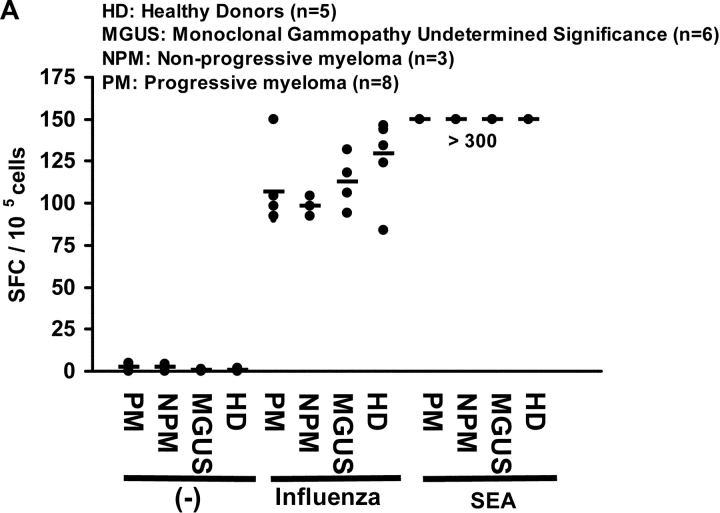
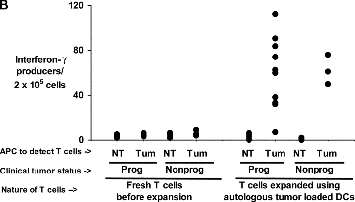
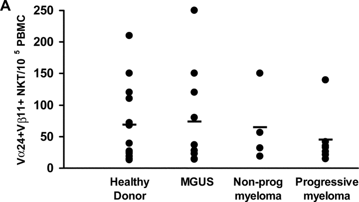
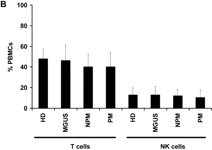
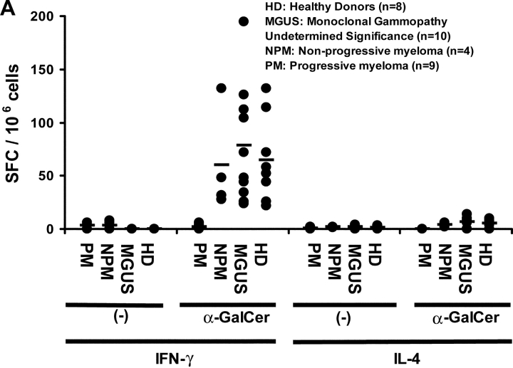
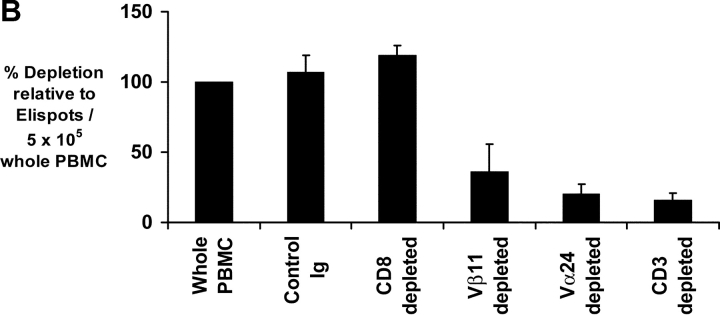
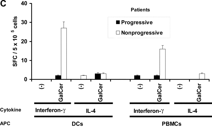
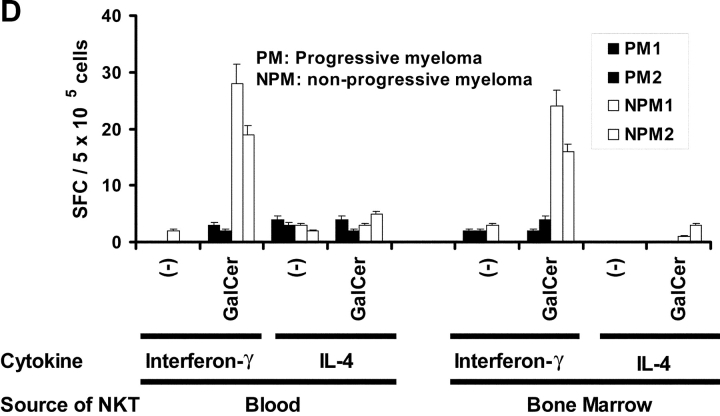
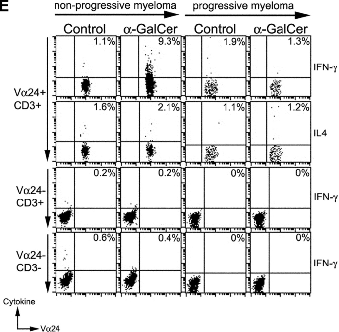
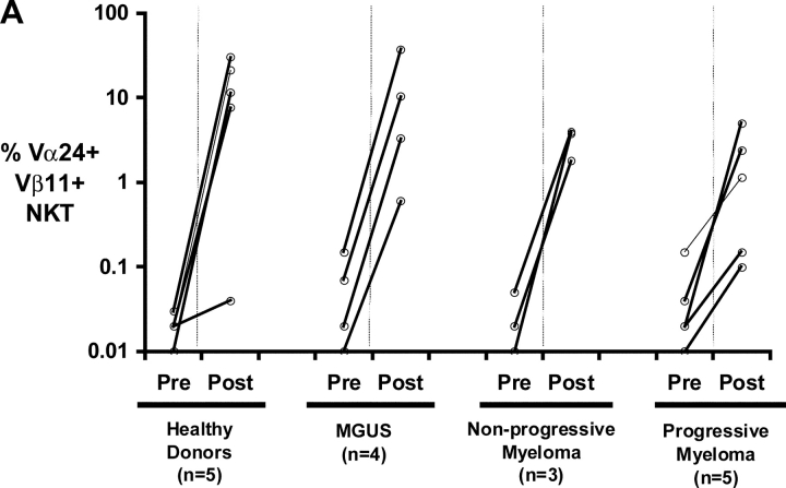
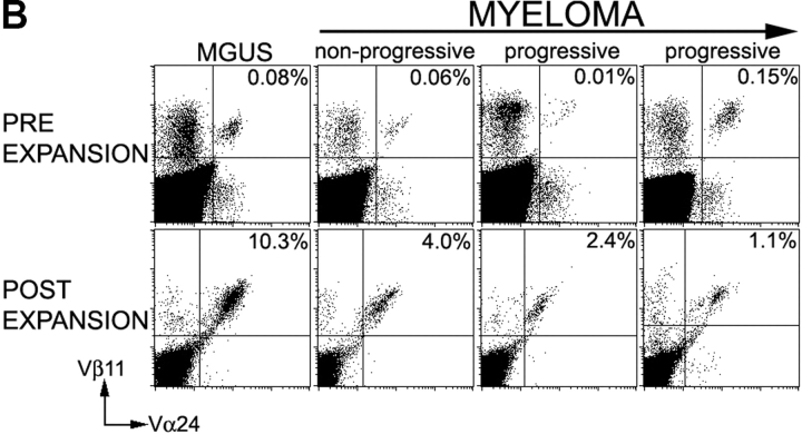
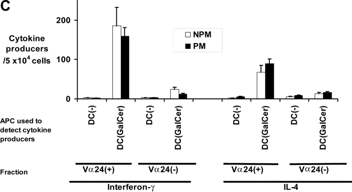
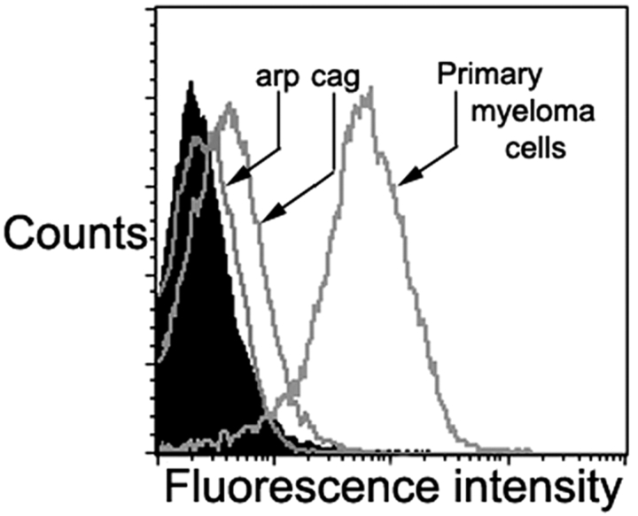
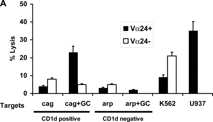
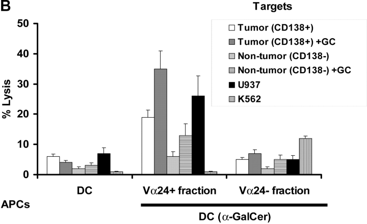
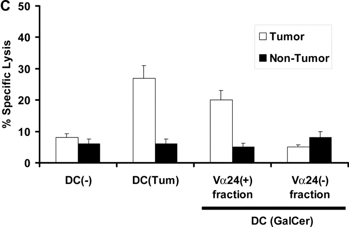
We studied the function of antitumor T and natural killer T (NKT) cells from the blood and tumor bed in 23 patients with premalignant gammopathy, nonprogressive myeloma, or progressive multiple myeloma. We show that antitumor killer T cells can be detected in patients with both progressive or nonprogressive myeloma. V alpha 24+V beta 11+ invariant NKT cells are detectable in the blood and tumor bed of all cohorts. However, freshly isolated NKT cells from both the blood and tumor bed of patients with progressive disease, but not nonprogressive myeloma or premalignant gammopathy, have a marked deficiency of ligand-dependent interferon-gamma production. This functional defect can be overcome in vitro using dendritic cells pulsed with the NKT ligand, alpha-galactosylceramide (alpha-GalCer). Fresh myeloma cells express CD1d, and can be efficiently killed by autologous NKT cells. We hypothesize that presentation of tumor derived glycolipids by myeloma cells leads to NKT dysfunction in vivo. These data demonstrate that clinical progression in patients with monoclonal gammopathies is associated with an acquired but potentially reversible defect in NKT cell function and support the possibility that these innate lymphocytes play a role in controlling the malignant growth of this incurable B cell tumor in patients.


Figures

Figure 1.
Figure 1.
(A) Influenza and SEA reactive T cell response in monoclonal gammopathies. ELISPOT assay quantifying the presence of influenza and SEA reactive T cells in patients with m MGUS (n = 6), NPM (n = 3), PM (n = 8), or healthy donors (HD, n = 5). Each dot represents an individual patient. (B) Antitumor T cell function in progressive or nonprogressive myeloma. ELISPOT assay testing IFN-γ production in response to autologous tumor (CD138+) or nontumor (CD138-ve) cells from bone marrow, by blood T cells tested either fresh immediately after isolation, or after in vitro expansion using autologous tumor loaded DCs. Each dot represents an individual patient.
Figure 1.
Figure 1.
(A) Influenza and SEA reactive T cell response in monoclonal gammopathies. ELISPOT assay quantifying the presence of influenza and SEA reactive T cells in patients with m MGUS (n = 6), NPM (n = 3), PM (n = 8), or healthy donors (HD, n = 5). Each dot represents an individual patient. (B) Antitumor T cell function in progressive or nonprogressive myeloma. ELISPOT assay testing IFN-γ production in response to autologous tumor (CD138+) or nontumor (CD138-ve) cells from bone marrow, by blood T cells tested either fresh immediately after isolation, or after in vitro expansion using autologous tumor loaded DCs. Each dot represents an individual patient.
Figure 2.
Figure 2.
(A and B) Quantitation of NKT (Vα24+ Vβ11+) (A), NK (CD3CD56+), and T cells (B) by flow cytometry in PBMCs of healthy donors (HD; n = 13), or patients with MGUS (n = 10), NPM (n = 4), or PM (n = 7).
Figure 2.
Figure 2.
(A and B) Quantitation of NKT (Vα24+ Vβ11+) (A), NK (CD3CD56+), and T cells (B) by flow cytometry in PBMCs of healthy donors (HD; n = 13), or patients with MGUS (n = 10), NPM (n = 4), or PM (n = 7).
Figure 3.
Figure 3.
Function of fresh uncultured NKT cells in monoclonal gammopathies. (A) Loss of α-GalCer–reactive IFN-γ production in fresh NKT cells in progressive myeloma. ELISPOT assay quantifying the presence of α-GalCer reactive IFN-γ/IL-4 producers in fresh PBMCs from healthy donors (HD; n = 13), or patients with MGUS (n = 10), NPM (n = 4), or PM (n = 9). (B) Effect of depletion of various cell populations on α-GalCer–reactive IFN-γ production in fresh PBMCs. PBMCs were depleted before the α-GalCer stimulated ELISPOT assay using anti-Vα24+, Vβ11+, CD3+, CD8+, or isotype control antibody, followed by magnetic bead depletion. Data shown are percentage of reactivity in whole PBMCs. (C) Fresh NKT cells from progressive myeloma do not react to ligand bearing DCs. ELISPOT assay quantifying the presence of α-GalCer–reactive IFN-γ/IL-4 producers in fresh PBMCs from a representative patient with progressive or nonprogressive myeloma, using PBMCs alone or autologous mature DCs (added to PBMCs at PBMC:DC ratio of 20:1) as APCs during the 16 h ELISPOT assay. Data represent two patients from each cohort. (D) Loss of effector NKT function in the tumor bed of patients with PM, but not those with NPM myeloma. ELISPOT assays quantifying the presence of α-GalCer–reactive IFN-γ/IL-4 producers in fresh mononuclear from blood or bone marrow (tumor bed) of two patients with progressive or nonprogressive myeloma. (E) Detection of ligand reactive cytokine production by uncultured NKT cells of patients with progressive or nonprogressive myeloma using flow cytometry. PBMCs were stimulated with α-GalCer + anti-CD28, or anti-CD28 alone (as a control), in the presence of monensin. FACS® plots show cytokine production by NKT cells from patients with progressive or nonprogressive myeloma. Data shown are representative of similar experiments on five additional patients.
Figure 3.
Figure 3.
Function of fresh uncultured NKT cells in monoclonal gammopathies. (A) Loss of α-GalCer–reactive IFN-γ production in fresh NKT cells in progressive myeloma. ELISPOT assay quantifying the presence of α-GalCer reactive IFN-γ/IL-4 producers in fresh PBMCs from healthy donors (HD; n = 13), or patients with MGUS (n = 10), NPM (n = 4), or PM (n = 9). (B) Effect of depletion of various cell populations on α-GalCer–reactive IFN-γ production in fresh PBMCs. PBMCs were depleted before the α-GalCer stimulated ELISPOT assay using anti-Vα24+, Vβ11+, CD3+, CD8+, or isotype control antibody, followed by magnetic bead depletion. Data shown are percentage of reactivity in whole PBMCs. (C) Fresh NKT cells from progressive myeloma do not react to ligand bearing DCs. ELISPOT assay quantifying the presence of α-GalCer–reactive IFN-γ/IL-4 producers in fresh PBMCs from a representative patient with progressive or nonprogressive myeloma, using PBMCs alone or autologous mature DCs (added to PBMCs at PBMC:DC ratio of 20:1) as APCs during the 16 h ELISPOT assay. Data represent two patients from each cohort. (D) Loss of effector NKT function in the tumor bed of patients with PM, but not those with NPM myeloma. ELISPOT assays quantifying the presence of α-GalCer–reactive IFN-γ/IL-4 producers in fresh mononuclear from blood or bone marrow (tumor bed) of two patients with progressive or nonprogressive myeloma. (E) Detection of ligand reactive cytokine production by uncultured NKT cells of patients with progressive or nonprogressive myeloma using flow cytometry. PBMCs were stimulated with α-GalCer + anti-CD28, or anti-CD28 alone (as a control), in the presence of monensin. FACS® plots show cytokine production by NKT cells from patients with progressive or nonprogressive myeloma. Data shown are representative of similar experiments on five additional patients.
Figure 4.
Figure 4.
Functional NKT cells can be expanded from patients with monoclonal gammopathies using α-GalCer pulsed mature DCs. (A) Expansion of Vα24+Vβ11+ NKT cells in culture using autologous α-GalCer pulsed mature DCs from healthy donors (HD; n = 5), or patients with MGUS (n = 4), NPM (n = 3), or PM (n = 5). Data shown are % NKT cells in PBMCs determined by flow cytometry before and after 2–3 wk of expansion. (B) Representative FACS plots showing detection of NKT cells (Vα24+ Vβ11+) in fresh PBMCs (pre) of patients with MGUS, nonprogressive or progressive myeloma, or after PBMC expansion (post) using α-GalCer pulsed mature DCs. Percentages refer to percent NKT cells in total recovered PBMCs. (C) Cytokine production by expanded NKT cells in response to ligand-bearing APCs. T cells expanded using autologous α-GalCer pulsed autologous mature DCs as in panel A, were separated into NKT (Vα24+) and non-NKT (Vα24-ve) fractions using magnetic beads. Individual fractions were then tested for reactivity to autologous α-GalCer pulsed mature DCs (DC: responder ratio of 1:10), using a 16 h Elispot assay for IFN-γ or IL-4 producing cells. Data shown are representative of two patients each with PM or NPM.
Figure 4.
Figure 4.
Functional NKT cells can be expanded from patients with monoclonal gammopathies using α-GalCer pulsed mature DCs. (A) Expansion of Vα24+Vβ11+ NKT cells in culture using autologous α-GalCer pulsed mature DCs from healthy donors (HD; n = 5), or patients with MGUS (n = 4), NPM (n = 3), or PM (n = 5). Data shown are % NKT cells in PBMCs determined by flow cytometry before and after 2–3 wk of expansion. (B) Representative FACS plots showing detection of NKT cells (Vα24+ Vβ11+) in fresh PBMCs (pre) of patients with MGUS, nonprogressive or progressive myeloma, or after PBMC expansion (post) using α-GalCer pulsed mature DCs. Percentages refer to percent NKT cells in total recovered PBMCs. (C) Cytokine production by expanded NKT cells in response to ligand-bearing APCs. T cells expanded using autologous α-GalCer pulsed autologous mature DCs as in panel A, were separated into NKT (Vα24+) and non-NKT (Vα24-ve) fractions using magnetic beads. Individual fractions were then tested for reactivity to autologous α-GalCer pulsed mature DCs (DC: responder ratio of 1:10), using a 16 h Elispot assay for IFN-γ or IL-4 producing cells. Data shown are representative of two patients each with PM or NPM.
Figure 5.
Figure 5.
Expression of CD1d on myeloma cells. CD1d expression on myeloma cell lines (arp and cag) as well as purified myeloma cells, as assessed by flow cytometry. Filled histogram reflects isotype control. Data on primary myeloma cells are representative of five different patients.
Figure 6.
Figure 6.
Cytolytic function of NKT cells from patients with gammopathies. (A) Lysis of CD1d positive (cag) or negative (arp) myeloma cell lines (the lines were tested with or without in vitro pulsing with α-GalCer [+GC]), by Vα24 positive or negative fractions of cultures after in vitro expansion with α-GalCer pulsed DCs. Additional positive control targets were U937 histiocytic lymphoma (NKT sensitive) and K562 erythroleukemia cells (NK sensitive). (B) Killing of autologous primary tumor cells (CD138 positive) versus nontumor cells (CD138 negative) in the tumor bed, by Vα24 positive or negative fraction of cells expanded using α-GalCer (GC) pulsed DCs (versus unpulsed DCs) from a patient with nonprogressive myeloma. The tumor cells were tested without or with supplementation with α-GalCer (GC) during the lytic assay. Additional control targets were U937 histiocytic lymphoma and K562 erythroleukemia cells. Data are representative of similar experiments on two patients. (C) Killing of autologous primary tumor cells (CD138+ve) versus nontumor cells (CD138-ve) from the tumor bed of a patient with progressive myeloma. Killing was measured with bulk T cells expanded using DC only (DC(−)) and DCs loaded with autologous tumor (DC-tum), and for the latter, Vα24 positive or negative fractions from cultures expanded using α-GalCer pulsed DCs (DC(Gal-Cer)).
Figure 6.
Figure 6.
Cytolytic function of NKT cells from patients with gammopathies. (A) Lysis of CD1d positive (cag) or negative (arp) myeloma cell lines (the lines were tested with or without in vitro pulsing with α-GalCer [+GC]), by Vα24 positive or negative fractions of cultures after in vitro expansion with α-GalCer pulsed DCs. Additional positive control targets were U937 histiocytic lymphoma (NKT sensitive) and K562 erythroleukemia cells (NK sensitive). (B) Killing of autologous primary tumor cells (CD138 positive) versus nontumor cells (CD138 negative) in the tumor bed, by Vα24 positive or negative fraction of cells expanded using α-GalCer (GC) pulsed DCs (versus unpulsed DCs) from a patient with nonprogressive myeloma. The tumor cells were tested without or with supplementation with α-GalCer (GC) during the lytic assay. Additional control targets were U937 histiocytic lymphoma and K562 erythroleukemia cells. Data are representative of similar experiments on two patients. (C) Killing of autologous primary tumor cells (CD138+ve) versus nontumor cells (CD138-ve) from the tumor bed of a patient with progressive myeloma. Killing was measured with bulk T cells expanded using DC only (DC(−)) and DCs loaded with autologous tumor (DC-tum), and for the latter, Vα24 positive or negative fractions from cultures expanded using α-GalCer pulsed DCs (DC(Gal-Cer)).

Similar articles

Cited by

References

    1. Bernards, R., and R.A. Weinberg. 2002. A progression puzzle. Nature. 418:823. - PubMed
    1. Rawstron, A.C., M.J. Green, A. Kuzmicki, B. Kennedy, J.A. Fenton, P.A. Evans, S.J. O'Connor, S.J. Richards, G.J. Morgan, A.S. Jack, and P. Hillmen. 2002. Monoclonal B lymphocytes with the characteristics of “indolent” chronic lymphocytic leukemia are present in 3.5% of adults with normal blood counts. Blood. 100:635–639. - PubMed
    1. Kyle, R.A., and S.V. Rajkumar. 1999. Monoclonal gammopathies of undetermined significance. Hematol. Oncol. Clin. North Am. 13:1181–1202. - PubMed
    1. Tricot, G. 2000. Multiple myeloma and related plasma cell disorders. Hematology: Principles and Practice. R. Hoffman, editor. Churchill Livingstone, New York. 1398–1415.
    1. Fonseca, R., R.J. Bailey, G.J. Ahmann, S.V. Rajkumar, J.D. Hoyer, J.A. Lust, R.A. Kyle, M.A. Gertz, P.R. Greipp, and G.W. Dewald. 2002. Genomic abnormalities in monoclonal gammopathy of undetermined significance. Blood. 100:1417–1424. - PubMed

Publication types

MeSH terms