. 2003 Jun 9;161(5):861-74.
doi: 10.1083/jcb.200301058.

Requirement of transcription factor NFAT in developing atrial myocardium

Affiliations

Requirement of transcription factor NFAT in developing atrial myocardium

William Schubert et al. J Cell Biol. .

Abstract

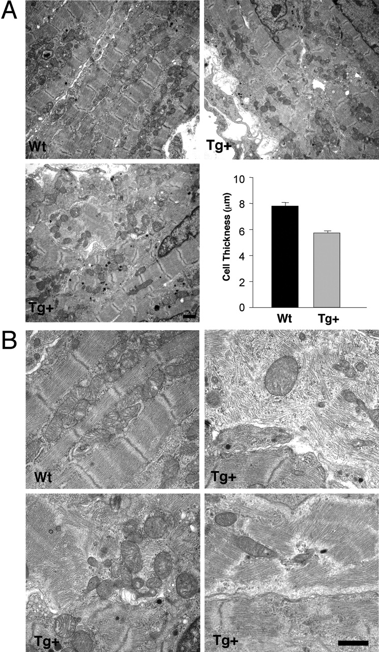
Nuclear factor of activated T cell (NFAT) is a ubiquitous regulator involved in multiple biological processes. Here, we demonstrate that NFAT is temporally required in the developing atrial myocardium between embryonic day 14 and P0 (birth). Inhibition of NFAT activity by conditional expression of dominant-negative NFAT causes thinning of the atrial myocardium. The thin myocardium exhibits severe sarcomere disorganization and reduced expression of cardiac troponin-I (cTnI) and cardiac troponin-T (cTnT). Promoter analysis indicates that NFAT binds to and regulates transcription of the cTnI and the cTnT genes. Thus, regulation of cytoskeletal protein gene expression by NFAT may be important for the structural architecture of the developing atrial myocardium.


Figures

Figure 1.
Figure 1.
Subcellular distribution of NFAT in developing atria. Subcellular distribution of NFAT in developing mouse atria was examined by immunofluorescence analysis (A). At E14, NFAT was not detected in the nucleus (arrowheads) as indicated by the separated staining of NFAT (green) and the nuclei (red; see inset for higher magnification). At E16, nuclear NFAT was observed, as indicated by the overlapped staining of NFAT and the nuclei (yellow). At E18, a weaker staining of nuclear NFAT was observed, suggesting that some nuclear export has occurred. At birth (P0), little nuclear NFAT was detected as indicated by the separated staining of NFAT (green) and the nuclei (red). The relative intensity of nuclear NFAT in the developing atria is presented (B). Bar, 30 μm.
Figure 2.
Figure 2.
Expression of dnNFAT blocks NFAT nuclear localization. (A) Schematic representation of constructs for the transgene creation. Expression of tetracycline regulator (rtTA) is driven by the heart-specific α-MHC promoter. Expression of FLAG epitope-tagged dnNFAT is regulated by the Tet-responsive promoter. Co-injection of both constructs generates heart-specific, tet-inducible expression of dnNFAT. Expression of dnNFAT in Wt and Tg+ hearts, in the presence (+) or absence (−) of Dox, was examined by immunoblotting analysis using the M2 mAb that recognizes the FLAG epitope. (B and C) Subcellular distribution of NFAT in Wt and Tg+ E16 heart was examined by immunofluorescence analysis. Expression of dnNFAT (blue) in Wt and Tg+ atria (A), in the presence (+) or absence (−) of Dox, was examined using the M2 mAb. Co-localization (yellow) of NFAT in the nuclei (arrowheads) was observed in Wt atria (B). Expression of dnNFAT blocks nuclear translocation, and thus separated staining of NFAT (green) and the nuclei (red) was observed in Dox-treated Tg+ atria. Localization of NFAT and the expression of dnNFAT in the ventricles (V) were also shown. The relative intensity of nuclear NFAT in Wt and Tg+ atria are presented (C). Bars, 30 μm.
Figure 3.
Figure 3.
Inhibition of NFAT causes thinning of developing atrial walls. (A and B) Histological analysis of E16 atria. Timed-pregnant mice were given Dox-treated water (+) or untreated, plain water (−) from the day of conception (E0.5). Wt littermate control and Tg+ embryos harvested from the same litter on E16 were fixed in 10% formalin and serial sectioned sagittally. Representative sections, collected from six embryos of multiple litters, were shown (A). Dox-treated Tg+ atria exhibited thinner atrial walls as compared with their Wt littermates. Enlarged images to illustrate thinning of atrial walls were shown (B). RA, right atrium; LA, left atrium. Bars: low magnification, 200 μm; high magnification, 60 μm. (C) Administration of cyclosporin A (CsA) disrupts myocardium development. Wt pregnant mice were given CsA daily starting on E14. Control and CsA treated embryos were harvested on day of birth (P0). Sagittal sections of developing atria and ventricles were shown. (D and E) Expression of calcineurin (Cn) attenuates thinning of the atrial walls in dnNFAT mice. Heterozygous Tg+ male mice expressing dnNFAT were crossed with Tg+ female mice expressing calcineurin. Dox-treated embryos were harvested on E16 and day of birth (P0). Immunofluorescence analysis indicated localization of NFAT in the nuclei (yellow) in calcineurin- expressing hearts (D). Histological analysis revealed that expression of calcineurin reduced thinning of myocardium in dnNFAT-expressing hearts (E). Bars: E16 hearts, 40 μm; P0 hearts, 75 μm.
Figure 3.
Figure 3.
Inhibition of NFAT causes thinning of developing atrial walls. (A and B) Histological analysis of E16 atria. Timed-pregnant mice were given Dox-treated water (+) or untreated, plain water (−) from the day of conception (E0.5). Wt littermate control and Tg+ embryos harvested from the same litter on E16 were fixed in 10% formalin and serial sectioned sagittally. Representative sections, collected from six embryos of multiple litters, were shown (A). Dox-treated Tg+ atria exhibited thinner atrial walls as compared with their Wt littermates. Enlarged images to illustrate thinning of atrial walls were shown (B). RA, right atrium; LA, left atrium. Bars: low magnification, 200 μm; high magnification, 60 μm. (C) Administration of cyclosporin A (CsA) disrupts myocardium development. Wt pregnant mice were given CsA daily starting on E14. Control and CsA treated embryos were harvested on day of birth (P0). Sagittal sections of developing atria and ventricles were shown. (D and E) Expression of calcineurin (Cn) attenuates thinning of the atrial walls in dnNFAT mice. Heterozygous Tg+ male mice expressing dnNFAT were crossed with Tg+ female mice expressing calcineurin. Dox-treated embryos were harvested on E16 and day of birth (P0). Immunofluorescence analysis indicated localization of NFAT in the nuclei (yellow) in calcineurin- expressing hearts (D). Histological analysis revealed that expression of calcineurin reduced thinning of myocardium in dnNFAT-expressing hearts (E). Bars: E16 hearts, 40 μm; P0 hearts, 75 μm.
Figure 4.
Figure 4.
Temporal requirement of NFAT in atrial development. (A) Timed-pregnant mice were given Dox-treated water (filled bars) from the day of conception (E0). Wt littermate control and Tg+ embryos were harvested (arrows) on E14, E16, or day of birth (P0). Harvested embryos were fixed in 10% formalin and serial sectioned sagittally. Representative sections, collected from six embryos of multiple litters, were shown. Thinning of atrial walls was only found in Dox-treated Tg+ hearts, harvested on E16, and P0 embryos. Tg+ embryos harvested from pregnant mice that drank untreated, plain water (shaded bars) exhibited similar atrial wall thickness as compared with the Wt control. The interval of nuclear NFAT was also indicated (red bar). Bar, 50 μm. (B and C) Morphometric analysis of the developing atria. Cross-sectional thickness of atrial (B) and ventricular (C) walls, from six embryos, was measured. Reduction in atrial wall thickness was found in Dox-treated Tg+ embryos on E16 or later. However, Wt and Tg+ ventricles exhibited similar thickness. Embryos that were not exposed to Dox also exhibited similar wall thickness. RA, right atria; LA, left atria; RV, right ventricles; LV, left ventricles.
Figure 4.
Figure 4.
Temporal requirement of NFAT in atrial development. (A) Timed-pregnant mice were given Dox-treated water (filled bars) from the day of conception (E0). Wt littermate control and Tg+ embryos were harvested (arrows) on E14, E16, or day of birth (P0). Harvested embryos were fixed in 10% formalin and serial sectioned sagittally. Representative sections, collected from six embryos of multiple litters, were shown. Thinning of atrial walls was only found in Dox-treated Tg+ hearts, harvested on E16, and P0 embryos. Tg+ embryos harvested from pregnant mice that drank untreated, plain water (shaded bars) exhibited similar atrial wall thickness as compared with the Wt control. The interval of nuclear NFAT was also indicated (red bar). Bar, 50 μm. (B and C) Morphometric analysis of the developing atria. Cross-sectional thickness of atrial (B) and ventricular (C) walls, from six embryos, was measured. Reduction in atrial wall thickness was found in Dox-treated Tg+ embryos on E16 or later. However, Wt and Tg+ ventricles exhibited similar thickness. Embryos that were not exposed to Dox also exhibited similar wall thickness. RA, right atria; LA, left atria; RV, right ventricles; LV, left ventricles.
Figure 5.
Figure 5.
Requirement of NFAT at later stages of atrial development. Pregnant mice were given and taken off Dox-treated water (filled bars) at various times during gestation. The duration that pregnant mice drank untreated, plain water (shaded bars) was also indicated. Wt littermate control and Tg+ embryos were harvested on date indicated (arrows). Harvested embryos were fixed in 10% formalin and serial sectioned sagittally. Representative sections, collected from six embryos of multiple litters, were shown. Thinning of atrial walls was found in E16 hearts that were exposed to Dox, from E14 to E16, for even 2 d (A). NFAT activity is required continuously from E16 to P0, and thinning of the atrial walls is irreversible (B). The interval of nuclear NFAT was also indicated (red bar). Bars, 50 μm.
Figure 6.
Figure 6.
Cell viability of the developing mouse atria. (A) TUNEL assays were performed to examine cell viability in Dox-treated developing mouse atria. As compared with control, which show an intense staining of fragmented DNA in apoptotic cells, TUNEL-positive cells were not detected at E14 and E16 Wt littermate and Tg+ atria, Bar, 50 μm. (B) The number of myocyte nuclei is similar. The number of intact nuclei in the Wt and the Tg+ atria was counted every 0.3 mm along the atrial wall and illustrated. RA, right atria; LA, left atria.
Figure 7.
Figure 7.
Cytoarchitecture of the developing mouse atria. Ultrastructure of P0 atria was examined by transmission electron microscopy. Electron micrographs revealed characteristic banding pattern of sarcomere in Dox-treated Wt littermate control. Mitochondria layered between myofilaments were also observed. However, Dox-treated Tg+ atria displayed severely perturbed sarcomere and mitochondria. Varying degrees of sarcomeric disorganization were also observed. Cross-sectional cell thickness of Wt and Tg+ atria was shown (A). Low (A) and high (B) magnifications of Wt and Tg+ atria are presented. Bar, 1 μm.
Figure 8.
Figure 8.
Expression of cytoskeletal and sarcomeric proteins in Tg+ and Wt hearts. Dox-treated embryos were harvested on P0, micro-dissected to separate atria and ventricles, and genotyped. 20 Wt or Tg+ atria (or 10 ventricles) were collected from multiple litters, homogenized, and extracts obtained were separated on SDS-PAGE. Immunoblots were performed with vinculin, desmin, α-actinin, actin, myosin, and caveolin-3 antibodies (A). Expression of tropomyosin, troponin-C, ssTnI, ssTnT, cTnT, and cTnT were also examined (B).
Figure 9.
Figure 9.
NFAT-binding elements are presented in the cTnT and cTnI genes. (A) Sequence comparison of the NFAT-binding sites found in IL-2, PPARγ2 (proximal and distal), cTnT (sites 1 and 2), and cTnI gene promoters. NFAT-binding elements from the mouse and rat cTnI are also shown. Canonical NFAT-binding site is illustrated (filled box). Adjacent NFAT partner binding site is also indicated (AP-1, shaded box; C/EBP, hatched box). Residues on the cTnT and cTnI NFAT sites that resemble the AP-1 (*) or C/EBP (^) binding sequence are also indicated. Similar nucleotides found in human, rat, and mouse cTnI are underlined. (B and C) NFAT interacts with the cTnT- and cTnI-binding elements. Gel mobility shift assays were performed, in the presence (+) or absence (−) of NFAT antibody, to demonstrate specific interaction of NFAT to the cTnT- and cTnI-binding elements (B). Antibody- supershifted complexes were also indicated. NFAT–DNA complexes were also competed by excess Wt, but not mutated, cTnT or cTnI oligonucleotides (C). (D) Formation of distinct NFAT complexes on the cTnT- and cTnI-binding elements. NFAT–DNA complexes were competed by using NFAT binding elements from the IL-2, the PPARγ2, and the cTnT genes as indicated. Differential competition by these canonical NFAT binding elements suggests formation of various NFAT–DNA complexes. (E) Comparison of NFAT complexes from the human, rat, and mouse cTnI NFAT elements. NFAT–DNA complexes were cross competed among different cTnI elements as indicated. Other canonical NFAT elements were also used to compete formation of NFAT complexes from the mouse cTnI NFAT element.
Figure 10.
Figure 10.
NFAT regulates the cTnI and the cTnT gene promoters. cTnI promoter (cTnI, −1 to −303 and −1 to −283) was subcloned upstream of a luciferase reporter gene (A). Mutation at the cTnI NFAT element was also shown. Constitutive active calcineurin (ΔCn, shaded bars) or constitutive nuclear NFATc4 (cnNFATc4, filled bars) was cotransfected with the cTnI luciferase reporter plasmid (B and C). Cells were harvested 36 h after transfection. Luciferase and β-galactosidase activities were measured. Luciferase reporter plasmid containing a triple repeat of the cTnT site 1 NFAT element was also examined similarly (D). Filled ovals represent NFAT binding sites.

Similar articles

Cited by

References

    1. Agarwal, S., O. Avni, and A. Rao. 2000. Cell-type-restricted binding of the transcription factor NFAT to a distal IL-4 enhancer in vivo. Immunity. 12:643–652. - PubMed
    1. Akagi, K., M. Kanai, H. Saya, T. Kozu, and A. Berns. 2001. A novel tetracycline-dependent transactivator with E2F4 transcriptional activation domain. Nucleic Acids Res. 29:E23. - PMC - PubMed
    1. Aramburu, J., F. Garcia-Cozar, A. Raghavan, H. Okamura, A. Rao, and P.G. Hogan. 1998. Selective inhibition of NFAT activation by a peptide spanning the calcineurin targeting site of NFAT. Mol. Cell. 1:627–637. - PubMed
    1. Ausoni, S., M. Campione, A. Picard, P. Moretti, M. Vitadello, C. De Nardi, and S. Schiaffino. 1994. Structure and regulation of the mouse cardiac troponin I gene. J. Biol. Chem. 269:339–346. - PubMed
    1. Barton, P.J., M.E. Cullen, P.J. Townsend, N.J. Brand, M.J. Mullen, D.A. Norman, P.K. Bhavsar, and M.H. Yacoub. 1999. Close physical linkage of human troponin genes: organization, sequence and expression of the locus encoding cardiac troponin I and slow skeletal troponin T. Genomics. 57:102–109. - PubMed

Publication types

MeSH terms