Comparative Study
. 2003 Jul;6(7):736-42.
doi: 10.1038/nn1073.

Brain-derived neurotrophic factor regulates energy balance downstream of melanocortin-4 receptor

Affiliations
Comparative Study

Brain-derived neurotrophic factor regulates energy balance downstream of melanocortin-4 receptor

Baoji Xu et al. Nat Neurosci. 2003 Jul.

Abstract

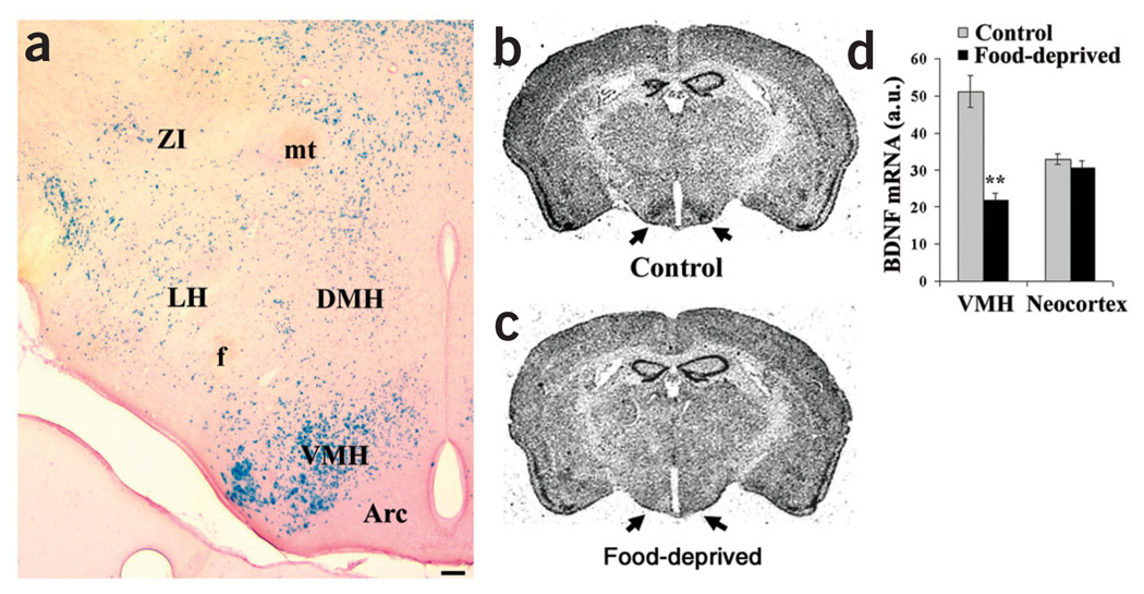
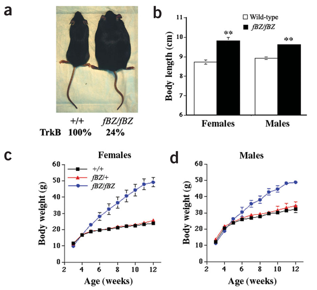
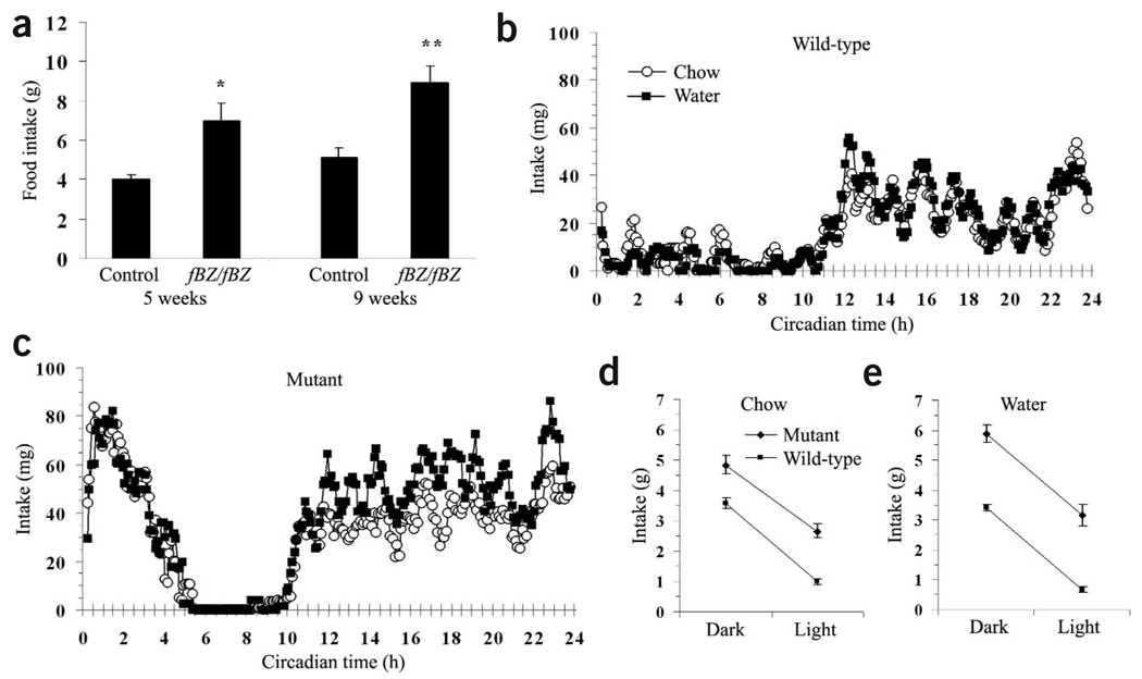
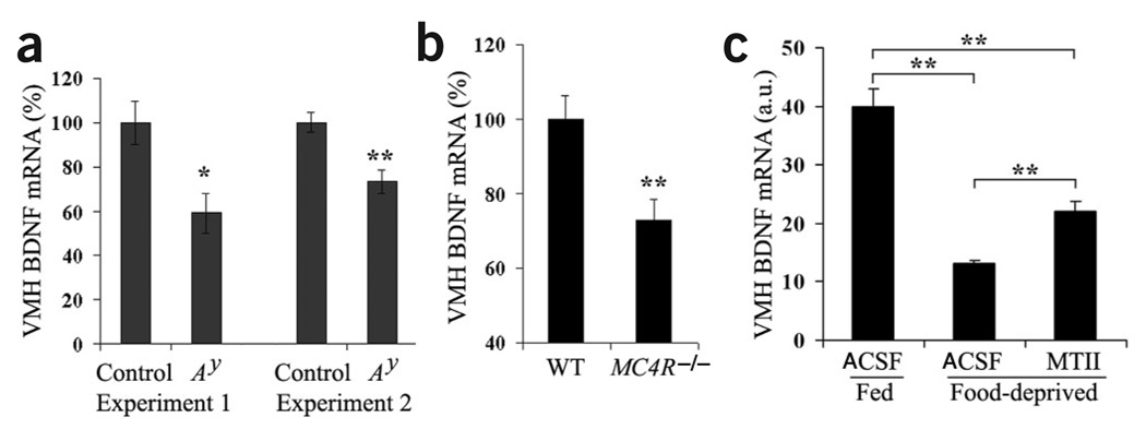
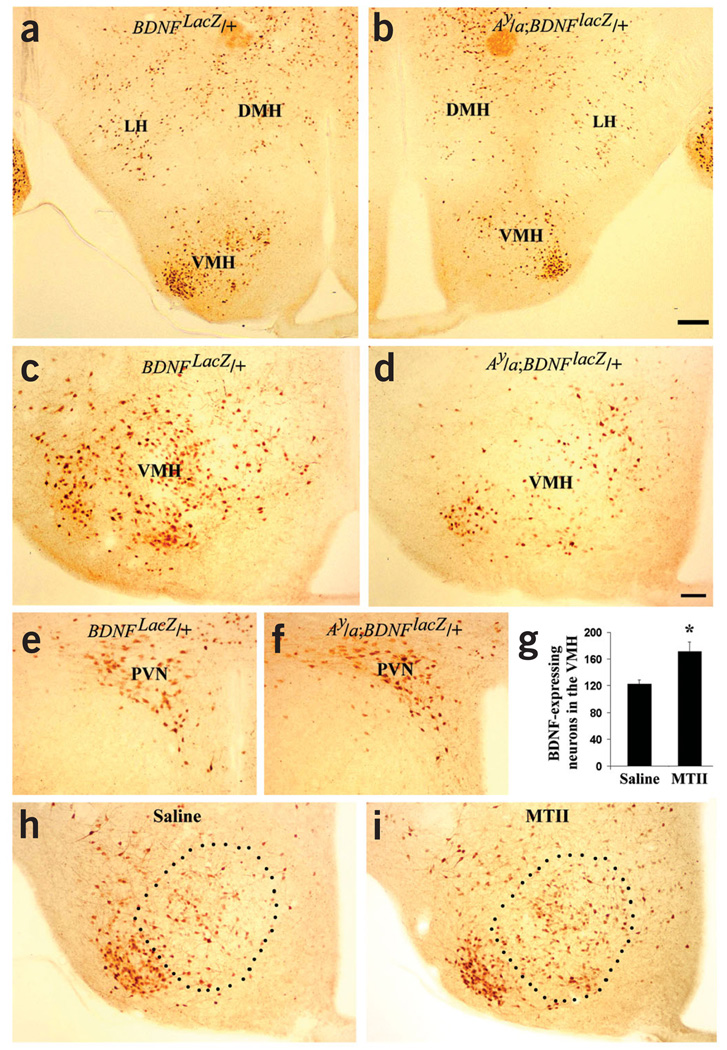
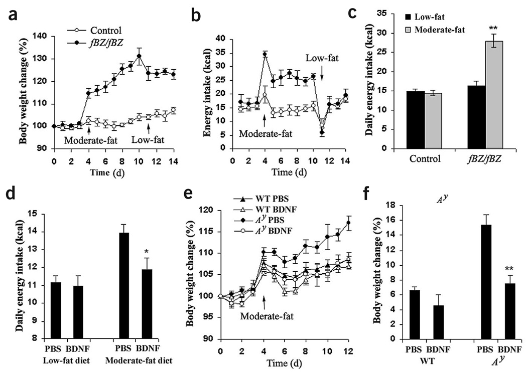
The melanocortin-4 receptor (MC4R) is critically involved in regulating energy balance, and obesity has been observed in mice with mutations in the gene for brain-derived neurotrophic factor (BDNF). Here we report that BDNF is expressed at high levels in the ventromedial hypothalamus (VMH) where its expression is regulated by nutritional state and by MC4R signaling. In addition, similar to MC4R mutants, mouse mutants that expresses the BDNF receptor TrkB at a quarter of the normal amount showed hyperphagia and excessive weight gain on higher-fat diets. Furthermore, BDNF infusion into the brain suppressed the hyperphagia and excessive weight gain observed on higher-fat diets in mice with deficient MC4R signaling. These results show that MC4R signaling controls BDNF expression in the VMH and support the hypothesis that BDNF is an important effector through which MC4R signaling controls energy balance.


Figures

Figure 1
Figure 1
Energy status regulates levels of BDNF in the VMH. (a) Expression of BDNF in the hypothalamus was examined by using an adult BDNFlacZ/+ mouse. Brain sections were stained for β-galactosidase with X-gal staining and counterstained with nuclear fast red. BDNF is highly expressed in the VMH. Arc, arcuate nucleus; DMH, dorsomedial hypothalamus; f, fornix; LH, lateral hypothalamus; mt, mammillothalamic tract; VMH, ventromedial hypothalamus; ZI, zona incerta. Scale bar, 100 µm. (b,c) Representative brain sections of BDNF in situ hybridization show that BDNF expression is specifically reduced in the VMH of food-deprived mice. The in situ probe was 35S-labeled antisense RNA complementary to the coding region of BDNF transcripts. Arrows indicate the location of the VMH. (d) The optical density of BDNF in situ signals was measured with NIH image software in two sections of each animal. Each group was composed of eight female C57BL/6J wild-type mice at 9 weeks of age. One group of mice had free access to mouse chow, and the other group was deprived of food for 48 h. The neocortex in the same section was used as a control. **P < 0.01 (two-tailed Student’s t-test).
Figure 2
Figure 2
Coexpression of TrkB and neuropeptides in the hypothalamus. Expression of TrkB was examined by using an adult trkBlacZ/+ mouse. Most TrkB-expressing neurons do not express MCH (a) or orexins (b) in the lateral hypothalamus and CART (c) or NPY (d) in the ARC. White arrows indicate representative neurons expressing MCH, orexin, CART or NPY. White arrowheads indicate representative TrkB-expressing cells. A yellow arrow denotes a cell expressing both TrkB and neuropeptides. Scale bar, 20 µm.
Figure 3
Figure 3
The trkB hypomorphic mutant shows maturity-onset obesity and increased linear growth. (a) The fBZ/fBZ mutant is obese. The two mice are female littermates at 3 months of age. (b) Both female and male trkB mutant mice show increased linear growth. Measurements were from four pairs of female and three pairs of male mice at about 3 months of age. Error bars are standard error of the mean (s.e.m.). **P < 0.01 by Student’s t-test. (c) Weight gain of female homozygous fBZ/fBZ mutant mice (n = 7), heterozygous fBZ/+ mice (n = 7) and wild-type (+/+) controls (n = 5). When the female mutant mice were 5 weeks or older, their body weights were significantly higher than those of their female +/+ or fBZ/+ littermates (P < 0.05 by two-tailed Student’s t-test). (d) Weight gain of male homozygous fBZ/fBZ mutant mice (n = 6), heterozygous fBZ/ mice (n = 4) and wild-type (+/+) controls (n = 5). When the male mutant mice were 7 weeks or older, their body weights were significantly higher than those of their male +/+ or fBZ/+ littermates (P < 0.05 by two-tailed Student’s t-test).
Figure 4
Figure 4
Hyperphagia and hyperdipsia of the trkB hypomorphic mutant mice. (a) The food intake of individually housed mice was measured every day over a one-week period. The bars represent mean of five measurements for each of five fBZ/fBZ mice (2 male, 3 female) and five sex-matched littermate controls (+/+ or fBZ/+). We used different mice for food intake assays at the 5-week and 9-week stages. Error bars are s.e.m. Two-tailed Student’s t-test, *P < 0.05, **P < 0.01. (b,c) Daily pattern of chow intake and water intake for a wild-type (b) and mutant littermate (c) in 6-min bins. Each bin represents the average of 8 d of intake at that circadian time. These averages were then smoothed by a centered five-bin moving average. (d) Average dark and light cycle chow intake during 8 d of home cage behavioral monitoring. For all home cage behavioral monitoring, error bars indicate s.e.m. for mutant (n = 9) and wild-type (n = 10) mice, and comparisons were made by 2 × 2 repeated-measures ANOVA (genotype, cycle). There was a significant effect of cycle (F1,17 = 109.4, P < 0.001) and genotype (F1,17 = 60.8, P < 0.001), but no interaction between cycle and genotype. (e) Average dark and light cycle water intake during 8 d of home cage behavioral monitoring. There was a significant effect of cycle (F1,17 = 181.9, P < 0.001) and of genotype (F1,17 = 98.0, P < 0.001) but no interaction between cycle and genotype.
Figure 5
Figure 5
Melanocortins regulate the levels of BDNF mRNAs in the VMH. (a) Expression of BDNF in the VMH is significantly reduced in the Ay mutant. Expression levels of BDNF in the VMH were determined with in situ hybridization on C57BL/6J–Ay/a mice and C57BL/6J wild-type littermates at 7–9 weeks of age. In the first experiment, we used four male wild-type mice (25.9 ± 1.7 g) and three male Ay/a mice (28.5 ± 1.3 g). In experiment 2, we used two males and one female for each genotype. (b) Deficiency in MC4R signaling reduces expression of BDNF in the VMH. Expression levels of BDNF in the VMH were determined with in situ hybridization on male MC4R−/− null mutants (n = 6) and C57BL/6J wild-type mice (n = 6) at 10 weeks of age. (c) Intracerebroventricular injection of MTII increased the levels of BDNF mRNAs in the VMH of food-deprived mice. ACSF (n = 5) or MTII (n = 4) was injected into the third ventricle of food-deprived female C57BL/6J wild-type mice at 10 weeks of age. As a control, ACSF was also injected into the third ventricle of normally fed female C57BL/6J mice (n = 5). The levels of BDNF in the VMH were determined with in situ hybridization on cryostat brain sections as described in Methods. Two-tailed Student’s t-test, *P < 0.05, **P < 0.01.
Figure 6
Figure 6
Melanocortins regulate expression of BDNF in selective populations of neurons in the VMH. (a,b) Expression of BDNF in the hypothalamus of BDNFlacZ/+ and Ay/a;BDNFlacZ/+ mice. Expression of BDNF is revealed with anti-β-galactosidase immunohistochemistry in BDNFlacZ/+ mice. Scale bar, 200 µm. (c,d) Expression of BDNF is reduced in selective populations of neurons in the VMH of the Ay mutant. Same magnification in cf, h and i; scale bar in d is 100 µm. (e,f) Expression of BDNF in the PVN is not affected in the Ay mutant. (g) MTII induced expression of BDNF in the VMH of Ay/a;BDNFlacZ/+ mice. BDNF-expressing neurons in the VMH were counted on sections (approximately bregma – 1.8 mm) from saline-injected (n = 5) and MTII-injected (n = 5) mice. Two-tailed Student’s t-test, *P < 0.05. (h,i) Representative sections of saline- and MTII-injected Ay/a;BDNFlacZ/+ mice. There are more intensely stained neurons in the outlined area of the VMH from the MTII-injected mouse. PVN, paraventricular hypothalamus.
Figure 7
Figure 7
TrkB acts downstream of MC4R to regulate feeding response to dietary fat. (a,b) Body weight and energy intake of female fBZ/fBZ (n = 4) and control littermates (+/+ or fBZ/+, n = 4). The average weights of the fBZ/fBZ and control mice at the start of the experiment were 21.7 ± 1.1 and 19.0 ± 0.6 g, respectively. (c) Average daily energy intake of fBZ/fBZ (n = 8) and control littermates (n = 9) on the low-fat and moderate-fat diets. The daily energy intake on two diets is an average from 7 d on the low-fat diet and 7 d on the moderate-fat diet, respectively. (d) BDNF infusion did not affect energy intake of the Ay mice on the low-fat diet, but did suppress their energy intake on the moderate-fat diet. The daily energy intake on the low-fat diet and moderate-fat diet is an average from the first 3 d and the last 8 d of the study, respectively. (e) Weight gain of wild-type mice treated with PBS (n = 5) or BDNF (n = 5) and Ay mice treated with PBS (n = 7) or BDNF (n = 5). Infusion of BDNF suppressed weight gain of Ay mice on the moderate-fat diet. (f) Body weight changes of wild-type and Ay mice treated with either PBS or BDNF over a 9-d period on the moderate-fat diet. Values represent mean ± s.e.m. Student’s t-test, *P < 0.05, **P < 0.01.

Comment in

  • The skinny on neurotrophins.
    Wisse BE, Schwartz MW. Wisse BE, et al. Nat Neurosci. 2003 Jul;6(7):655-6. doi: 10.1038/nn0703-655. Nat Neurosci. 2003. PMID: 12830151 No abstract available.

Similar articles

Cited by

References

    1. Huang EJ, Reichardt LF. Neurotrophins: roles in neuronal development and function. Annu. Rev. Neurosci. 2001;24:677–736. - PMC - PubMed
    1. Lyons WE, et al. Brain-derived neurotrophic factor-deficient mice develop aggressiveness and hyperphagia in conjunction with brain serotonergic abnormalities. Proc. Natl. Acad. Sci. USA. 1999;96:15239–15244. - PMC - PubMed
    1. Kernie SG, Liebl DJ, Parada LF. BDNF regulates eating behavior and locomotor activity in mice. EMBO J. 2000;19:1290–1300. - PMC - PubMed
    1. Rios M, et al. Conditional deletion of brain-derived neurotrophic factor in the postnatal brain leads to obesity and hyperactivity. Mol. Endocrinol. 2001;15:1748–1757. - PubMed
    1. Ono M, et al. Brain-derived neurotrophic factor reduces blood glucose level in obese diabetic mice but not in normal mice. Biochem. Biophys. Res. Commun. 1997;238:633–637. - PubMed

Publication types