Comparative Study
. 2003 Jul;6(7):708-16.
doi: 10.1038/nn1074.

Neurexin mediates the assembly of presynaptic terminals

Affiliations
Comparative Study

Neurexin mediates the assembly of presynaptic terminals

Camin Dean et al. Nat Neurosci. 2003 Jul.

Abstract

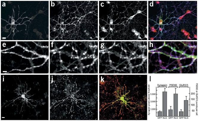
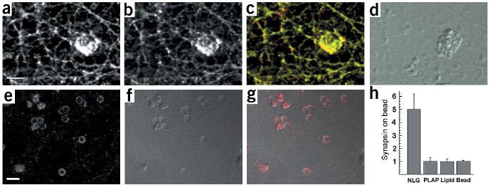
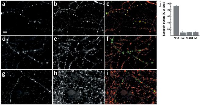
Neurexins are a large family of proteins that act as neuronal cell-surface receptors. The function and localization of the various neurexins, however, have not yet been clarified. Beta-neurexins are candidate receptors for neuroligin-1, a postsynaptic membrane protein that can trigger synapse formation at axon contacts. Here we report that neurexins are concentrated at synapses and that purified neuroligin is sufficient to cluster neurexin and to induce presynaptic differentiation. Oligomerization of neuroligin is required for its function, and we find that beta-neurexin clustering is sufficient to trigger the recruitment of synaptic vesicles through interactions that require the cytoplasmic domain of neurexin. We propose a two-step model in which postsynaptic neuroligin multimers initially cluster axonal neurexins. In response to this clustering, neurexins nucleate the assembly of a cytoplasmic scaffold to which the exocytotic apparatus is recruited.


Figures

Figure 1
Figure 1
Synaptogenic activity of neuroligin. (a) Schematic representation of inactive neuroligin mutants. Within the sequences mutated in the chimeric mutant NLG/AChE-6, two inactive alanine substitution mutants were isolated (K578A/V579A and E584A/L585A). See Supplementary Fig. 1 online for details. (b) Binding of a neurexin–alkaline phosphatase fusion protein to HEK293 cells expressing neuroligin mutants. NLG/AChE-2 shows background levels of neurexin-binding, as do mock-transfected cells. All other constructs show calcium-dependent neurexin binding at levels similar to wild-type neuroligin-1.
Figure 2
Figure 2
Inactive neuroligin mutants are impaired in neuroligin/neurexin mediated cell adhesion. (a) Aggregates of HA-tagged neuroligin-1 expressing cells (green) and VSV-tagged neurexin-1β expressing PC12 cells (red) show localization of neuroligin-1 and neurexin at cell contacts. The asterisks indicate three HA–neuroligin expressing cells forming contacts with adjacent VSV–neurexin expressing cells. Scale bar is 7 μm. (b) An aggregation index N0 (number of particles at time 0) divided by Nt (number of particles at time t) was determined after 0, 20, 40, 60 and 80 min of incubation. Mutants E584A/L585A and K578A/V579A both showed a significant loss of aggregation with neurexin-1β expressing cells. NRXsp4 indicates neurexin-1β with an insertion at site 4 that abolishes the binding to neuroligin. (c) Surface expression of wild-type neuroligin-1 and neuroligin mutants. Western blots of an aliquot of the total cell lysates and the surface biotinylated neuroligin proteins are shown. The biotin-modified NLG/swap reproducibly showed an aberrant migration on SDS-PAGE; the lower band is most likely due to proteolytic cleavage that occurred during the isolation of the biotinylated protein. In the graph, means and standard errors of the mutant neuroligin protein levels at the cell surface are compared to wild-type neuroligin surface levels (n =4 independent experiments). See Supplementary Fig. 2 online for additional data.
Figure 3
Figure 3
Oligomerization of the neuroligin-1 AChE-homologous domain. (a) Mouse AChE crystal structure (blue) and modeled neuroligin-1 structure (green). Regions mutated in NLG/AChE-4 and NLG/AChE-6 are part of a four-helix bundle at the base of the AChE-homologous domain of neuroligin-1 (located at the left of the drawing). (b) In the crystal structure, AChE forms tetramers assembled from two dimers. Two views of analogous, predicted neuroligin-1 dimers. Regions mutated in NLG/AChE-4 (highlighted in light blue) and NLG/AChE-6 are part of a four-helix bundle; residues E584/L585 and K578/V579 are marked in purple. (c) Analysis of neuroligin oligomers. Left panel, purified recombinant extracellular domain of neuroligin-1 could be cross-linked into dimers and tetramers. Higher-order complexes of wild-type neuroligin with different electrophoretic mobility were detected by native gel electrophoresis and western blotting (middle panel, arrowheads). Right panel, oligomerization of neuroligin was also detected by co-immunoprecipitation of differentially tagged neuroligin forms. Wild-type neuroligin was precipitated with myc antibodies and any co-precipitating HA-tagged wild-type or mutant neuroligin protein was detected by immunoblotting with anti-HA antibodies. Expression levels of all transfected constructs were monitored by western blotting of total cell lysates and were similar (data not shown and Supplementary Fig. 1).
Figure 4
Figure 4
Neurexins are concentrated in growth cones and at synaptic junctions. (a) Western blot of total cerebellar lysate probed with anti-neurexin antibodies. (b–d) Pontine explants were grown in vitro for 3 d and immunostained with affinity-purified anti-neurexin antibodies (b, green in the overlay) and Alexa-conjugated phalloidin (c, red in the overlay) to reveal filamentous actin. (e–g) Dissociated cultures of hippocampal neurons (24 d in vitro) were immunostained for neurexin (e, green in overlay) and synaptobrevin (f, red in overlay). (h–j) Cryostat sections (18 μm) of mouse cerebellum (P21) were immunostained for neurexin (h, green in the overlay) and synaptobrevin (i, red in the overlay). Scale bars are 10 μm (a,e) and 20 μm (h).
Figure 5
Figure 5
Overexpression of neuroligin-1 induces recruitment of neurexin to newly forming synapses. (a–d) Cerebellar granule cells were transfected with HA-tagged neuroligin-1 at the time of plating and were fixed after 3 d in culture. The localization of HA–neuroligin-1 (a, green in overlay), synaptobrevin (b, blue in overlay) and neurexin (c, red in overlay) was analyzed by immunostaining. The strong peri-nuclear neurexin immunoreactivity was consistently observed for young cultures and decreased after maintaining cells for 5–10 d in vitro (data not shown). (e–h) High-magnification view of neuroligin-induced cell–cell contacts showing HA–neuroligin-1 (e, green in overlay), synaptobrevin (f, blue in overlay) and neurexin (g, red in overlay). (i–k) Hippocampal neurons were transfected with HA–neuroligin-1 after 12 d in vitro, maintained for 2 d and immunostained with antibodies against the HA tag to detect neuroligin (i, green in overlay) and antibodies against synapsin (j, red in overlay). (l) Quantitation of synapsin-, PSD95- and GluR2/3-positive puncta in neuroligin-1 overexpressing cells. Hippocampal neurons were transfected at 12 d.i.v. and were analyzed 2 d later. Synaptic puncta were quantitated as described in the Methods. Means and standard errors are given (n ≥ 10 cells each). Scale bars are 15 μm (a,i) and 5 μm (e). See Supplementary Fig. 3 online for additional data.
Figure 6
Figure 6
Induction of neurexin clustering and presynaptic differentiation by purified neuroligin. (a–d) Beads coated with lipid bilayers containing purified GPI–neuroligin were added to 12-d.i.v. hippocampal cultures. After 24 h, cultures were fixed and stained with antibodies for neurexin (a, green in the overlay) and synaptobrevin (b, red in the overlay). (d) A differential interference contrast (DIC) image of the lipid-coated beads. Note that these beads coated with a lipid bilayer have a more irregular, rough surface, whereas uncoated beads (without lipid bilayer) appear as discreet particles with a smooth outline (see Supplementary Fig. 4 online for more details). (e–g) Uptake of synaptotagmin antibodies at axonal specializations induced by neuroligin-coated beads. Cultures were incubated with beads as above and depolarized in the presence of antibodies directed against the luminal domain of synaptotagmin. (e) Staining for bound synaptotagmin antibodies (red in the overlay). (f) A DIC image of the beads. (g) Overlay of synaptotagmin staining and DIC image. (h) Quantitation of synapsin clustering on silica beads. The average intensity of synapsin staining on the bead and in a neighboring area of equal size was measured and the enrichment of staining on beads over the neighboring area was calculated. The graph shows the mean ± s.d. (n = 10 beads each). Beads coated with GPI-anchored neuroligin-1 reconstituted into lipid bilayers (NLG) show a five-fold enrichment of synapsin staining. No enrichment is observed for beads coated with GPI-anchored placental alkaline phosphatase reconstituted into lipid bilayers (PLAP), beads coated only with lipid bilayers (lipid) or uncoated beads (bead). Scale bars are 5 μm (a) and 10 μm (e). See Supplementary Fig. 4 online for more details.
Figure 7
Figure 7
Clustering of neurexin is sufficient to induce recruitment of synaptic vesicles. (a–i) Pre-clustered anti-VSV multimers were added to 12-day-old hippocampal cultures transfected with VSV–neurexin (a–c), VSV–neurexin-delta-C (d–f) and VSV–L1 (g–i). Antibody clustered proteins (a, d and g, green in the overlay) and the distribution of synapsin (b, e and h, red in overlay) are shown. The integrity of all cells and the cellular processes was controlled by DIC microscopy to ensure that the cells remained intact during the procedure. Scale bar, 10μm. (j) Quantitation of synapsin accumulation in response to clustering of membrane proteins with antibodies. The percentage of recognizable antibody-induced clusters that showed accumulation of synapsin was determined (n ≥ 140 puncta from ≥ 4 cells for each construct). The mean ± s.d. for cells expressing VSV–neurexin (NRX), VSV–neurexin lacking the cytoplasmic tail (ΔC), VSV–N-cadherin (N-cad) and VSV–L1CAM (L1) are shown. See Supplementary Fig. 5 online for additional data.

References

    1. Sanes JR, Lichtman JW. Development of the vertebrate neuromuscular junction. Annu. Rev. Neurosci. 1999;22:389–442. - PubMed
    1. Garner CC, Zhai RG, Gundelfinger ED, Ziv NE. Molecular mechanisms of CNS synaptogenesis. Trends Neurosci. 2002;25:243–251. - PubMed
    1. Scheiffele P. Cell-cell signaling during synapse formation in the CNS. Annu. Rev. Neurosci. 2003;26:485–508. - PubMed
    1. Ichtchenko K, et al. Neuroligin 1: a splice site-specific ligand for beta-neurexins. Cell. 1995;81:435–443. - PubMed
    1. Song JY, Ichtchenko K, Sudhof TC, Brose N. Neuroligin 1 is a postsynaptic cell-adhesion molecule of excitatory synapses. Proc. Natl. Acad. Sci. USA. 1999;96:1100–1105. - PMC - PubMed

Publication types

MeSH terms

Substances