. 2003 Jun 15;31(12):3016-26.
doi: 10.1093/nar/gkg431.

Identification of novel co-repressor molecules for Interferon Regulatory Factor-2

Affiliations

Identification of novel co-repressor molecules for Interferon Regulatory Factor-2

Kay S Childs et al. Nucleic Acids Res. .

Abstract

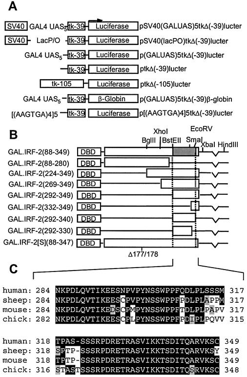
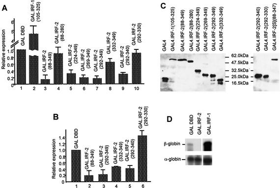
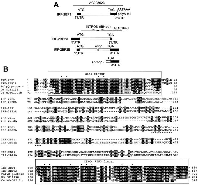
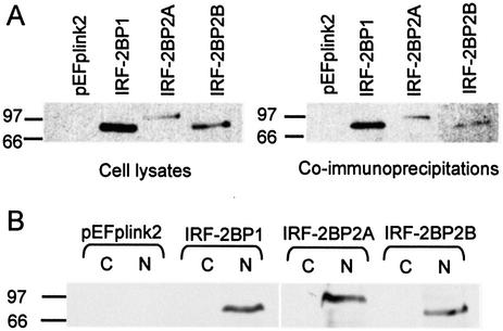
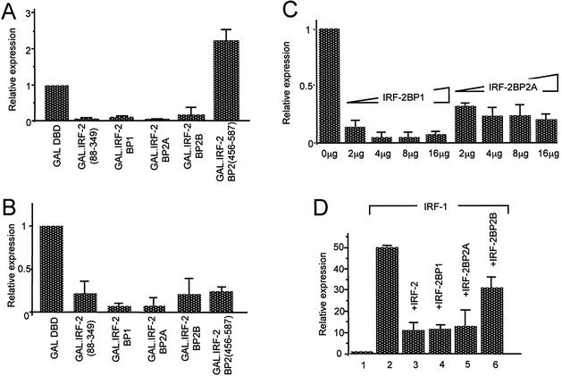
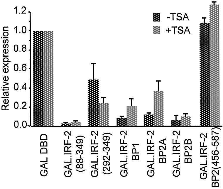
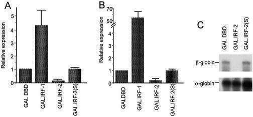
We have identified two novel proteins that interact specifically with the C-terminal repression domain of Interferon Regulatory Factor-2 (IRF-2). These proteins, which we term IRF-2 binding proteins 1 and 2 (IRF-2BP1 and IRF-2BP2, the latter having two splicing isoforms, A and B), are nuclear proteins, and have the properties of IRF-2-dependent transcriptional co-repressors that can inhibit both enhancer-activated and basal transcription in a manner that is not dependent upon histone deacetylation. IRF-2BP1 and IRF-2BP2A/B contain an N-terminal zinc finger and a C-terminal RING finger domain of the C3HC4 subclass, but show no homology to other known transcriptional regulators; they therefore define a new family of co- repressor proteins. An alternatively spliced form of IRF-2 that lacks two amino acids (valines 177 and 178) in the central portion of the protein (IRF-2[S]) cannot bind to these co-repressors and cannot mediate repression despite having the same C- terminal repression domain as IRF-2, suggesting that the relative conformation of the DNA binding domain and the C-terminal region of IRF-2 is crucial for transcriptional repression.


Figures

Figure 1
Figure 1
Reporter and effector gene constructs. (A) Reporter constructs used in this report. (B) GAL.IRF-2 fusion effector constructs used in this report. The GAL4 DBD is fused in-frame to the indicated fragments of IRF-2 (white boxes). The hatched area corresponds to the repression domain defined in this study. (C) Alignment of the extreme C-termini of human IRF-2 (amino acids 284–349) with the equivalent amino acids from sheep, mouse and chicken. Amino acids highlighted in black represent residues conserved with human IRF-2, and grey highlighting indicates functional conservation.
Figure 2
Figure 2
IRF-2 contains a C-terminal domain that can repress both activated and basal transcription. (A) pSV40(GALUAS)5tkΔ(–39)lucter was co-transfected into HeLa cells with a mammalian expression plasmid driving over-expression of the GAL4 DBD (pEF.GAL147–GAL DBD) or a fusion between the GAL4 DBD and the putative effector domains of IRF-1 (pEF.GAL.IRF-1[105–325]) or fragments of IRF-2 (amino acids as indicated), and the β-galactosidase expression vector, pJATlac. (B) p(GALUAS)5tkΔ(–39)lucter was co-transfected into HeLa cells with the indicated effector plasmid and pJATlac. For both (A) and (B), luciferase and β-galactosidase activities were determined from cellular extracts and relative expression values calculated accordingly (expressed relative to the level of the vector-only sample = 1.0). Values shown represent data from between three and eight independent experiments and averages with error bars are shown. (C) Expression levels of GAL4 DBD fusions were determined by western blotting (see Materials and Methods). (D) p(GALUAS)5tkΔ(–39)β-globin] was co-transfected into HeLa cells with either pEF.GAL147 (GAL DBD), pEF.GAL.IRF-2[88–349]) (GAL.IRF-2) or pEF.GAL.IRF-1[105–325] (GAL.IRF-1), and πSVHSα118 as a co-transfection control. RNA was prepared from transfections and mapped by RNase protection with α- or β-globin-specific probes.
Figure 3
Figure 3
The repression domain of IRF-2 interacts with two novel factors, IRF-2BP1 and IRF-2BP2. (A) Organisation of clones isolated for IRF-2BP1, IRF-2BP2A and IRF-2BP2B. The yeast two-hybrid cDNA library screen generated five independent isolates of a 2579 bp insert that encodes IRF-2BP1. This sequence derives from an uncharacterised gene on chromosome 19 (GenBank accession number AC008623, nucleotides 134839–137352). The first in-frame ATG is located 348 bp from the 5′ end of the insert; this was determined to be the initiating methionine, as an in-frame stop codon is present 600 bp upstream from this in the genomic sequence, and no EST clones have been identified containing upstream sequences. The ATG is followed by an uninterrupted open reading frame of 584 amino acids followed by a 3′UTR, polyadenylation signal (AATAAA) and a poly(A) tail. A single isolate of the insert shown for IRF-2BP2A was obtained. The insert comprises ∼2500 bp derived from an uncharacterised gene on chromosome 1 (GenBank accession numbers AL161640, nucleotides 28362–30847, and AL160408 nucleotides 4723–2239). Due to the high G-C content of the 5′ end of this insert (and that for IRF-2BP2B; see below) we were unable to obtain accurate sequence data at the 5′ end and we note that the available sequences of both genomic clones and the ESTs stop in the same place. We have tentatively assigned the initiator ATG for IRF-2BP2A on the basis that: (i) the sequence is identical to IRF-2BP2B and very similar to IRF-2BP1 in this region; (ii) the encoded protein initiating from this ATG is highly homologous to proteins found in Drosophila and C.elegans proteins (see text and B); (iii) the assigned initiator ATG conforms to the consensus Kozak sequence; (iv) such G-C-rich sequences are frequently found in 5′UTRs. The ATG is followed by an open reading frame of 587 amino acids, which is terminated by a 3′ TAG stop codon; this cDNA clone does not have a poly(A) tail. Two independent isolates of the 2600 bp insert encoding IRF-2BP2B were obtained. IRF-2BP2A and IRF-2BP2B differ in the size of the single intron, and thus represent alternative splice forms of a single gene. IRF-2BP2B lacks 48bp, representing 16 amino acids, that are present in IRF-2BP2A. The presumptive splice acceptor site is the same in both inserts (AL161640 nucleotide 30026) and conforms to the consensus of 10 pyrimidines followed by NCAG. The presumptive donor sites are AGGT at nucleotide 29433 for IRF-2BP2A and AGGC at nucleotide 29385 for IRF-2BP2B. We also recovered 28 independent isolates of the indicated insert of 775 bp that encodes a 3′ region common to both IRF-2BP2A and IRF-2BP2B. (B) The primary amino acid sequences of IRF-2BP1 and IRF-2BP2 are aligned with each other using the BLAST pairwise programme and common residues are highlighted in black (functionally conserved residues are highlighted in grey). The asterisk-underlined sequence shown in IRF-2BP2A is not present in IRF-2BP2B. The conserved zinc finger and C3HC4 RING domains at the N- and C-terminus, respectively, are boxed and the cysteines and histidines are indicated. Homologies with the hypothetical human C14orf4 (polyQ) protein, and the C.elegans MO4G12 and the D.melanogaster CG11138 and CG1855 gene products in these regions are indicated.
Figure 4
Figure 4
IRF-2BP1, IRF-2BP2A and IRF-2BP2B interact with IRF-2 in vivo and are nuclear proteins. (A) HeLa cells were transiently transfected with pEF.plink2, pEF.IRF-2BP1, pEF.IRF-2BP2A or pEF.IRF-2BP2B and whole cell lysates prepared. An aliquot of each lysate was fractionated on a SDS–PAGE gel and analysed by western blotting using a monoclonal antibody against the SV5 epitope tag (left hand panel). To determine association with cellular IRF-2, whole cell extracts were immunoprecipitated with rabbit polyclonal anti-human IRF-2 serum and the immunoprecipitates were analysed by western blotting with SV5 epitope tag antibody (right hand panel). (B) HeLa cells were transiently transfected with pEF.plink2, pEF.IRF-2BP1, pEF.IRF-2BP2A or pEF.IRF-2BP2B and extracts from cytoplasmic (C) and nuclear (N) subcellular fractions were prepared. An aliquot of each extract was fractionated on an SDS–PAGE gel and analysed by western blotting using a monoclonal antibody against the SV5 epitope tag.
Figure 5
Figure 5
IRF-2BP1, IRF-2BP2A and IRF-2BP2B can act as transcriptional co-repressors. (A) pSV40(GALUAS)5tkΔ(–39)lucter or (B) p(GALUAS)5tkΔ(–39)lucter were co-transfected into HeLa cells with either pEF.GAL147, pEF.GAL.IRF-2BP1, pEF.GAL.IRF-2BP2A, pEF.GAL.IRF-2BP2B or pEF.GAL.IRF-2BP2[456–587]), and the β-galactosidase expression vector, pJATlac. (C) Four micrograms of pSV40(GALUAS)5tkΔ(–39)lucter were co-transfected using calcium phosphate co-precipitation into HeLa cells with 4 µg of pEF.GAL.IRF-2[88–349], 4 µg of the β-galactosidase expression vector, pJATlac and increasing amounts of either full-length IRF-2BP1 (pEF.IRF-2BP1) or IRF-2BP2A (pEF.IRF-2BP2A) as indicated (the total amount of plasmid DNA being made up to a total of 30 µg with the ‘empty’ effector vector, pEFplink2). Reporter activity was determined, and corrected for squelching by dividing the results by the values obtained for an identical experiment using pSV40(lacP/O)tkΔ(–39)lucter as the reporter. (D) The IRF-dependent reporter, p[(AAGTGA)4]5tkΔ(–39)lucter was co-transfected into HeLa cells with either pEF.plink2 (lane 1), pEF.IRF-1 (lane 2), pEF.IRF-1 plus pEF.IRF-2 (lane 3), pEF.IRF-1 plus pEF.IRF-2BP1 (lane 4), pEF.IRF-1 plus pEF.IRF-2BP2A (lane 5) or pEF.IRF-1 plus pEF.IRF-2.BP2B (lane 6), and the β-galactosidase expression vector, pJATlac. For each of (A) to (D), luciferase and β-galactosidase activities were determined from cellular extracts and relative expression values calculated accordingly (expressed relative to the level of the vector-only sample = 1.0). Values shown represent data from three independent experiments and averages with error bars are shown.
Figure 6
Figure 6
IRF-2BP1- and IRF-2BP2-mediated repression does not require histone deacetylase activity. pSV40(GALUAS)5tkΔ(–39)lucter was co- transfected into HeLa cells with either pEF.GAL147, pEF.GAL.IRF-2[88–349], pEF.GAL.IRF-2(292–349), pEF.GAL.IRF-2BP1, pEF.GAL.IRF-2BP2A, pEF.GAL.IRF-2BP2B or pEF.GAL.IRF-2BP2[456–587]), and the β-galactosidase expression vector, pJATlac. Cells were incubated with the histone deacetylase inhibitor trichostatin A (200 nM) where indicated, for 26 h prior to harvesting. Control cells not treated with trichostatin A were treated with 0.01% v/v DMSO. Luciferase and β-galactosidase activities were determined from cellular extracts and relative expression values calculated accordingly (expressed relative to the level of the vector-only sample = 1.0). In the presence of trichostatin A both luciferase and β-galactosidase activities were increased relative to the DMSO control. This global effect has been observed by others (57). Therefore, repression is expressed relative to the GAL4 DBD alone in each case. Values shown represent data from three independent experiments.
Figure 7
Figure 7
An isoform of IRF-2, IRF-2[S], which lacks two amino acids, cannot repress transcription. (A) pSV40(GALUAS)5tkΔ(–39)lucter or (B) p(GALUAS)5tkΔ(–39)lucter was co-transfected into HeLa cells with either pEF.GAL147, pEF.GAL.IRF-1[105–325], pEF.GAL.IRF-2[88–349] or pEF.GAL.IRF-2(S)[88–347], and the β-galactosidase expression vector, pJATlac. Luciferase and β-galactosidase activities were determined from cellular extracts and relative expression values calculated accordingly (expressed relative to the vector only sample = 1.0). Values shown represent data from three separate experiments and averages with error bars are shown. (C) p(GALUAS)5tkΔ(–39)β-globin was co-transfected into HeLa cells with either pEF.GAL147, pEF.GAL.IRF-2[88–349] or pEF.GAL.IRF-2(S)[88–347], and πSVHSα118 as a co-transfection control. RNA was prepared from transfections and mapped by RNase protection with α- or β-globin-specific probes.

Similar articles

Cited by

References

    1. Taniguchi T., Ogasawara,K., Takaoka,A. and Tanaka,N. (2001) IRF family of transcription factors as regulators of host defense. Annu. Rev. Immunol., 19, 623–655. - PubMed
    1. Harada H., Fujita,T., Miyamoto,M., Kimura,Y., Maruyama,M., Furia,A., Miyata,T. and Taniguchi,T. (1989) Structurally similar but functionally distinct factors, IRF-1 and IRF-2, bind to the same regulatory elements of IFN and IFN-inducible genes. Cell, 58, 729–739. - PubMed
    1. Hida S., Ogasawara,K., Sato,K., Abe,M., Takayanagi,H., Yokochi,T., Sato,T., Hirose,S., Shirai,T., Taki,S. and Taniguchi,T. (2000) CD8(+) T cell-mediated skin disease in mice lacking IRF-2, the transcriptional attenuator of interferon-alpha/beta signaling. Immunity, 13, 643–655. - PubMed
    1. Harada H., Kitagawa,M., Tanaka,N., Yamamoto,H., Harada,K., Ishihara,M. and Taniguchi,T. (1993) Anti-oncogenic and oncogenic potentials of interferon regulatory factors-1 and -2. Science, 259, 971–974. - PubMed
    1. Nguyen H., Mustafa,A., Hiscott,J. and Lin,R. (1995) Transcription factor IRF-2 exerts its oncogenic phenotype through the DNA binding/transcription repression domain. Oncogene, 11, 537–544. - PubMed

Publication types

MeSH terms