. 2003 Jun 15;31(12):3038-49.
doi: 10.1093/nar/gkg434.

The C-terminal domain of Escherichia coli MutY is involved in DNA binding and glycosylase activities

Affiliations

The C-terminal domain of Escherichia coli MutY is involved in DNA binding and glycosylase activities

Lina Li et al. Nucleic Acids Res. .

Abstract

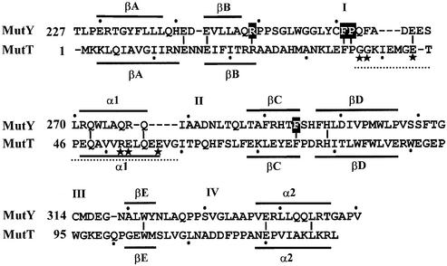
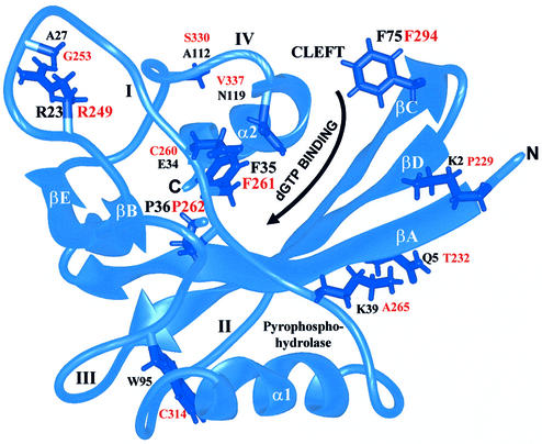
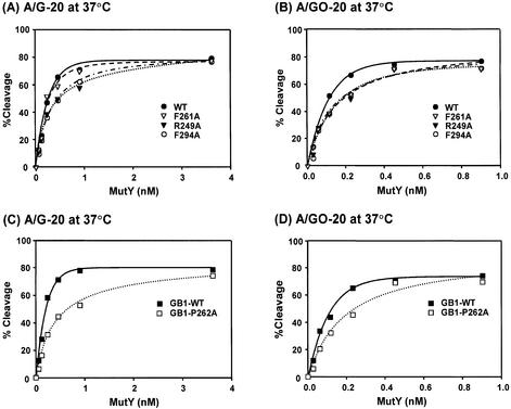
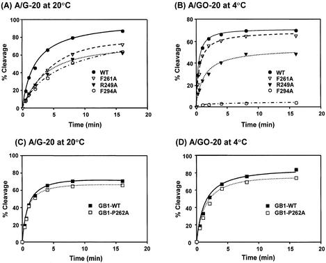
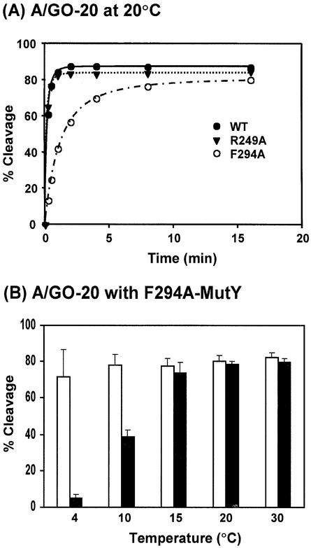
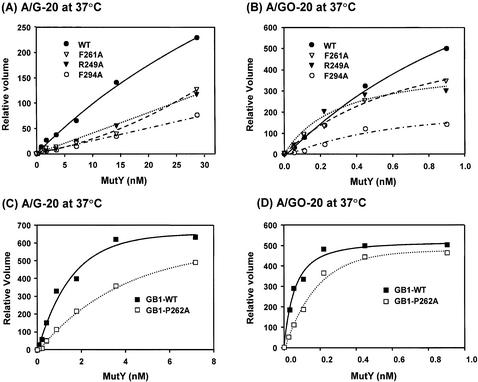
Escherichia coli MutY is an adenine and a weak guanine DNA glycosylase involved in reducing mutagenic effects of 7,8-dihydro-8-oxo-guanine (8-oxoG). The C-terminal domain of MutY is required for 8-oxoG recognition and is critical for mutation avoidance of oxidative damage. To determine which residues of this domain are involved in 8-oxoG recognition, we constructed four MutY mutants based on similarities to MutT, which hydrolyzes specifically 8-oxo-dGTP to 8-oxo-dGMP. F294A-MutY has a slightly reduced binding affinity to A/G mismatch but has a severe defect in A/8-oxoG binding at 20 degrees C. The catalytic activity of F294A-MutY is much weaker than that of the wild-type MutY. The DNA binding activity of R249A-MutY is comparable to that of the wild-type enzyme but the catalytic activity is reduced with both A/G and A/8-oxoG mismatches. The biochemical activities of F261A-MutY are nearly similar to those of the wild-type enzyme. The solubility of P262A-MutY was improved as a fusion protein containing streptococcal protein G (GB1 domain) at its N-terminus. The binding of GB1-P262A-MutY with both A/G and A/8-oxoG mismatches are slightly weaker than those of the wild-type protein. The catalytic activity of GB1-P262A-MutY is weaker than that of the wild-type enzyme at lower enzyme concentrations. Importantly, all four mutants can complement mutY mutants in vivo when expressed at high levels; however, F294A, R249A and P262A, but not F261A, are partially defective in vivo when they are expressed at low levels. These results strongly support that the C-terminal domain of MutY is involved not only in 8-oxoG recognition, but also affects the binding and catalytic activities toward A/G mismatches.


Figures

Figure 1
Figure 1
Structure-based sequence alignment of the C-terminal domain of MutY and MutT. Boxed residues were mutated to alanines. Lines above or underneath of the sequences represent the α-helixes (α1 and α2) or β-sheets (βA–βE). The loops connecting α-helixes or β-sheets are marked with I–IV. The region G37–G59 (marked by a dotted line) of MutT is the pyrophosphohydrolase module with essential residues marked with stars. Identical residues between the C-terminal domain of MutY and MutT are marked with short vertical lines.
Figure 2
Figure 2
MutT structure (34) showing residues (labeled in black) corresponding to MutY residues (labeled in red). The ribbon diagram was produced with the graphic program MIDAS (58). Four conserved residues of MutY (R249, F261, P262 and F294) were mutated to Ala. The α-helixes (α1 and α2), β-strands (βA–βE) and loops (I–IV) connecting the α-helix or β-sheet are marked. The nucleotide binding site is located in a cleft defined by β-strands A, C and D on one side, and loop I, the end of loop IV and the beginning of helix 2 on the other side. The reaction center of pyrophosphohydrolase consists of residues from loop I, helix 1 and loop III.
Figure 3
Figure 3
Glycosylase activities of the wild-type and mutant MutY proteins at different enzyme concentrations. (A) and (C), A/G-containing DNA (1.8 fmol) was incubated with MutY proteins at concentrations of 0.056, 0.1125, 0.225, 0.45, 0.9 and 3.6 nM in a 10 µl reaction at 37°C for 30 min. (B) and (D), A/GO-containing DNA (1.8 fmol) was incubated with MutY proteins at concentrations of 0.028, 0.056, 0.1125, 0.225, 0.45 and 0.9 nM at 37°C for 30 min. After reaction, the products were dried, resuspended in formamide dye, heated at 90°C for 2 min, and analyzed on a 14% denaturing sequencing gel. Data were from PhosphorImager quantitative analyses of gel images over three experiments. Percentages of DNA cleaved were plotted versus protein concentrations. Wild-type MutY (WT, solid circle), F261A (open triangle), R249A-MutY (solid triangle), F294A-MutY (open circle), GB-tagged MutY, (GB1-WT, solid rectangle), and GB-F261A-MutY (open rectangle).
Figure 4
Figure 4
Time course studies of glycosylase activities of the wild-type and mutant MutY proteins. (A) and (C), A/G-containing DNA (1.8 fmol) was incubated with 3.6 nM proteins in a 10 µl reaction at 20°C. (B) and (D), A/GO-containing DNA (1.8 fmol) was incubated with 0.45 nM proteins in a 10 µl reaction at 4°C. After reaction, the products were treated with 0.1 M NaOH as described in Materials and Methods and analyzed on a 14% denaturing sequencing gel. Data were from PhosphorImager quantitative analyses of gel images over three experiments. Percentages of DNA cleaved were plotted as a function of time. Wild-type MutY (WT, solid circle), F261A (open triangle), R249A-MutY (solid triangle), F294A-MutY (open circle), GB-tagged MutY, (GB1-WT, solid rectangle), and GB-F261A-MutY (open rectangle).
Figure 5
Figure 5
(A) Time course studies of glycosylase activities of the wild-type and mutant MutY proteins with A/GO-containing DNA at 20°C. Reactions were similar to Figure 4B with 0.45 nM proteins but were performed at 20°C. Wild-type MutY (WT, solid circle), R249A-MutY (solid triangle) and F294A-MutY (open circle). (B) The temperature effect on glycosylase activities of the wild-type MutY and F294A-MutY with A/GO-containing DNA. Reactions were performed at 4, 10, 15, 20 and 30°C for 30 min with 0.45 nM proteins.
Figure 6
Figure 6
Trapping activities of the wild-type and mutant MutY proteins. A/G- (A and C) and A/GO- (B and D) containing DNA (1.8 fmol) was incubated with various amounts of proteins in the presence of NaBH4 in a 10 µl reaction at 37°C for 30 min. The samples were heated at 90°C for 2 min and separated on a 12% polyacrylamide gel in the presence of SDS. Data were from PhosphorImager quantitative analyses of gel images over three experiments. Volumes of covalent complexes were plotted versus protein concentrations. The percentages of covalent complexes could not be calculated because the free DNA and free [α-32P]dCTP were not separated far enough in the gel. Wild-type MutY (WT, solid circle), F261A (open triangle), R249A-MutY (solid triangle), F294A-MutY (open circle), GB-tagged MutY (GB1-WT, solid rectangle), and GB-F261A-MutY (open rectangle).

Similar articles

Cited by

References

    1. Ames B.N. and Shigenaga,M.K. (1993) Oxidants are a major contributor to cancer and aging. In Halliwell,B. and Aruoma,O. (eds), DNA and Free Radicals. Ellis Horwood, New York, NY, pp. 1–18.
    1. Wiseman H. and Halliwell,B. (1996) Damage to DNA by reactive oxygen and nitrogen species: role in inflammatory disease and progression to cancer. Biochem. J., 313, 17–29. - PMC - PubMed
    1. Halliwell B. and Gutteridge,J.M.C. (1989) Free radicals in Biology and Medicine. Oxford University Press, New York.
    1. Michaels M.L. and Miller,J.H. (1992) The GO system protects organisms from the mutagenic effect of the spontaneous lesion 8-hydroxyguanine (7,8-dihydro-8-oxo-guanine). J. Bacteriol., 174, 6321–6325. - PMC - PubMed
    1. Tchou J. and Grollman,A.P. (1993) Repair of DNA containing the oxidatively-damaged base 8-hydroxyguanine. Mutat. Res., 299, 277–287. - PubMed

Publication types