

Specific interaction of heterogeneous nuclear ribonucleoprotein A1 with the -219T allelic form modulates APOE promoter activity
- PMID: 12799433
- PMCID: PMC162339
- DOI: 10.1093/nar/gkg435
Specific interaction of heterogeneous nuclear ribonucleoprotein A1 with the -219T allelic form modulates APOE promoter activity
Abstract
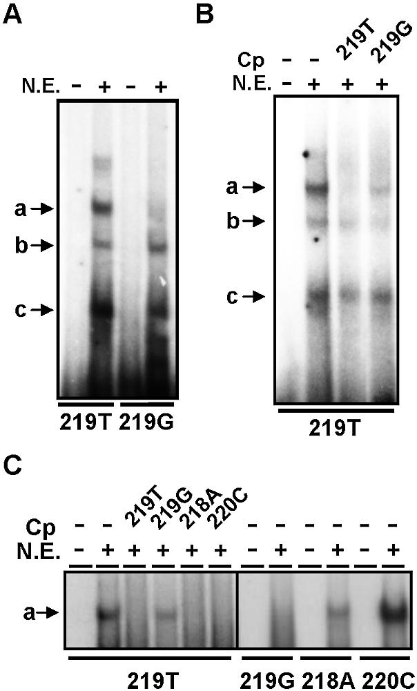
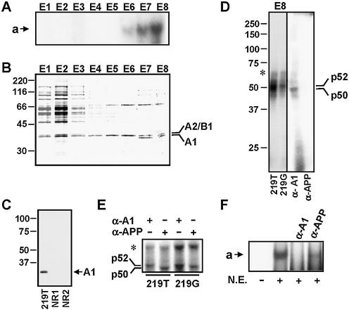
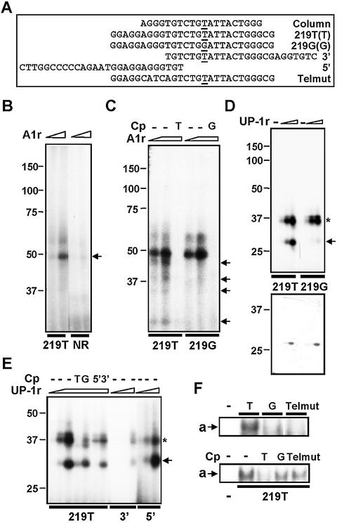
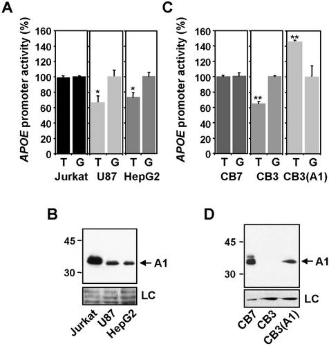
The polymorphic -219T/G variant in the APOE promoter has been associated with variations in basal transcriptional activity as well as with the risk of developing Alzheimer's disease, myocardial infarction and early-onset coronary heart disease. The molecular mechanisms underlying these effects are presently unknown. In this report, we show that nuclear extracts from Jurkat cells form a T-specific complex with a motif including the -219 site within the APOE promoter. By DNA-affinity chromatography and mass spectrometry, the human heterogeneous nuclear ribonucleoprotein hnRNPA1(A1) was identified as one component of the complex. In vitro binding analysis indicated that a fragment of A1 had a marked binding specificity for the T form. Interaction of A1 with this region is driven by an adjacent telomeric-like sequence; however, the presence of G, but not T, at -219 position inhibited this interaction. The differences in transcriptional activity between the -219T and -219G promoter allelic forms correlated with the expression levels of A1 in several cell lines; also, over-expression of A1 increased the activity of the T form relative to that of the G form. These results indicate that A1 transactivates APOE promoter activity by direct and specific interaction with the -219T site.
Figures
Similar articles
-
Heterogeneous nuclear ribonucleoproteins as regulators of gene expression through interactions with the human thymidine kinase promoter.J Cell Biochem. 2000 Sep 7;79(3):395-406. doi: 10.1002/1097-4644(20001201)79:3<395::aid-jcb50>3.0.co;2-m. J Cell Biochem. 2000. PMID: 10972977
-
Phenylalanines that are conserved among several RNA-binding proteins form part of a nucleic acid-binding pocket in the A1 heterogeneous nuclear ribonucleoprotein.J Biol Chem. 1988 Mar 5;263(7):3307-13. J Biol Chem. 1988. PMID: 2830282
-
Interactions of the A1 heterogeneous nuclear ribonucleoprotein and its proteolytic derivative, UP1, with RNA and DNA: evidence for multiple RNA binding domains and salt-dependent binding mode transitions.Biochemistry. 1991 Mar 19;30(11):2968-76. doi: 10.1021/bi00225a034. Biochemistry. 1991. PMID: 1848781
-
Heterogeneous nuclear ribonucleoprotein A1 in health and neurodegenerative disease: from structural insights to post-transcriptional regulatory roles.Mol Cell Neurosci. 2013 Sep;56:436-46. doi: 10.1016/j.mcn.2012.12.002. Epub 2012 Dec 14. Mol Cell Neurosci. 2013. PMID: 23247072 Review.
-
Critical role of hnRNP A1 in activating KRAS transcription in pancreatic cancer cells: A molecular mechanism involving G4 DNA.Biochim Biophys Acta Gen Subj. 2017 May;1861(5 Pt B):1389-1398. doi: 10.1016/j.bbagen.2016.11.031. Epub 2016 Nov 22. Biochim Biophys Acta Gen Subj. 2017. PMID: 27888145 Review.
Cited by
-
hnRNP A1 in RNA metabolism regulation and as a potential therapeutic target.Front Pharmacol. 2022 Oct 21;13:986409. doi: 10.3389/fphar.2022.986409. eCollection 2022. Front Pharmacol. 2022. PMID: 36339596 Free PMC article. Review.
-
HnRNPA1 interacts with G-quadruplex in the TRA2B promoter and stimulates its transcription in human colon cancer cells.Sci Rep. 2019 Jul 16;9(1):10276. doi: 10.1038/s41598-019-46659-x. Sci Rep. 2019. PMID: 31311954 Free PMC article.
-
Tandem hnRNP A1 RNA recognition motifs act in concert to repress the splicing of survival motor neuron exon 7.Elife. 2017 Jun 26;6:e25736. doi: 10.7554/eLife.25736. Elife. 2017. PMID: 28650318 Free PMC article.
-
Compensatory expression regulation of highly homologous proteins HNRNPA1 and HNRNPA2.Turk J Biol. 2021 Apr 20;45(2):187-195. doi: 10.3906/biy-2010-29. eCollection 2021. Turk J Biol. 2021. PMID: 33907500 Free PMC article.
-
RNA-binding proteins with prion-like domains in health and disease.Biochem J. 2017 Apr 7;474(8):1417-1438. doi: 10.1042/BCJ20160499. Biochem J. 2017. PMID: 28389532 Free PMC article. Review.
References
-
- Mahley R.W. (1988) Apolipoprotein E: cholesterol transport protein with expanding role in cell biology. Science, 240, 622–630. - PubMed
-
- Weisgraber K.H. (1994) Apolipoprotein E: structure–function relationships. Adv. Protein Chem., 45, 249–302. - PubMed
-
- Masliah E., Mallory,M., Aford,M., Veinbergs,I. and Roses,A.D. (1996) In Roses,A.D., Weisgraber,K.H. and Christen,Y. (eds), Apolipoprotein E and Alzheimer’s Disease. Springer Verlag, Berlin, Germany, pp. 59–73.
-
- Das H.K., McPherson,J., Bruns,G.A., Karathanasis,S.K. and Breslow,J.L. (1985) Isolation, characterization and mapping to chromosome 19 of the human apolipoprotein E gene. J. Biol. Chem., 260, 6240–6247. - PubMed
-
- Davignon J., Gregg,R.E. and Sing,C.F. (1988) Apolipoprotein E polymorphism and atherosclerosis. Arteriosclerosis, 8, 1–21. - PubMed
