. 2003 Jun 15;31(12):3166-73.
doi: 10.1093/nar/gkg427.

Characterization of the 2'-5'-oligoadenylate synthetase ubiquitin-like family

Affiliations

Characterization of the 2'-5'-oligoadenylate synthetase ubiquitin-like family

Signe Eskildsen et al. Nucleic Acids Res. .

Abstract

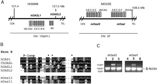
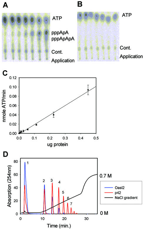
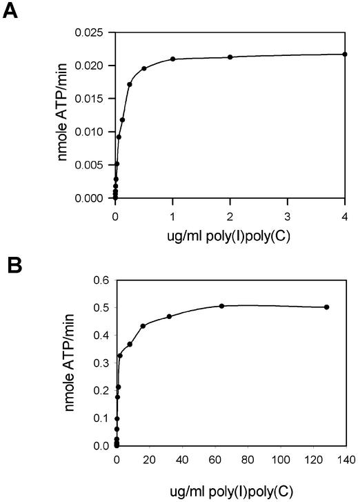
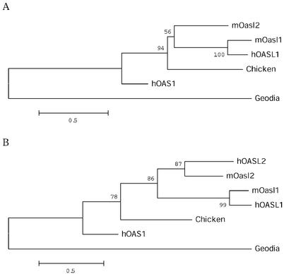
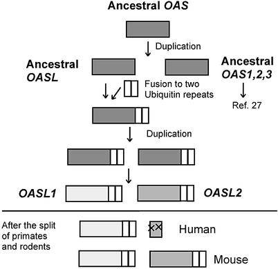
The interferon-induced 2'-5'-oligoadenylate synthetases (OAS) are important for the antiviral activity of interferons. The human and murine OAS gene families each contain four genes: OAS1, OAS2, OAS3 and OASL, all having one or more conserved OAS units composed of five translated exons. The OASL gene has both an OAS unit and a C-terminus of two ubiquitin-like repeats. In this study, we demonstrate that murine Oasl1 protein is inactive while murine Oasl2 is active as an OAS. Further more, murine Oasl2 requires double-stranded RNA as co-factor. The affinity of murine Oasl2 for the double-stranded RNA activator is higher than that of human OAS1 (p42 isoform). We propose a model for the evolutionary origin of the murine Oasl1 and Oasl2 genes. The identification of a human orthologue (hOASL2) to the murine Oasl2 gene establishes that the OASL gene was duplicated prior to the radiation of the rodent and primate groups. We suggest that murine Oasl2, which has both enzymatic activity and a ubiquitin-like domain, is a functional intermediate between the active OAS species and the inactive human OASL1/murine Oasl1 proteins. In addition, we propose that murine Oasl1 appears to have gained a hitherto uncharacterized function independent of 2'-5'-linked oligoadenylate synthesis.


Figures

Figure 1
Figure 1
(A) Schematic drawing of the hOASL and mOasl genes on human chromosome 12 and mouse chromosome 5. Genes with reading frames in the same direction as the annotated nucleotide sequence are indicated by squares above the lines; genes below are in the reverse direction. The genes are not drawn to scale. CEN, centromere; TEL, telomere. (B) Amino acid alignments of hOAS1 p42 (D00068), ChOASL (AB037592), hOASL1 (AJ22089), hOASL2 (NT_028327), mOasl1 (AY089728) and mOasl2 (AK010034) (numbers in parentheses indicate the GenBank accession no. for each protein). Black boxes indicate conserved residues, grey boxes related sequences. Gaps are indicated by –. The middle part of exon B has been cut out as indicated by black dots. The three catalytic aspartate residues are indicated by asterisks. (C) Transcription of mOasl2 (lanes 1–3) and mOasl1 (lanes 4–6) genes. Products of PCRs (30 cycles) with cDNA from untreated (lanes 1 and 4), IFN-α-induced (lanes 2 and 5) and IFN-γ-induced (lanes 3 and 6) mouse L929 cells, analysed by gel electrophoresis (1% agarose). The mOasl reaction and the control reaction (murine β-actin) were mixed in equal amounts before gel electrophoresis.
Figure 2
Figure 2
Purification of recombinant mOasl2 protein. (A) The elution profile of mOasl2 using the heparin–Sepharose column for purification. The graphs show the absorption at 280 nm and the NaCl gradient from 0 to 1 M. (B) A 10% SDS–polyacrylamide gel with 5 µM of pooled His tag fractions (lane 1) and 5 µM of pooled fractions from the heparin column (lane 2) was stained with Coomassie brilliant blue. The size of the molecular markers is indicated. (C) Western blot analysis of the samples loaded in (B). They were visualised using an anti-His tag antibody and a secondary goat anti-mouse HRP-conjugated antibody.
Figure 3
Figure 3
mOasl2 is active whereas mOasl1 is not. (A) ATP was separated from the produced 2–5A products (pppApA and pppApApA) synthesised by mOasl2 using TLC. Increasing amounts of mOasl2 protein were added (0, 0.0007, 0.0014, 0.0028, 0.0056, 0.0112 and 0.0224 µg/µl) with a final concentration of 100 µg/ml poly(I)·poly(C). Cont., contaminations present in all reactions. (B) 2–5A synthetase activity assay using mOasl1 protein, and nucleotides separated by TLC. The same experimental conditions as in (A). (C) The amount of 2–5A produced by mOasl2 in (A) was quantified and normalised to the ATP spot. A linear correlation exists between the activity (nmol ATP/min) and the concentration of mOasl2 (µg). The experiment was repeated twice. (D) The heat-inactivated 2–5A synthetase reaction [final concentration of 100 µg/ml poly(I)·poly(C)] was treated with alkaline phosphatase and separated using a Mono Q column. The elution profiles of the products produced by mOasl2 (2.5 ng/µl) (blue) and p42 (2.5 ng/µl) (red) are superimposed. 1, adenylate; 2, pppApA; 3, pppA(pA)2; 4, pppA(pA)3; 5, pppA(pA)4; 6, pppA(pA)5; and 7, pppA(pA)6.
Figure 4
Figure 4
mOasl2 is induced by poly(I)·poly(C). (A) Dose–response curve for poly(I)·poly(C) induction of mOasl2 (2.5 ng/µl). (B) Dose–response curve for poly(I)·poly(C) induction of hOAS1 (2.5 ng/µl).
Figure 5
Figure 5
Phylogenetic analyses. (A) Phylogenetic tree of exons A–E. Bootstrap values are based on 1000 bootstrap replications. Sequences from mOasl2 (AK010034), mOasl1 (AY089725), hOAS1 p42 (D00068), hOASL (AJ22089), hOASL2 (NT_028327), ChOASL (AB037592) and Geodia OAS1 (Y18497) are aligned (numbers in parentheses indicate the GenBank accession no. for each protein). (B) Phylogenetic tree of exon B.
Figure 6
Figure 6
Model for the evolutionary origin of the OASL1 and OASL2 genes.

References

    1. Stark G.R., Kerr,I.M., Williams,B.R., Silverman,R.H. and Schreiber,R.D. (1998) How cells respond to interferons. Annu. Rev. Biochem., 67, 227–264. - PubMed
    1. Der S.D., Zhou,A., Williams,B.R. and Silverman,R.H. (1998) Identification of genes differentially regulated by interferon alpha, beta, or gamma using oligonucleotide arrays. Proc. Natl Acad. Sci. USA, 95, 15623–15628. - PMC - PubMed
    1. Roberts W.K., Hovanessian,A., Brown,R.E., Clemens,M.J. and Kerr,I.M (1976) Interferon-mediated protein kinase and low-molecular-weight inhibitor of protein synthesis. Nature, 264, 477–480. - PubMed
    1. Hovanessian A., Brown,R.E. and Kerr,I.M. (1977) Synthesis of low molecular weight inhibitor of protein synthesis with enzyme from interferon-treated cells. Nature, 268, 537–540. - PubMed
    1. Chebath J., Benech,P., Revel,M. and Vigneron,M. (1987) Constitutive expression of (2′–5′) oligo A synthetase confers resistance to picornavirus infection. Nature, 330, 587–588. - PubMed

Publication types

MeSH terms