. 2003 Jun 15;31(12):3194-207.
doi: 10.1093/nar/gkg419.

Alternatively spliced isoforms of the human constitutive androstane receptor

Affiliations

Alternatively spliced isoforms of the human constitutive androstane receptor

Scott S Auerbach et al. Nucleic Acids Res. .

Abstract

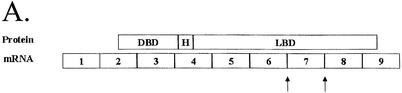
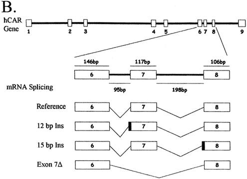
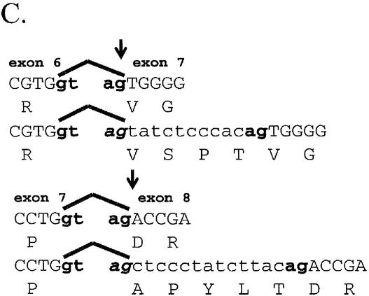
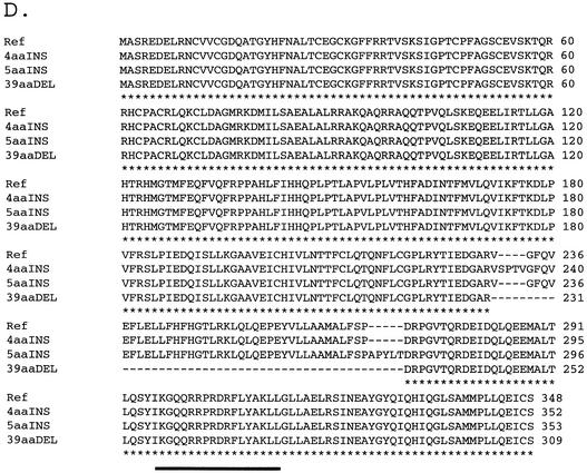
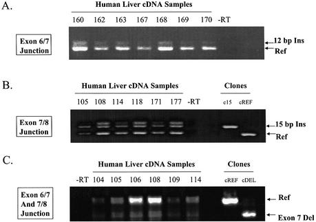
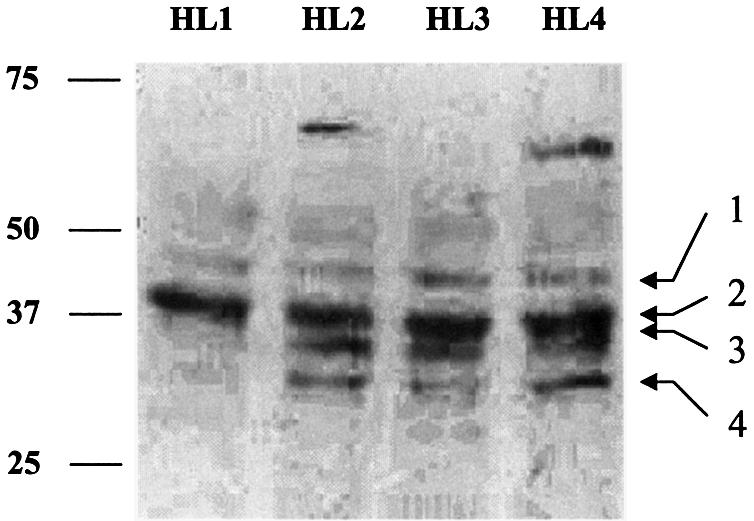
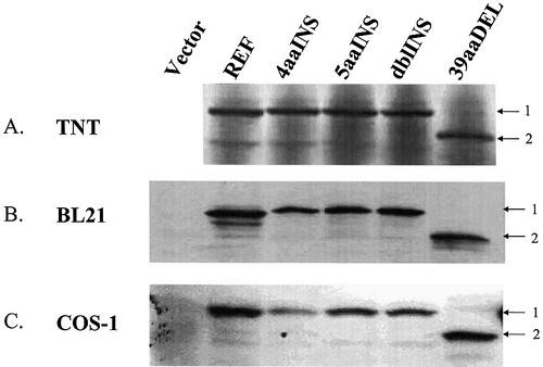
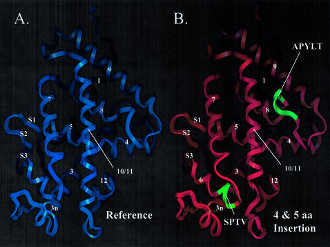
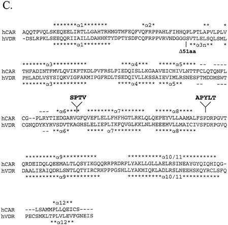
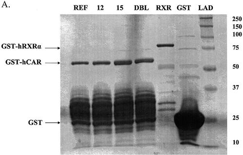
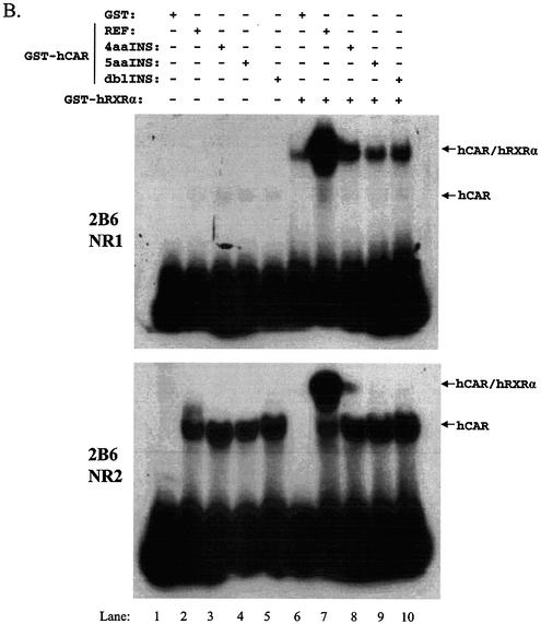
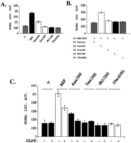
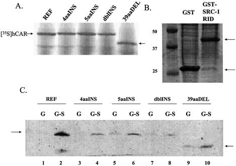
The nuclear receptor CAR (NR1I3) regulates transcription of genes encoding xenobiotic- and steroid-metabolizing enzymes. Regulatory processes that are mediated by CAR are modulated by a structurally diverse array of chemicals including common pharmaceutical and environmental agents. Here we describe four in-frame splice variants of the human CAR receptor gene. The variant mRNA splice transcripts were expressed in all human livers evaluated. Molecular modeling of the splice variant proteins predicts that the structural effects are localized within the receptor's ligand-binding domain. Assays to assess function indicate that the variant proteins, when compared with the reference protein isoform, exhibit compromised activities with respect to DNA binding, transcriptional activation and coactivator recruitment.


Figures

Figure 1
Figure 1
Description of hCAR isoforms. (A) Diagram detailing the hCAR protein structure aligned to the exonic regions of the corresponding mRNA, the DNA-binding domain (DBD), the hinge domain (H) and the ligand-binding domain (LBD), respectively. (B) A schematic representation (top) of hCAR exons 1–9 in the context of the gene (our unpublished observations and Locuslink ID no. 9970/genomic contig: NT_026945). The region of mRNA that demonstrates the alternative splicing events identified in this study, exons 7, 8 and 9, is depicted in greater detail. The sizes of the introns and exons noted above and below the amplified schematic are derived from the reference isoforms, obtained from the Locuslink data (as above). The mRNA isoform containing the 12 bp insertion is generated by an alternative splice acceptor site within intron 6 leading to a 5′ extension of exon 7. Incorporation of these nucleotides leads to an in-frame 4 amino acid insertion. The mRNA isoform containing the 15 bp insertion is generated by an alternative splice acceptor site within intron 7 leading to a 5′ extension of exon 8. Incorporation of these 15 nucleotides leads to an in-frame 5 amino acid insertion. Individual clones that contained both the 12 and 15 bp insertion were also identified. An additional mRNA isoform is generated by the complete removal of exon 7, leading to an in-frame deletion of 39 amino acids. (C) Detail of the nucleotide sequence surrounding the splice junctions between exons 6 and 7 (top) and exons 7 and 8 (bottom). Reference sequences are represented in upper case letters, and incorporated nucleotides due to alternative splicing are represented by lower case letters. Canonical splice donor and splice acceptor sites present in the genomic DNA are shown in bold lower case. The reading frame of each sequence is shown below each sequence. Splice acceptor sites are indicated by arrows. (D) ClustalW alignment of the predicted hCAR isoform amino acid sequences, Reference, 4aaINS, 5aaINS and 39aaDEL (53).
Figure 1
Figure 1
Description of hCAR isoforms. (A) Diagram detailing the hCAR protein structure aligned to the exonic regions of the corresponding mRNA, the DNA-binding domain (DBD), the hinge domain (H) and the ligand-binding domain (LBD), respectively. (B) A schematic representation (top) of hCAR exons 1–9 in the context of the gene (our unpublished observations and Locuslink ID no. 9970/genomic contig: NT_026945). The region of mRNA that demonstrates the alternative splicing events identified in this study, exons 7, 8 and 9, is depicted in greater detail. The sizes of the introns and exons noted above and below the amplified schematic are derived from the reference isoforms, obtained from the Locuslink data (as above). The mRNA isoform containing the 12 bp insertion is generated by an alternative splice acceptor site within intron 6 leading to a 5′ extension of exon 7. Incorporation of these nucleotides leads to an in-frame 4 amino acid insertion. The mRNA isoform containing the 15 bp insertion is generated by an alternative splice acceptor site within intron 7 leading to a 5′ extension of exon 8. Incorporation of these 15 nucleotides leads to an in-frame 5 amino acid insertion. Individual clones that contained both the 12 and 15 bp insertion were also identified. An additional mRNA isoform is generated by the complete removal of exon 7, leading to an in-frame deletion of 39 amino acids. (C) Detail of the nucleotide sequence surrounding the splice junctions between exons 6 and 7 (top) and exons 7 and 8 (bottom). Reference sequences are represented in upper case letters, and incorporated nucleotides due to alternative splicing are represented by lower case letters. Canonical splice donor and splice acceptor sites present in the genomic DNA are shown in bold lower case. The reading frame of each sequence is shown below each sequence. Splice acceptor sites are indicated by arrows. (D) ClustalW alignment of the predicted hCAR isoform amino acid sequences, Reference, 4aaINS, 5aaINS and 39aaDEL (53).
Figure 2
Figure 2
Alternatively spliced mRNAs encoding the reference, 12 bp insertion, 15 bp insertion and the exon 7 deletion of hCAR are present in human liver. cDNAs derived from different individuals are noted above each the gel. (A) Amplicons from PCR of human liver cDNA using the primers Exon 6 FP and Exon 7 RP. The amplicon from the mRNA isoform containing the 12 bp insertion runs slightly higher than the reference isoform. Identical amplifications were performed on clones of these two isoforms and used as markers (not shown). (B) Amplicons from PCR of human liver cDNA using the primers Exon 7 FP and Exon 8 RP. The amplicon from the mRNA isoform containing the 15 bp insertion runs slightly higher than the reference isoform. Identical amplifications were performed on clones of these two isoforms in order to run as markers; these lanes are noted as cREF (reference clone) and c15 (15 bp insertion clone). (C) Amplicons from PCR of human liver cDNA using the primers Exon 6 FP and Exon 8 RP. The amplicon from the mRNA isoform containing the deletion of exon 7 runs lower than the reference isoform. Identical amplifications were performed on clones of two isoforms to use as markers, noted as cREF (reference clone) and cDEL (exon 7 deletion).
Figure 3
Figure 3
Multiple hCAR immunoreactive species are detected in whole-cell liver extracts. A western immunoblot of 200 µg/lane, whole-cell lysate from four different human livers is presented. Proteins were separated by electrophoresis on a 7.5% polyacrylamide gel and transferred to a PDVF membrane. The blot was probed with an antibody preparation generated in our laboratory (described in Materials and Methods). A number of immunoreactive bands (labeled 1–4) are apparent within the size range predicted for hCAR isoforms.
Figure 4
Figure 4
hCAR protein isoforms are stable when transiently expressed. (A) hCAR isoforms were expressed using a rabbit reticulocyte lysate system as described in Materials and Methods. Product proteins were visualized by autoradiography of 35S-labeled proteins. (B) hCAR isoforms expressed in bacteria [BL21 (DE3)]. A total of 20 µg/lane of crude bacterial lysate was applied. (C) COS-1 cells were transfected with 2 µg of empty expression plasmid or expression plasmid containing different hCAR isoforms. Whole-cell lysates were prepared from the transfected cells and 30 µg/lane of total lysate was applied to the gel. hCAR antibody developed in our laboratory was used to detect immunoreactive species.
Figure 5
Figure 5
hCAR structure model suggests that hCAR isoforms possess altered ligand-binding domain structure. (A) hCAR-REF was modeled using the hVDR X-ray crystallographic structure [PDB accession code 1DB1(31)] as a template. α-Helices are labeled 1–12. β-Strands are labeled S1, 2 or 3, according to the VDR structure. (B) hCAR-dblINS was generated using the same procedure as for the hCAR-REF model. Secondary structure is labeled as in the reference model. Inserted amino acid sequences are shown in green and are labeled with their corresponding amino acid sequences. The 4 amino acid insertion (SPTV) is predicted to extend α-helix 6 in the C-terminal direction. The 5 amino acid insertion (APYLT) is predicted to extend the loop between α-helices 8 and 9. (C) hCAR (upper sequence) was aligned with the sequence of hVDR (lower sequence) used in the generation of the 1DB1 structure. α-Helices are indicated by asterisks, and β-strands are indicated by dashes. The locations of amino acid insertions are indicated by arrows and shown as text in bold font above the sequences. The location of the 51 amino acid deletion in the VDR model is also indicated.
Figure 5
Figure 5
hCAR structure model suggests that hCAR isoforms possess altered ligand-binding domain structure. (A) hCAR-REF was modeled using the hVDR X-ray crystallographic structure [PDB accession code 1DB1(31)] as a template. α-Helices are labeled 1–12. β-Strands are labeled S1, 2 or 3, according to the VDR structure. (B) hCAR-dblINS was generated using the same procedure as for the hCAR-REF model. Secondary structure is labeled as in the reference model. Inserted amino acid sequences are shown in green and are labeled with their corresponding amino acid sequences. The 4 amino acid insertion (SPTV) is predicted to extend α-helix 6 in the C-terminal direction. The 5 amino acid insertion (APYLT) is predicted to extend the loop between α-helices 8 and 9. (C) hCAR (upper sequence) was aligned with the sequence of hVDR (lower sequence) used in the generation of the 1DB1 structure. α-Helices are indicated by asterisks, and β-strands are indicated by dashes. The locations of amino acid insertions are indicated by arrows and shown as text in bold font above the sequences. The location of the 51 amino acid deletion in the VDR model is also indicated.
Figure 6
Figure 6
hCAR-REF and hCAR-4aaINS bind to the CYP2B6 PBREM NR1 and NR2 elements. EMSA studies were performed using recombinantly expressed proteins (A). (B) GST alone did not bind the respective DNA elements (lane 1, top and bottom gel). All hCAR isoforms appeared to interact with the tested DNA elements in the absence of RXRα, forming a more rapidly migrating complex (lanes 2, 3, 4 and 5). This effect was more pronounced with the NR2 element. RXRα bound weakly to the NR1, but not the NR2 element (lane 6, top gel). The hCAR-REF–RXRα complex bound to both of the previously characterized hCAR response elements (lane 7, top and bottom gels). The hCAR-4aaINS–RXRα complex also bound to both elements, although this interaction appeared to be weaker when compared with the hCAR-REF complex. In the top gel, it appears that the hCAR-dblINS–RXRα complex bound DNA (lane 10, top gel); however, subsequent attempts to reproduce this result were unsuccessful.
Figure 6
Figure 6
hCAR-REF and hCAR-4aaINS bind to the CYP2B6 PBREM NR1 and NR2 elements. EMSA studies were performed using recombinantly expressed proteins (A). (B) GST alone did not bind the respective DNA elements (lane 1, top and bottom gel). All hCAR isoforms appeared to interact with the tested DNA elements in the absence of RXRα, forming a more rapidly migrating complex (lanes 2, 3, 4 and 5). This effect was more pronounced with the NR2 element. RXRα bound weakly to the NR1, but not the NR2 element (lane 6, top gel). The hCAR-REF–RXRα complex bound to both of the previously characterized hCAR response elements (lane 7, top and bottom gels). The hCAR-4aaINS–RXRα complex also bound to both elements, although this interaction appeared to be weaker when compared with the hCAR-REF complex. In the top gel, it appears that the hCAR-dblINS–RXRα complex bound DNA (lane 10, top gel); however, subsequent attempts to reproduce this result were unsuccessful.
Figure 7
Figure 7
hCAR-REF and hCAR-4aaINS transactivate a reporter containing the PBREM sequence. Transfections were performed in COS-1 cells. All data are presented as values normalized to Renilla luciferase. (A) Transfection of hCAR-REF in combination with hRXRα resulted in an ∼3-fold activation of the PBREM reporter. Identical transfections with hCAR-4aaINS led to 2-fold activation of the same reporter. Transfection of hRXRα with empty expression vector or any of the other variants isoforms of hCAR did not result in detectable transactivation. (B) A 100 ng aliquot of hCAR-REF and 50 ng of hRXRα (1× REF/RXR) were co-transfected with either 200 ng (2×) of empty vector (V), 4aaINS, 5aaINS, dblINS or 39aaDEL hCAR expression constructs. None of the hCAR variant isoforms exhibited dominant-negative activity. However, the 4aaINS isoform appears to cooperate with hCAR-REF in transactivating the reporter. (C) Cells were transfected as before and treated with either vehicle (acetone) or 10 µM clotrimazole. As expected, hCAR-REF transactivation activity was diminished by addition of clotrimazole. The transactivation by the 4aaINS isoform was also suppressed by clotrimazole. Each bar represents the mean ± SD of four separate measurements.
Figure 8
Figure 8
All isoforms of hCAR interact specifically with the SRC-1 RID. (A) 35S-Labeled hCAR protein isoforms expressed in rabbit reticulocyte lysate used for the GST pull-down experiments. Bands representing the [35S]hCAR isoforms are indicated by arrows. (B) Bacterially expressed GST and GST–SRC-1-RID used in the GST pull-down experiments. Arrows indicate bands of the expected size. (C) GST pull-down experiments using immobilized GST–SRC-1-RID (even lanes) or GST (odd lanes) proteins and reticulocyte lysate expressed [35S]hCAR protein isoforms. hCAR-REF interacted robustly and specifically with GST–SRC-1-RID (lane 2 versus lane 1). The other hCAR isoforms interacted specifically with GST–SRC-1-RID, but the interactions were weaker than that obtained with the reference isoform. Equal input c.p.m. levels were applied to each pull-down reaction.

References

    1. Goodwin B., Hodgson,E., D’Costa,D.J., Robertson,G.R. and Liddle,C. (2002) Transcriptional regulation of the human CYP3A4 gene by the constitutive androstane receptor. Mol. Pharmacol., 62, 359–365. - PubMed
    1. Kast H.R., Goodwin,B., Tarr,P.T., Jones,S.A., Anisfeld,A.M., Stoltz,C.M., Tontonoz,P., Kliewer,S., Willson,T.M. and Edwards,P.A. (2002) Regulation of multidrug resistance-associated protein 2 (ABCC2) by the nuclear receptors pregnane X receptor, farnesoid X-activated receptor and constitutive androstane receptor. J. Biol. Chem., 277, 2908–2915. - PubMed
    1. Sueyoshi T. and Negishi,M. (2001) Phenobarbital response elements of cytochrome P450 genes and nuclear receptors. Annu. Rev. Pharmacol. Toxicol., 41, 123–143. - PubMed
    1. Sugatani J., Kojima,H., Ueda,A., Kakizaki,S., Yoshinari,K., Gong,Q.H., Owens,I.S., Negishi,M. and Sueyoshi,T. (2001) The phenobarbital response enhancer module in the human bilirubin UDP-glucuronosyltransferase UGT1A1 gene and regulation by the nuclear receptor CAR. Hepatology, 33, 1232–1238. - PubMed
    1. Xiong H., Yoshinari,K., Brouwer,K.L. and Negishi,M. (2002) Role of constitutive androstane receptor in the in vivo induction of Mrp3 and CYP2B1/2 by phenobarbital. Drug Metab. Disposition, 30, 918–923. - PubMed

Publication types

MeSH terms