. 2003 Jun 15;31(12):3274-86.
doi: 10.1093/nar/gkg405.

DNA binding properties of the adenovirus DNA replication priming protein pTP

Affiliations

DNA binding properties of the adenovirus DNA replication priming protein pTP

R N de Jong et al. Nucleic Acids Res. .

Abstract

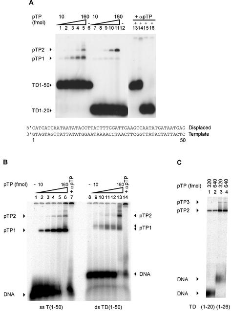
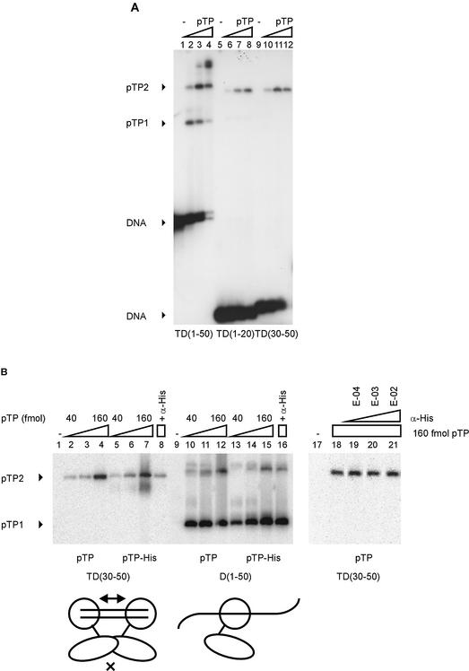
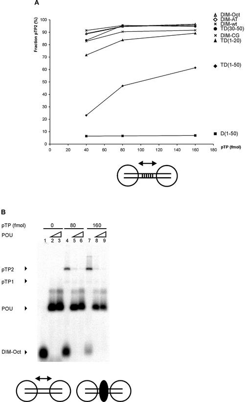
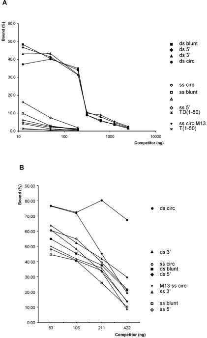
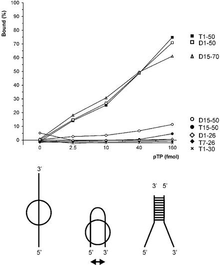
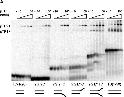
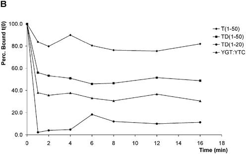
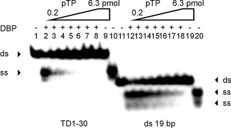
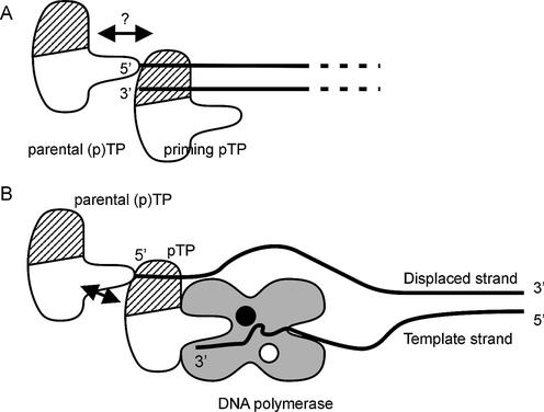
The precursor terminal protein pTP is the primer for the initiation of adenovirus (Ad) DNA replication and forms a heterodimer with Ad DNA polymerase (pol). Pol can couple dCTP to pTP directed by the fourth nucleotide of the viral genome template strand in the absence of other replication proteins, which suggests that pTP/pol binding destabilizes the origin or stabilizes an unwound state. We analyzed the contribution of pTP to pTP/pol origin binding using various DNA oligonucleotides. We show that two pTP molecules bind cooperatively to short DNA duplexes, while longer DNA fragments are bound by single pTP molecules as well. Cooperative binding to short duplexes is DNA sequence independent and most likely mediated by protein/protein contacts. Furthermore, we observed that pTP binds single-stranded (ss)DNA with a minimal length of approximately 35 nt and that random ssDNA competed 25-fold more efficiently than random duplex DNA for origin binding by pTP. Remarkably, short DNA fragments with two opposing single strands supported monomeric pTP binding. pTP did not stimulate, but inhibited strand displacement by the Ad DNA binding and unwinding protein DBP. These observations suggest a mechanism in which the ssDNA affinity of pTP stabilizes Ad pol on partially unwound origin DNA.


Figures

Figure 1
Figure 1
pTP binds both ssDNA and dsDNA. Increasing amounts of pTP were incubated with radiolabeled DNA probes containing Ad5 origin sequences and analyzed using EMSA. T(emplate) or D(isplaced) strand oligonucleotides were hybridized to form the double-stranded TD probes as indicated at the bottom of (A). DNA probes and DNA/protein complexes are indicated by arrowheads. (A) pTP binding to double-stranded TD(1–50) (lanes 1–6 and 13–14) and TD(1–20) (lanes 7–12 and 15–16). Lanes 1–5 and 7–11, 10–160 fmol (0.5–8 nM) pTP in 2-fold increments; lanes 6 and 12, free probes; lanes 13–16, 160 fmol pTP with α-pTP polyclonal antibody at 1:1000 (lanes 13 and 15) or 1:100 dilutions (lanes 14 and 16). (B) pTP binding to single-stranded T(1–50) (lanes 1–7) and double-stranded TD(1–50) (8–14). Lanes 1 and 8, free probe; lanes 2–6 and 9–13, 10–160 fmol pTP in 2-fold increments; lanes 7 and 14, 160 fmol pTP with α-pTP polyclonal antibody at 1:100. The different percentage of bound DNA in (B) when compared to (A) is caused by a lower specific activity of the DNA probes in (A), which allows the visualization of the pTP1 complex at TD(1–20). (C) pTP binding to TD(1–20) (lanes 1 and 2) and TD(1–26) (lanes 3 and 4). pTP3 marks the formation of a third pTP complex (arrowhead). Lanes 1 and 3, 160 fmol pTP; lanes 2 and 4, 320 fmol pTP.
Figure 2
Figure 2
pTP dimerizes specifically on small DNA duplexes. (A) pTP binding to TD(30–50) was studied using EMSA. Lanes 1–4, TD(1–50); lanes 5–8, TD(1–20); lanes 9–12, TD(30–50). Lanes 1, 5 and 9, free probe; lanes 2, 6 and 10, 40 fmol pTP; lanes 3, 7 and 11, 80 fmol pTP; lanes 4, 8 and 12, 160 fmol pTP. Free DNA, pTP1 and pTP2 complexes are indicated. (B) Comparison of pTP and pTP-His binding to TD(30–50) and D(1–50). Lanes 1–8 and 17–21, TD(30–50); lanes 9–16, D(1–50). Lanes 2–4 and 10–12, 40, 80 and 160 fmol pTP; lanes 5–7 and 13–15, 40, 80 and 160 fmol pTP-His. Lanes 4, 16 and 18–21, 160 fmol pTP + α-His antibody added during preincubation at E-04 (lane 19), E-03 (lane 20) or E-02 dilution (lanes 8, 16 and 21). pTP1 and pTP2 complexes are indicated; free DNA was run off the gel to obtain large separation of the pTP1 and pTP2 (and potential pTP-His/antibody) complexes. Schematically indicated at the bottom of the figure are two pTP-His (large circle with extending line)/α-His (small oval) complexes binding to the short DNA duplex TD(30–50) or a single complex bound to D(1–50). E-02, 1:100 dilution; E-03, 1:1000 dilution; E-04, 1:10 000 dilution.
Figure 3
Figure 3
Cooperative pTP binding is most likely mediated by protein/protein contacts. (A) Increasing amounts of pTP were incubated with different DNA duplexes consisting of TD(1–20) with the following 10 bp long insertions between bp 10 and 11. DIM-wt, Ad ori bp 11–20; DIM-AT, AT-rich; DIM-CG, CG-rich; DIM-Oct, octamer site. Amount of pTP2 complex as fraction of the total amount of shifted pTP/DNA complex is plotted against pTP concentration. Values are averaged over two experiments; error margins were <10% fraction pTP2. Indicated schematically at the bottom of the figure are two pTP molecules (large circles) binding to the DIM-wt probe with the variable insertion sequence indicated by parallel lines. (B) Binding of pTP to DIM-Oct was studied using EMSA. 0, (lanes 1–3), 80 (4–6) or 160 (7–9) fmol pTP was incubated with DNA in the presence of 5 (lanes 2, 5 and 8) or 10 (lanes 3, 6 and 9) ng Oct-1 POU domain. Indicated schematically at the bottom of the figure are two pTP molecules bound to DIM-Oct in the absence (left) and presence (right) of Oct-1 POU (black oval) binding the insertion sequence in the middle of the DIM-Oct probe. (C) EDC crosslinking analysis of protein/protein interaction. An aliquot of 5 µM of each protein was incubated without (– lanes) or with (+ lanes) 10 mM EDC, separated by SDS–PAGE (lanes 1 and 2, 10%; lanes 3–7, 7.5%) and detected by silver-staining (lanes 1–3) or western blotting using an α-pTP polyclonal antibody (lanes 4–7). Lane 1, ovalbumin; lane 2, NFI DNA binding domain; lane 3, Ad5 pTP; lane 4, Ad5 pTP; lane 5, Ad5 pTP + pol; lane 6, Ad4 pTP; lane 7, Ad5 pTP + 7 µM TD(1–20). Filled triangles indicate monomers, open triangles indicate dimers formed after EDC treatment.
Figure 3
Figure 3
Cooperative pTP binding is most likely mediated by protein/protein contacts. (A) Increasing amounts of pTP were incubated with different DNA duplexes consisting of TD(1–20) with the following 10 bp long insertions between bp 10 and 11. DIM-wt, Ad ori bp 11–20; DIM-AT, AT-rich; DIM-CG, CG-rich; DIM-Oct, octamer site. Amount of pTP2 complex as fraction of the total amount of shifted pTP/DNA complex is plotted against pTP concentration. Values are averaged over two experiments; error margins were <10% fraction pTP2. Indicated schematically at the bottom of the figure are two pTP molecules (large circles) binding to the DIM-wt probe with the variable insertion sequence indicated by parallel lines. (B) Binding of pTP to DIM-Oct was studied using EMSA. 0, (lanes 1–3), 80 (4–6) or 160 (7–9) fmol pTP was incubated with DNA in the presence of 5 (lanes 2, 5 and 8) or 10 (lanes 3, 6 and 9) ng Oct-1 POU domain. Indicated schematically at the bottom of the figure are two pTP molecules bound to DIM-Oct in the absence (left) and presence (right) of Oct-1 POU (black oval) binding the insertion sequence in the middle of the DIM-Oct probe. (C) EDC crosslinking analysis of protein/protein interaction. An aliquot of 5 µM of each protein was incubated without (– lanes) or with (+ lanes) 10 mM EDC, separated by SDS–PAGE (lanes 1 and 2, 10%; lanes 3–7, 7.5%) and detected by silver-staining (lanes 1–3) or western blotting using an α-pTP polyclonal antibody (lanes 4–7). Lane 1, ovalbumin; lane 2, NFI DNA binding domain; lane 3, Ad5 pTP; lane 4, Ad5 pTP; lane 5, Ad5 pTP + pol; lane 6, Ad4 pTP; lane 7, Ad5 pTP + 7 µM TD(1–20). Filled triangles indicate monomers, open triangles indicate dimers formed after EDC treatment.
Figure 4
Figure 4
pTP binds dsDNA and ssDNA with overlapping protein surfaces. DNA bound by 160 fmol pTP as a fraction of the total amount of DNA was used as the index (100%); the signals of pTP1 and pTP2 were summed. Error margins for individual points are less than 5% of binding. (A) Binding of 160 fmol pTP to TD(1–50) was challenged with increasing amounts of competitor DNA. Circular plasmid DNA (ds circ) was digested using AluI, Sau3A or CfoI to create blunt (ds blunt), 5′ overhang (ds 5′) or 3′ overhang (ds 3′) fragments. Single-stranded competitors were generated by heat denaturation followed by rapid cooling for 1 min to keep the single strands separated, after which pTP was immediately added. As a control on heat denaturation, single-stranded circular M13 DNA was used as a competitor. Controls for specific binding were unlabeled TD(1–50) probe and single-stranded T(1–50). (B) pTP binding to T(1–50) was challenged with increasing amounts of dsDNA and ssDNA competitors as described in (A).
Figure 5
Figure 5
pTP binds ssDNA with a minimal length of 35 nt without sequence dependence. pTP binding to different origin sequences was analyzed in EMSA. The DNA bound by pTP as a fraction of the total amount of DNA was determined (%). Results are averaged over two experiments with individually labeled probes with error margins <10% binding. At the bottom of the figure, a model is indicated that might explain the size of ssDNA needed for stable pTP binding. The ssDNA strand (left) could loop back to allow the simultaneous interaction of two DNA binding regions within pTP (middle), a situation similar to a partially unwound replication origin DNA structure (right).
Figure 6
Figure 6
pTP binds as a monomer to short DNA duplexes in the presence of two opposing single strands. At the bottom of (A), the structures of the DNA probes examined are schematically indicated. (A) pTP ranges from 0 to 160 fmol in 2-fold increments were incubated with the indicated DNA probes in an EMSA. YG:YC, CG-rich stem; YG:YTC, CG-stem with 5′ 8 nt T overhang; YGT:YC, CG-stem with 3′ 8 nt T overhang; YGT:YTC, CG-stem with two opposing 8 nt T overhangs. (B) Time course analysis of 8 nM pTP binding in EMSA before (0 min) and after addition of 31 ng competitor vector dsDNA (1–16 min). The fraction pTP bound to DNA was indexed at 100% to allow comparison of the relative stability of pTP binding.
Figure 6
Figure 6
pTP binds as a monomer to short DNA duplexes in the presence of two opposing single strands. At the bottom of (A), the structures of the DNA probes examined are schematically indicated. (A) pTP ranges from 0 to 160 fmol in 2-fold increments were incubated with the indicated DNA probes in an EMSA. YG:YC, CG-rich stem; YG:YTC, CG-stem with 5′ 8 nt T overhang; YGT:YC, CG-stem with 3′ 8 nt T overhang; YGT:YTC, CG-stem with two opposing 8 nt T overhangs. (B) Time course analysis of 8 nM pTP binding in EMSA before (0 min) and after addition of 31 ng competitor vector dsDNA (1–16 min). The fraction pTP bound to DNA was indexed at 100% to allow comparison of the relative stability of pTP binding.
Figure 7
Figure 7
pTP inhibits strand displacement by DBP in a DNA unwinding assay. TD(1–30) DNA (lanes 1–10, ds) or a probe unrelated to the origin (lanes 11–20, ds) were incubated with DBP (lanes 2 and 12), pTP (lanes 9 and 19) or DBP mixed with 0.2–6.3 pmol pTP in 2-fold increments (lanes 3–8 and 13–18). Displaced single strands are indicated by arrowheads (ss). Lanes 1 and 11, free probe; lanes 10 and 20, probe boiled to separate individual strands (ss).
Figure 8
Figure 8
Model of pTP interactions during replication initiation. (A) A parental (p)TP molecule stays covalently bound to the 5′ end of the Ad genome (lines) after having primed the preceding replication round. The observed cooperative binding to short DNA duplexes could mimic the interaction of an incoming priming pTP with the parental (p)TP (arrow). The ds/ssDNA binding region of pTP is indicated by hatching. (B) The ssDNA binding affinity of pTP could contribute to pTP/pol binding stability on an unwound origin structure, while the pTP region providing the priming Ser580 should present the Ser hydroxyl group to the catalytic center (black circle) in the Ad DNA pol. Potential stabilizing contacts can be made with the parental (p)TP (arrow) and two ssDNA contact points spaced by the dimensions of pTP: the displaced strand (bound in the DNA binding region) and the three 3′ terminal nucleotides of the template strand (3′).

Similar articles

Cited by

References

    1. de Jong R.N. and van der Vliet,P.C. (1999) Mechanism of DNA replication in eukaryotic cells: cellular host factors stimulating adenovirus DNA replication. Gene, 236, 1–12. - PubMed
    1. Hay R.T., Freeman,A., Leith,I., Monaghan,A. and Webster,A. (1995) Molecular interactions during adenovirus DNA replication. Curr. Top. Microbiol. Immunol., 199, 31–48. - PubMed
    1. Temperley S.M. and Hay,R.T. (1992) Recognition of the adenovirus type 2 origin of DNA replication by the virally encoded DNA polymerase and preterminal proteins. EMBO J., 11, 761–768. - PMC - PubMed
    1. Dekker J., Kanellopoulos,P.N., Loonstra,A.K., van Oosterhout,J.A., Leonard,K., Tucker,P.A. and van der Vliet,P.C. (1997) Multimerization of the adenovirus DNA-binding protein is the driving force for ATP-independent DNA unwinding during strand displacement synthesis. EMBO J., 16, 1455–1463. - PMC - PubMed
    1. Nagata K., Guggenheimer,R.A. and Hurwitz,J. (1983) Adenovirus DNA replication in vitro: synthesis of full-length DNA with purified proteins. Proc. Natl Acad. Sci. USA, 80, 4266–4270. - PMC - PubMed