. 2003 Jun 24;100(13):7465-70.
doi: 10.1073/pnas.1330328100. Epub 2003 Jun 10.

Suppressed catalytic activity of base excision repair enzymes on rotationally positioned uracil in nucleosomes

Affiliations

Suppressed catalytic activity of base excision repair enzymes on rotationally positioned uracil in nucleosomes

Brian C Beard et al. Proc Natl Acad Sci U S A. .

Abstract

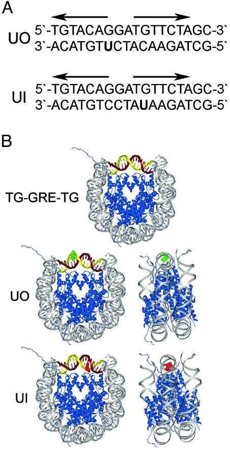
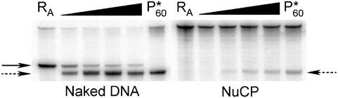
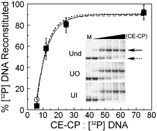
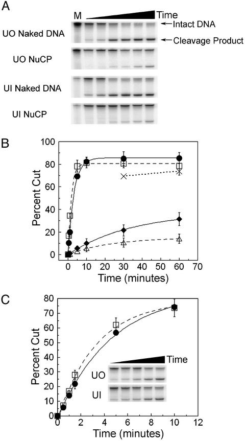
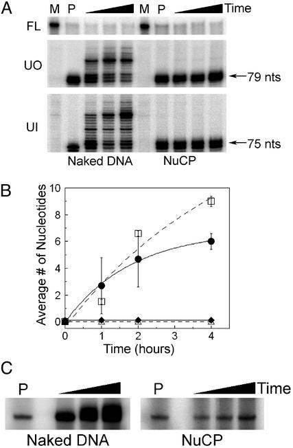
The majority of DNA in eukaryotic cells exists in the highly condensed structural hierarchy of chromatin, which presents a challenge to DNA repair enzymes in that recognition, incision, and restoration of the original sequence at most sites must take place within these structural constraints. To test base excision repair (BER) activities on chromatin substrates, an in vitro system was developed that uses human uracil DNA glycosylase (UDG), apyrimidinic/apurinic endonuclease (APE), and DNA polymerase beta (pol beta) on homogeneously damaged, rotationally positioned DNA in nucleosomes. We find that UDG and APE carry out their combined catalytic activities with reduced efficiency on nucleosome substrates ( approximately 10% of that on naked DNA). Furthermore, these enzymes distinguish between two different rotational settings of the lesion on the histone surface, showing a 2- to 3-fold difference in activity between uracil facing "toward" and "away from" the histones. However, UDG and APE will digest such substrates to completion in a concentration-dependent manner. Conversely, the synthesis activity of pol beta is inhibited completely by nucleosome substrates and is independent of enzyme concentration. These results suggest that the first two steps of BER, UDG and APE, may occur "unassisted" in chromatin, whereas downstream factors in this pathway (i.e., pol beta) may require nucleosome remodeling for efficient DNA BER in at least some regions of chromatin in eukaryotic cells.


Figures

Fig. 1.
Fig. 1.
Modified sequence element and assembled nucleosome structure. (A) GRE sequence for UO and UI substrates. Bold arrows are the pseudo-palindrome binding site for the glucocorticoid receptor. (B Top) Nucleosome schematic of the TG motif (gray) flanking the GRE (maroon and gold). (Middle and Bottom) UO and UI (red) substrates (B was adapted from ref. 4). The rotational setting of DNA in these NuCPs has been determined by hydroxyl radical footprinting (10).
Fig. 5.
Fig. 5.
RsaI digestion of UI nucleosome during UDG/APE digestion. Denaturing gel of naked DNA and NuCPs after RsaI and UDG/APE digestion. RA lanes are for naked DNA and NuCP samples digested by RsaI only (solid arrow), and P*60 lanes are for these samples digested for 60 min by UDG and APE only (dashed arrow). Aliquots of samples digested with all three enzymes were taken from 30 sec to 60 min.
Fig. 2.
Fig. 2.
Nucleosome reconstitutions. Percent of NuCPs present for different CE-CP to [32P] DNA ratios. Data represent the mean ± 1 SD (three independent reconstitutions for each substrate) of the ratio of band intensities for naked DNA and NuCPs on native gels (Inset). Undamaged DNA, solid line, ○; UO DNA, dashed line, ▪, and UI DNA, dotted line, ⋄. (Inset) Native gels of nucleosome reconstitutions at varying CE-CP concentration (naked DNA, dashed arrow; NuCP, solid arrow). Lane M is naked DNA only; the black triangle indicates increasing CE-CP concentration.
Fig. 3.
Fig. 3.
UDG and APE digestion of UO and UI nucleosomes. (A) Denaturing gels of UO and UI naked DNA and NuCPs after incubation with UDG and APE, each at 1 nM. Time is from 30 sec to 1 h and M is mock-treated. (B) Plot of the mean ± 1 SD (three independent experiments) of the percent DNA cut by 1 nM UDG and APE for UO DNA (solid line, •), UI DNA (dashed line, □), UO NuCP (solid line, ⋄), and UI NuCP (dashed line, ▵). Dotted line (×) represents nucleosome samples digested with 10 nM UDG and APE. (C) Plot of the mean ± 1 SD (three independent experiments) of the percent of DNA cut by 100 pM UDG and APE for UO DNA (solid line, □) and UI DNA (dashed line, □). (Inset) Representative gel for digestions of UO and UI naked DNA for times from 30 sec to 10 min.
Fig. 4.
Fig. 4.
pol β synthesis activity on UO and UI nucleosomes. (A) Denaturing gels of UO and UI naked DNA and NuCPs. Shown is the uncut fraction remaining (Top) and increasing size of fragment (Middle and Bottom) after incorporation of nucleotides by pol β into naked DNA or NuCPs. Lanes M are for mock-treated samples (no enzyme present), and lanes P are for samples digested with UDG and APE only. Incubation times are from 1 to 4 h with all three enzymes. (B) Plot of the mean ± 1 SD (three independent experiments) of the average number of nucleotides inserted by pol β into UO DNA (solid line, •), UI DNA (dashed line, □), UO NuCP (solid line, ⋄), and UI NuCP (dashed line, ▵). (C) Denaturing gels of UI naked DNA and NuCPs showing the 5′ end-labeled cleavage product and subsequent addition of radionucleotides to the 3′ terminus. P represents incubation with UDG and APE alone, and the black triangles represent increasing incubation time (30 min to 2 h) with UDG, APE, pol β, and [α-32P]dCTP.

Similar articles

Cited by

References

    1. Hoeijmakers, J. H. J. (2001) Nature 411, 366–374. - PubMed
    1. Wood, R. D., Mitchell, M., Sgouros, J. & Lindahl, T. (2001) Science 291, 1284–1289. - PubMed
    1. Hanawalt, P. C. (1998) Mutat. Res. 400, 117–125. - PubMed
    1. Luger, K., Mader, A. W., Richmond, R. K., Sargent, D. F. & Richmond, T. J. (1997) Nature 389, 251–260. - PubMed
    1. Wolffe, A. P. (1998) Chromatin: Structure and Function (Academic, San Diego).

Publication types