. 2003 Jun 16;88(12):1971-8.
doi: 10.1038/sj.bjc.6600986.

Zoledronic acid induces antiproliferative and apoptotic effects in human pancreatic cancer cells in vitro

Affiliations

Zoledronic acid induces antiproliferative and apoptotic effects in human pancreatic cancer cells in vitro

P Tassone et al. Br J Cancer. .

Abstract

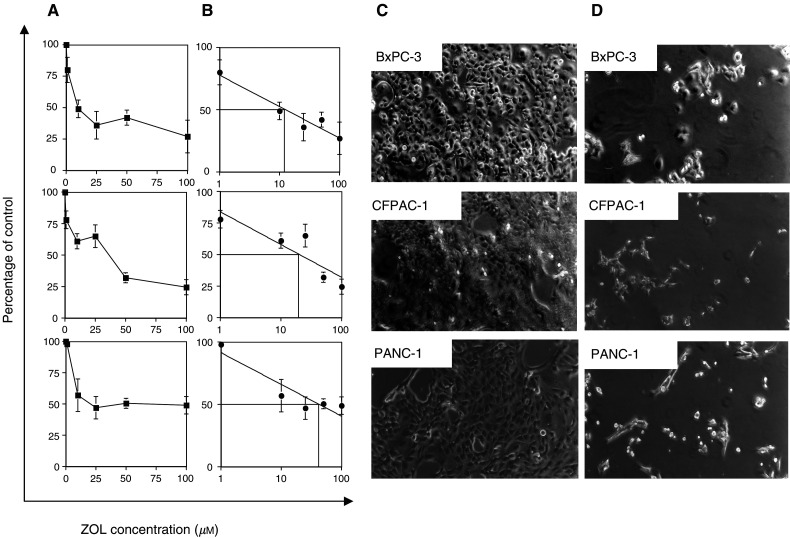
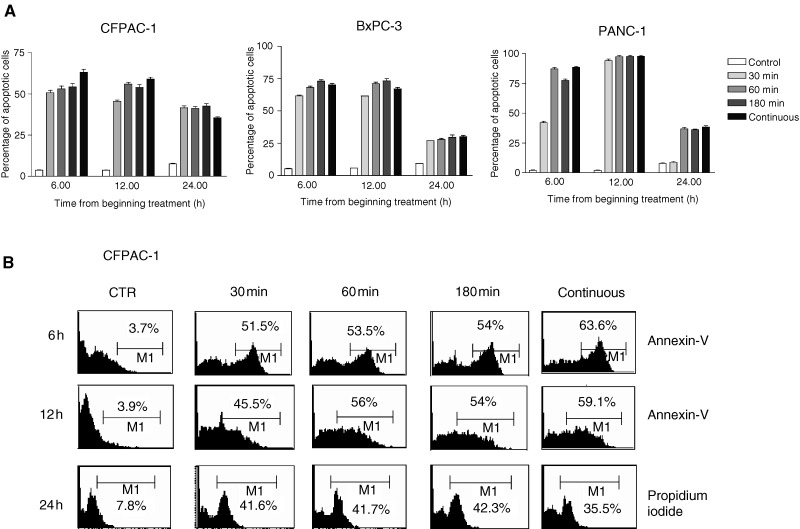
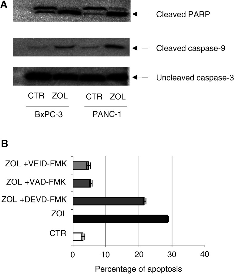
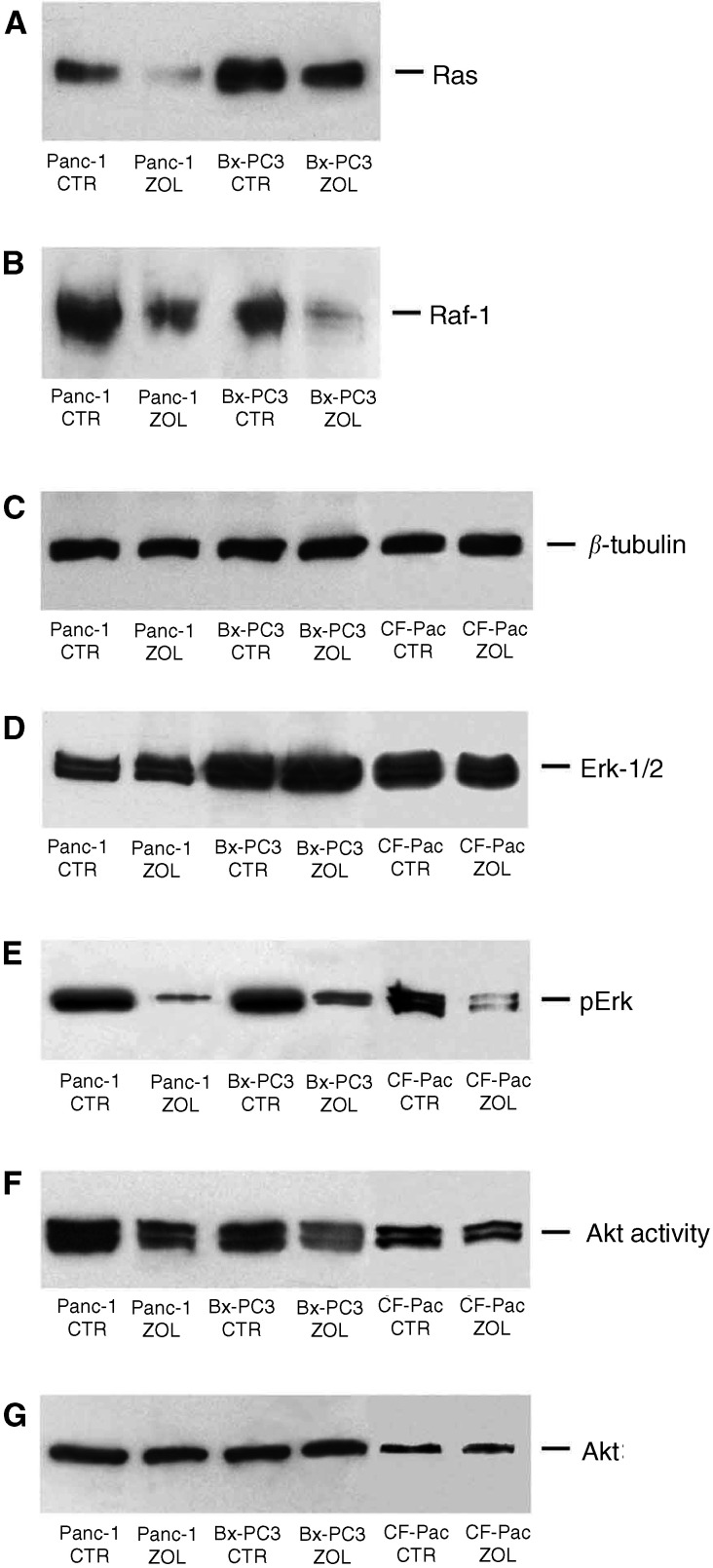
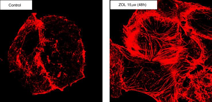
Bisphosphonates (BPs) are an emerging class of drugs mostly used in the palliative care of cancer patients. We investigated the in vitro activity of the most potent antiresorptive BP, zoledronic acid (ZOL), on the growth and survival of three human pancreatic cancer (PC) cell lines (BxPC-3, CFPAC-1 and PANC-1). Pancreatic cancer frequently has a dysregulated p21(ras) pathway and therefore appears to be a suitable target for BPs that interfere with the prenylation of small GTP-binding proteins such as p21(ras). We found that ZOL induces growth inhibition (IC(50):10-50 micro M) and apoptotic death of PC cells. The proapoptotic effect was correlated to cleavage/activation of caspase-9 and poly(ADP)-ribose polymerase, but not of caspase-3. Moreover, we studied the p21(ras) signalling in cells exposed to ZOL and detected a reduction of p21(ras) and Raf-1 content and functional downregulation of the terminal enzyme ERK/MAPkinase and of the pKB/Akt survival pathway. Finally, we observed that ZOL induces significant cytoskeletal rearrangements. In conclusion, we demonstrated that ZOL induces growth inhibition and apoptosis on PC cells and interferes with growth and survival pathways downstream to p21(ras). These findings might be relevant for expanding application of BPs in cancer treatment.


Figures

Figure 1
Figure 1
Zoledronic acid inhibits the growth of PC cells. (A) The cell lines BxPC-3, CFPAC-1 and PANC-1 (3 × 10⁴ ml−1) were seeded in 96-well plates and incubated for 24 h. After 24 h, medium was removed and replaced with medium containing increasing concentrations of ZOL (1–100 μM). After 72 h of incubation, cell proliferation was determined by the MTT assay. A value of 100% was assigned to untreated control cultures, and the antiproliferative effects of ZOL were calculated as percentage of residual growth. Each point is the mean value of at least four replicate experiments ±s.e. (B) The IC50 drug concentration for each cell line was measured by a logarithmic interpolate curve on the percentage of residual growth at each concentration point (C and D) Morphological changes induced in cell cultures after 72 h exposure to ZOL. Analysis was performed by inverted phase-contrast microscopy. (C) Untreated cultures; (D) treated cultures with ZOL (50 μM).
Figure 2
Figure 2
(A) Apoptotic effects induced by ZOL on CFPAC-1, PANC-1, BxPC-3 PC cancer cells. The analysis of apoptosis was performed after exposure to 50 μM of ZOL by flow cytometric detection of Annexin-V immunostaining. Cells were pulse exposed for 30, 60 and 180 min or continuously exposed to the drug. The PI method was used when apoptosis was evaluated 24 h after beginning treatment. Data are expressed as the mean value of at least four replicate experiments ±s.e. (B) Flow cytometric profiles from a representative experiment performed on CFPAC-1 cells. The percentage of apoptotic stained cells (%) is indicated in each quadrant.
Figure 3
Figure 3
Role of caspase-9/-3 and PARP in apoptotic cell death induced by ZOL. (A) Detection of caspase-9/-3 and PARP in PC cells after ZOL treatment. Western blotting analysis was performed on protein extracts from solubilized whole cell pellets from BxPC-3 and PANC1 PC cell lines after 48 h exposure to ZOL (50 μM) demonstrating abundant cleaved species of both caspase-9 and PARP in lysates for drug-exposed cells, while caspase-3 remained almost completely uncleaved. The experiments were performed at least three times and the results were always similar. (B) Apoptotic effects induced by ZOL in the presence of caspase inhibitors. Analysis was performed by flow cytometric immunodetection of Annexin-V staining on BxPC-3 cells after 48 h exposure to ZOL (50 μM). VAD is a caspase-9 inhibitor, VEID is a specific caspase-6 inhibitor and DEVD is a caspase-3 inhibitor. Pretreatment with 15 μM VAD and 15 μM VEID significantly antagonised ZOL-induced apoptosis (P<0.005).
Figure 4
Figure 4
Effects of ZOL treatment on p21ras/Raf-1/MEK1/ERK signalling and PK-B/Akt pathways in PC cells. Western blotting analysis of p21ras (A), raf-1 (B), Erk-1/2 (D) and pErk (E). The immunoblotting shows downregulation of p21ras (A) and raf-1 (B) levels after 48 h of exposure to 50 μM ZOL. While no significant effects were detected on Erk-1/2 expression after exposure of cells to the drug (D), the active phosphorylated Erk-1/2 (recognised by an anti-pERK Mab) levels were greatly reduced in ZOL-treated cells (E). (F and G) Akt expression and functional analysis in BxPC-3, PANC-1 and CFPAC-1 pancreatic adenocarcinoma cells. For detection of Akt activity, immunoprecipitation was performed from cell lysates with anti-Akt antibody, and an in vitro kinase assay was performed using GSK3α/β as a substrate, followed by Western blotting analysis with antiphospho-GSK3α/β. GSK3α/β phosphorylation by immunoprecipitated Akt was downregulated after 48 h treatment of PC cells with 50 μM ZOL (F), whereas the Akt protein levels remained unmodified. The experiments were performed at least three times and the results were always similar. CTR: untreated cells; ZOL: ZOL-treated cells.
Figure 5
Figure 5
Analysis of cytoskeletal reorganisation by confocal microscopy on PANC-1 PC cells after 48 h exposure to ZOL (15 μM). The figure shows actin architecture rearrangements in cortical rings. The cells were examined under a confocal microscope at a magnification of × 100. The experiments were performed at least three times and the results were always similar.

Similar articles

Cited by

References

    1. Almoguera C, Shibata D, Forrester K, Martin J, Arnheim N, Perucho M (1988) Most human carcinomas of the exocrine pancreas contain mutant c-Kras gene. Cell 53: 549–554 - PubMed
    1. Aparicio A, Gardner A, Tu Y, Savage A, Berenson J, Lichtenstein A (1998) In vitro cytoreductive effects on multiple myeloma cells induced by bisphosphonates. Leukemia 12: 220–229 - PubMed
    1. Benford HL, Frith JC, Auriola S, Monkkonen J, Rogers MJ (1999) Farnesol and geranylgeraniol prevent activation of caspases by aminobisphosphonates: biochemical evidence for two distinct pharmacological classes of bisphosphonate drugs. Mol Pharmacol 56: 131–140 - PubMed
    1. Benford HL, McGowan NW, Helfrich MH, Nuttall ME, Rogers MJ (2001) Visualization of bisphosphonate-induced caspase-3 activity in apoptotic osteoclasts in vitro. Bone 28: 465–473 - PubMed
    1. Berenson JR, Lichtenstein A, Porter L, Dimopoulos MA, Bordoni R, George S, Lipton A, Keller A, Ballester O, Kovacs M, Blacklock H, Bell R, Simeone JF, Reitsma DJ, Heffernan M, Seaman J, Knight RD (1998) Long-term pamidronate treatment of advanced multiple myeloma patients reduces skeletal events. Myeloma Aredia Study Group. J Clin Oncol 16: 593–602 - PubMed

Publication types

MeSH terms