. 2003 May;82(5):209-21.
doi: 10.1078/0171-9335-00316.

Loss of function and impaired degradation of a cataract-associated mutant connexin50

Affiliations

Loss of function and impaired degradation of a cataract-associated mutant connexin50

Viviana M Berthoud et al. Eur J Cell Biol. 2003 May.

Abstract

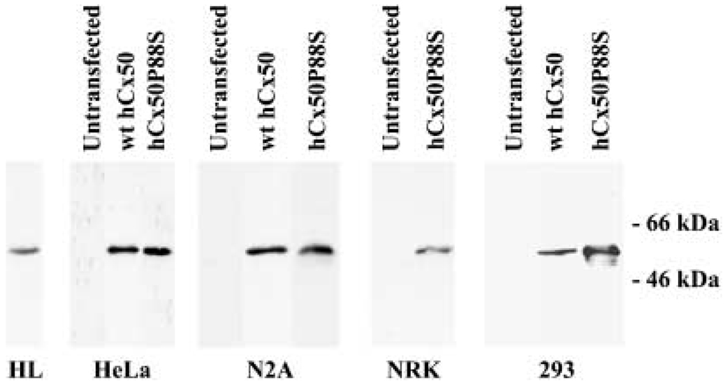
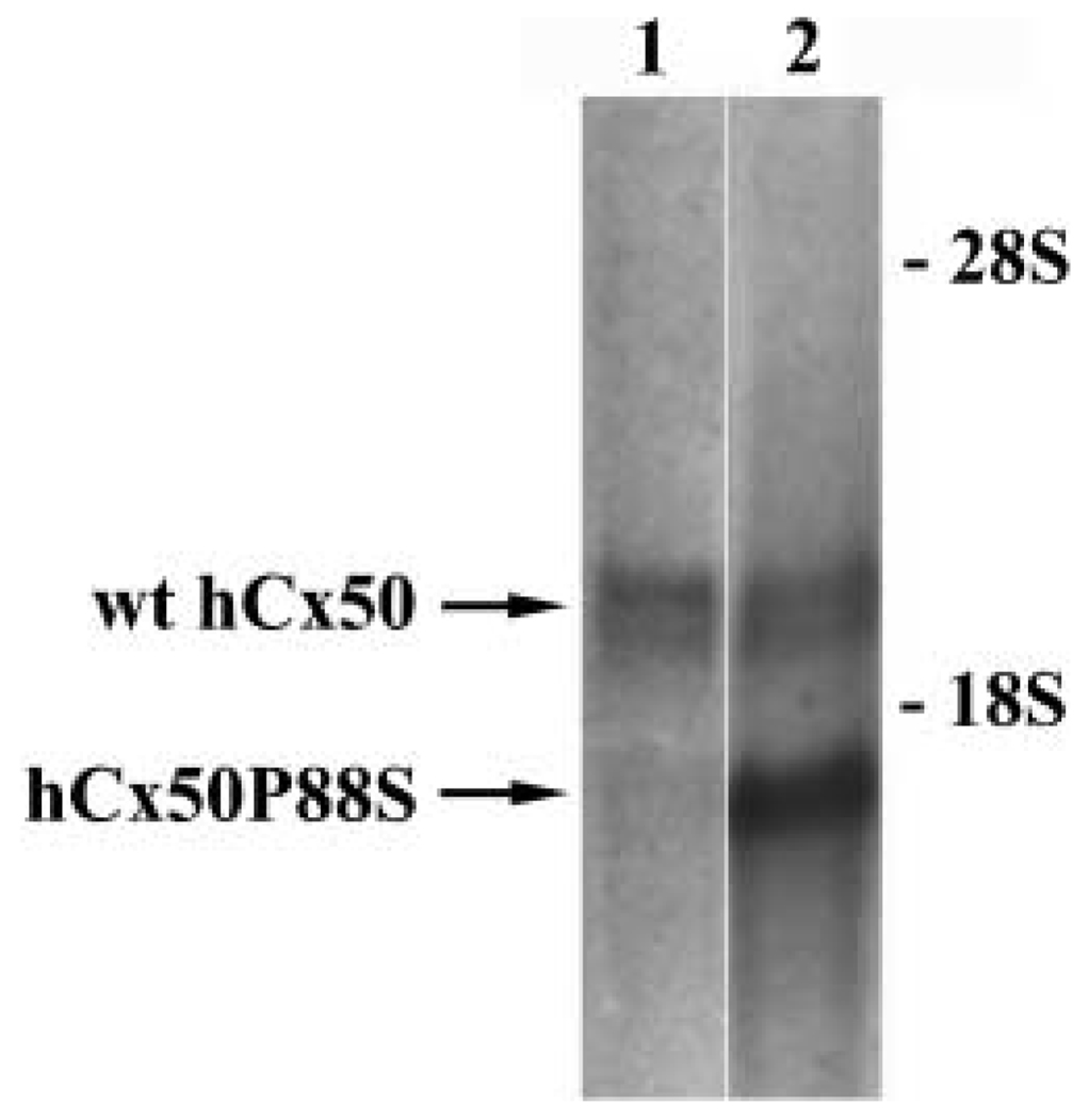
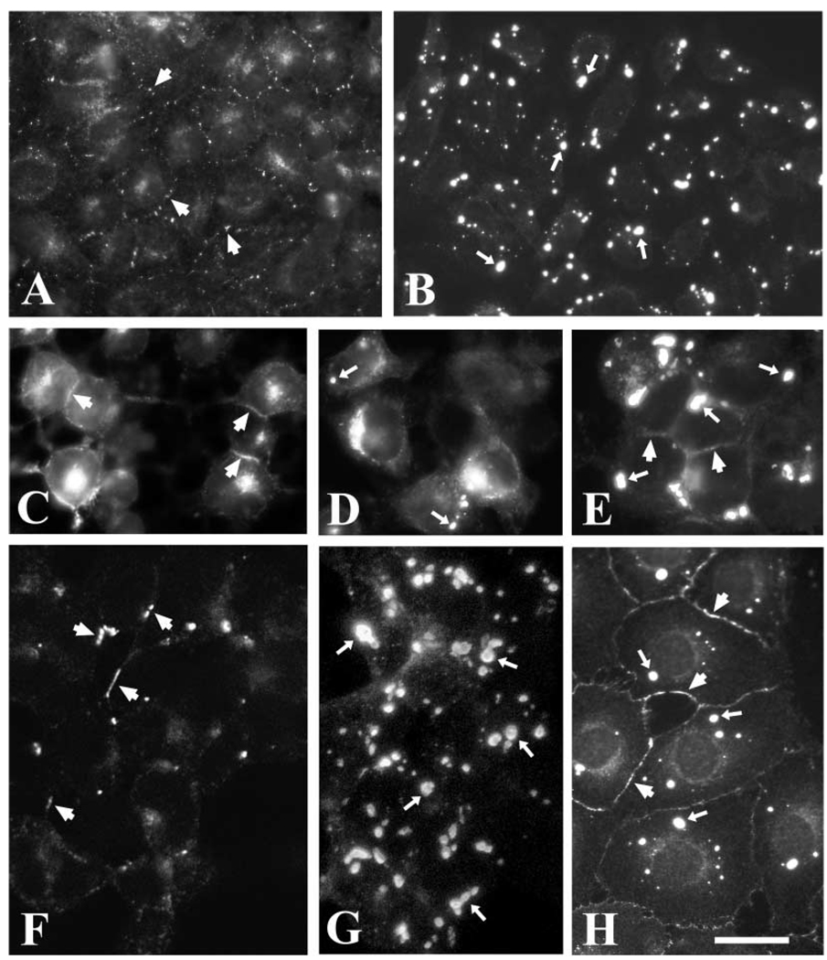
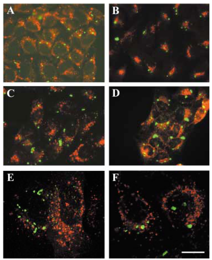
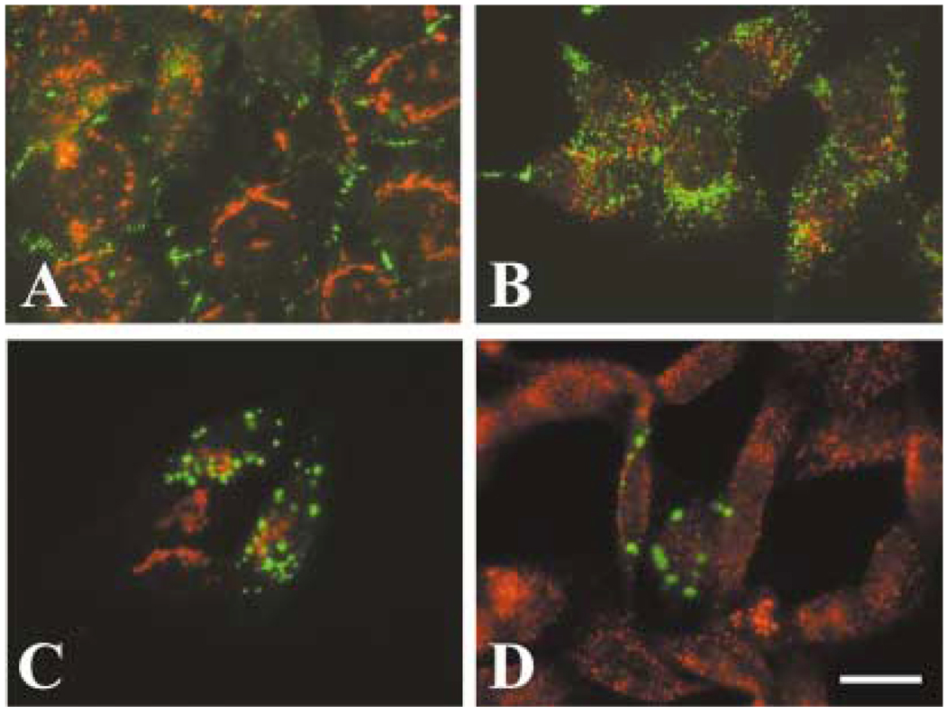
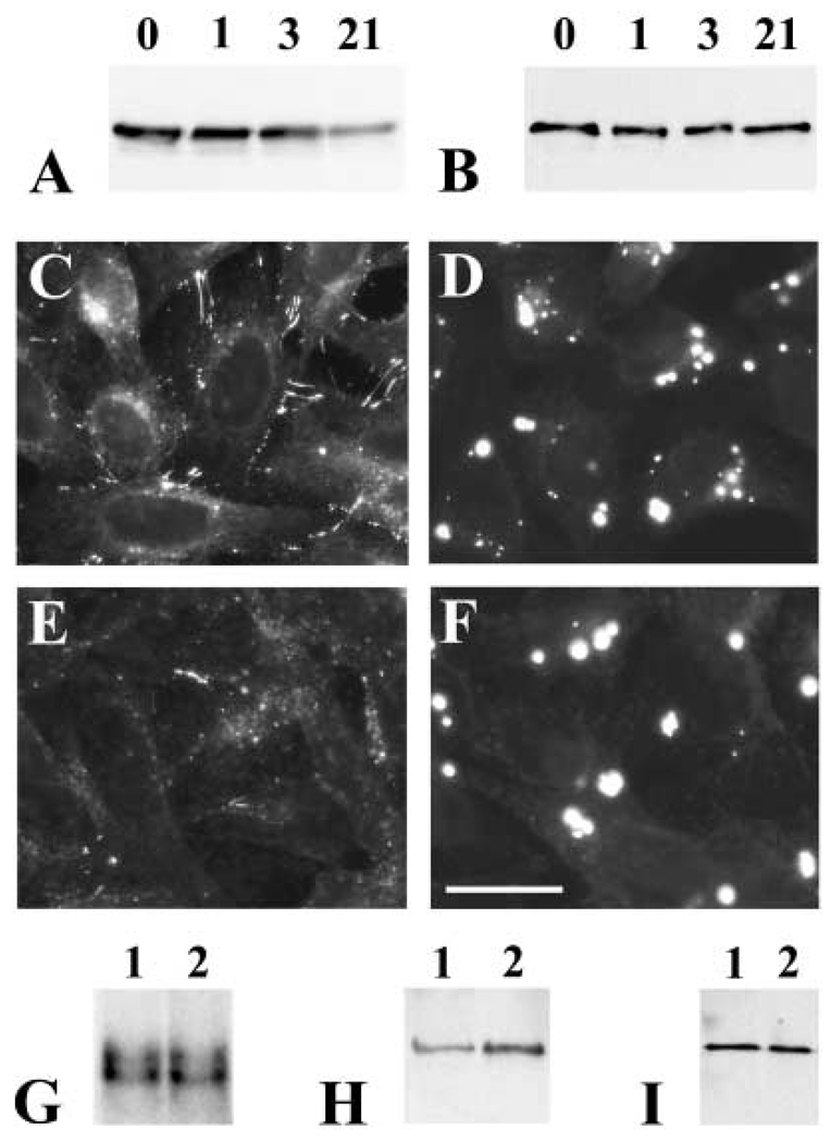
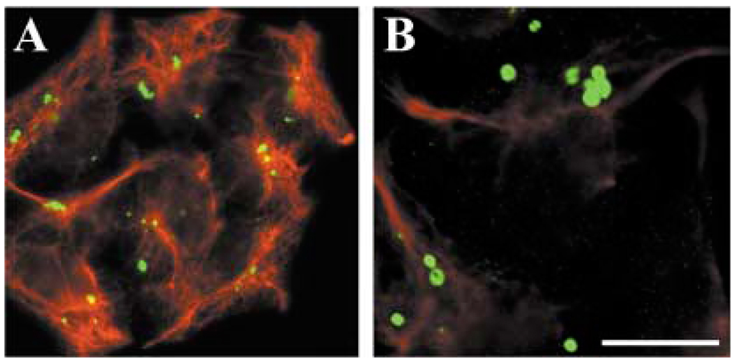
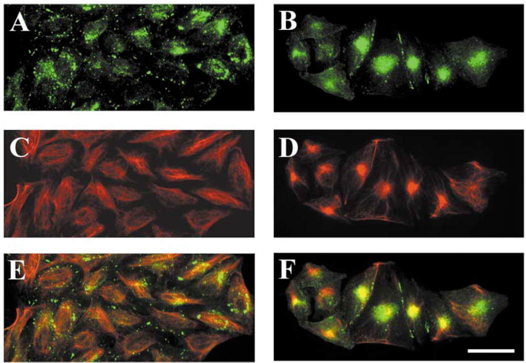
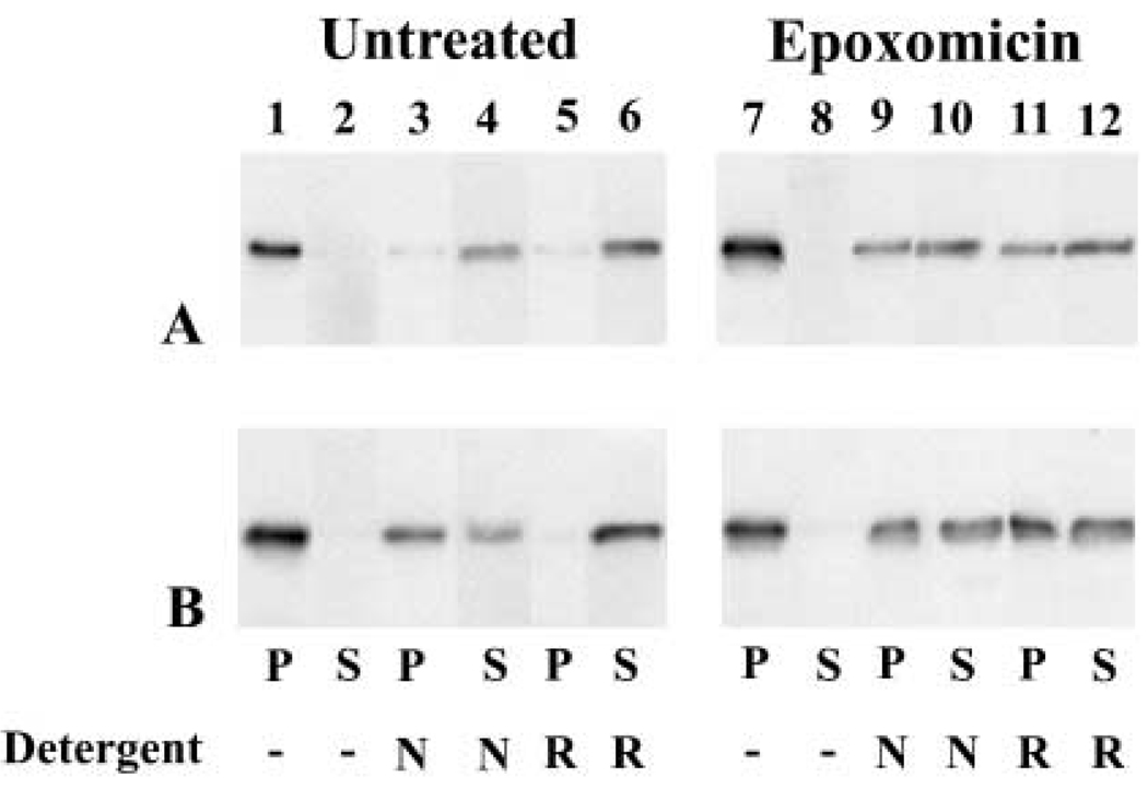
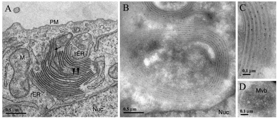
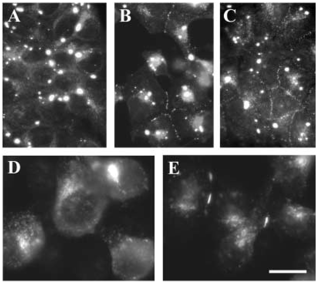
A mutant human connexin50 (hCx50), hCx50P88S, has been linked to cataracts inherited as an autosomal dominant trait. The functional, biochemical and cellular behavior of wild-type and mutant hCx50 were examined in transfected cells. hCx50P88S was unable to induce gap junctional currents by itself, and it abolished gap junctional currents when co-expressed with wild-type (wt) hCx50. Cells transfected with hCx50P88S showed cytoplasmic accumulations of Cx50 immunoreactivity in addition to staining at appositional membranes; these accumulations did not significantly co-localize with markers for the endoplasmic reticulum, Golgi apparatus, lysosomes, endosomes or vimentin filaments. Immunoelectron microscopy studies localized hCx50P88S to cytoplasmic membrane stacks in close vicinity to the endoplasmic reticulum. In contrast, aggresome-like accumulations were induced by treatment of wt hCx50-transfected cells with proteasomal inhibitors. The formation of hCx50P88S accumulations in transiently transfected cells was not blocked by treatment with Brefeldin A suggesting that they form before Cx50 transits through the Golgi apparatus to the plasma membrane. Treatment of HeLa-hCx50P88S cells with cycloheximide demonstrated the presence of a very stable pool of hCx50P88S. Taken together, these results suggest that the P to S mutation at amino acid residue 88 causes a defect that leads to decreased degradation and subsequent accumulation of hCx50P88S in a cellular structure different from aggresomes.


Figures

Fig. 1
Fig. 1
Detection of wild-type human Cx50 or Cx50P88S protein in transfected cells. Proteins from whole cell homogenates of untransfected N2A, HeLa, NRK or 293 cells or cells transfected with wt hCx50 or hCx50P88S were resolved by SDS-PAGE and subjected to immunoblotting using rabbit polyclonal anti-human Cx50 antibodies. A sample from a human lens homogenate (HL) was used as a positive control. The positions of the 66- and 46-kDa molecular mass standards are indicated on the right.
Fig. 2
Fig. 2
Co-expression of wild-type hCx50 and hCx50P88S. Total RNA from N2A cells transfected with wild-type hCx50 (lane 1) or both human wild-type hCx50 and hCx50P88S (lane 2) was hybridized with a hCx50 probe. The identities of the hybridizing bands are indicated by arrows. Because cells were transfected with wt hCx50 in the pSFFV-neo vector and hCx50P88S in the pcDNA3.1/Hygro(+) vector, the sizes of the mRNAs differ. The positions of the 18S and 28S ribosomal RNAs are indicated.
Fig. 3
Fig. 3
Distribution of anti-Cx50 immunoreactivity in transfected cells. Photomicrographs show the distribution of anti-Cx50 immunoreactivity in HeLa (A, B), N2A (C, D, E), 293 (F, G) and NRK (H) cells transfected with human wild-type Cx50 (A, C, F), hCx50P88S (B, D, G, H), or both wild-type and mutant hCx50 (E). Panel F is a confocal image and panel G is a reconstruction of a confocal z series. Staining at membrane appositions (short arrows) and cytoplasmic accumulations (long arrows) is indicated. Bar represents 35 µm in A, 37 µm in B, 21 µm in C and D, 31 µm in E, F and G, and 33 µm in H.
Fig. 4
Fig. 4
Lack of co-localization of anti-Cx50 immunoreactivity with several cellular compartments. (A–D) Photomicrographs of double-immunofluorescence staining of HeLa cells stably transfected with hCx50P88S using rabbit polyclonal anti-Cx50 antibodies and a mouse monoclonal antibody directed against (A) protein disulfide isomerase (an endoplasmicretic reticulum-resident protein), (B) Golgi 58K protein (a Golgi-resident protein), (C) the lysosome-associated membrane protein 1 (LAMP1), or (D) transferrin receptor (a marker for the endocytic pathway). (E, F) Photomicrographs of HeLa cells transiently (E) or stably (F) transfected with hCx50P88S showing the lack of co-localization of Cx50 immunoreactivity with tetramethylrhodamine-dextran (shown in red) used as a different marker for the endocytic pathway. Anti-Cx50 immunoreactivity is shown in green; immunoreactivity to the antibodies against proteins from specific cellular compartments is shown in red. Bar represents 31 µm in A, 27 µm in B and C, 47 µm in D, 14 µm in E and 12 µm in F.
Fig. 5
Fig. 5
Accumulations of hCx50P88S form before the protein transits through the Golgi to the plasma membrane. Photomicrographs showing the distribution of Cx50 (green) and Golgi 58K protein (red) immunoreactivities in HeLa cells stably transfected with wt hCx50 (A, B) or transiently transfected with hCx50P88S (C, D) under control conditions (A, C) or after treatment with BFA (B, D). For the transient transfection experiments, BFA treatment started 1 h after DNA transfection; cells were fixed 24 h after transfection and then processed for immunofluorescence. Co-localization of the two signals appears yellow. Bar represents 21 µm.
Fig. 6
Fig. 6
Presence of a stable pool of immunoreactive Cx50 in cells transfected with hCx50P88S. (A, B) Immunoblots of homogenates from HeLa-wt hCx50 (A) or HeLa-hCx50P88S (B) cells that were left untreated (lanes 0) or treated with 40 µg/ml cycloheximide for 1, 3 or 21 h (lanes 1, 3, 21, respectively) using anti-Cx50 antibodies. (Exposure times for A and B were different). (C–F) Photomicrographs showing Cx50 immunoreactivity in HeLa cells transfected with wild-type hCx50 (C, E) or hCx50P88S (D, F) that were left untreated (C, D) or treated with cycloheximide for 12 hours (E, F). (G) Northern blot of Cx50 RNA from HeLa-wt hCx50 (lane 1) and HeLa-hCx50P88S (lane 2) cells hybridized with a hCx50 probe. (Because cells were transfected with DNAs encoding wt hCx50 or hCx50P88S in the same vector, pSFFV-neo, the mRNA sizes of wt hCx50 and hCx50P88S are identical). (H, I) Immunoblots of homogenates from HeLa-wt hCx50 (lanes 1) and HeLa-hCx50P88S (lanes 2) cells reacted with anti-Cx50 (H) or antivimentin (I) antibodies. Bar represents 29 µm in C, and 26 µm in D, E and F.
Fig. 7
Fig. 7
Absence of co-localization of hCx50P88S accumulations with vimentin. Anti-Cx50 (green) and anti-vimentin (red) immunoreactivities after double-immunofluorescence staining of HeLa cells transfected with hCx50P88S. An epifluorescence photomicrograph is shown in A; a confocal image is shown in B. Bar represents 33 µm in A and 13 µm in B.
Fig. 8
Fig. 8
Induction of accumulations of wt hCx50 following treatment of cultured cells with proteasomal inhibitors. Photomicrographs of HeLa cells transfected with wt hCx50 cultured under control conditions (A, C, E) or treated with 10 µM clasto-lactacystin β-lactone (B, D, F) after double-label immunostaining using anti-Cx50 (green; A, B) and anti-vimentin (red; C, D) antibodies. Superposition of the two signals is shown in E and F; the overlap between the two signals appears yellow. Bar represents 46 µm.
Fig. 9
Fig. 9
Solubility of Cx50 in HeLa cells and effects of proteasomal inhibitor treatment. Homogenates prepared from HeLa cells transfected with wt hCx50 (A) or hCx50P88S (B) under control conditions (lanes 1 – 6) or after treatment with 0.5 µM epoxomicin (lanes 7 – 12) were incubated in the absence or in the presence of 1% NP40 (N) or RIPA buffer (R). After 30 min, the samples were centrifuged at 13 000g and the pellets (P) were separated from the supernatants (S) and subjected to immunoblotting.
Fig. 10
Fig. 10
Electron micrographs showing the fine structure and immunogold labeling of HeLa-hCx50P88S cells. (A) Transmission electron micrograph showing the presence of membrane stacks in the cytoplasm of these cells; in most areas, membranes are closely apposed (arrow-heads) similar to the appearance of normal gap junctions, but other regions of the membrane show a widened intermembrane space (arrow). Rough ER (rER) is seen associated with membrane stacks. These individual membranes curve around to make contact with other membranes about 5 – 10 stacks away (*). (B and C) Cryosections of immunogold stained specimens showing membrane stacks. Membrane stacks showed intense labeling with anti-Cx50 antibodies; these labeled rings of membranes correspond to those identified by transmission electron microscopy (different orientation of profile). An enlarged view is shown in C. (D) Cryo-immunolocalization was used to examine classic multi-vesicular bodies (Mvb) in these cells. In no case, were any Mvbs seen to react with anti-Cx50 antibodies.
Fig. 11
Fig. 11
Increased hCx50P88S immunoreactivity at appositional membranes after incubation at lower temperatures or in the presence of chemical chaperones. Photomicrographs of anti-Cx50 immunoreactivity from HeLa (A–C) or N2A (D, E) cells transfected with hCx50P88S grown at 37 °C and kept at 37 °C (A, C, D) or shifted to 30°C (E) or 25°C (B) grown under control conditions (A, B, D, E) or treated with 1 mM sodium 4-phenylbutyrate for 24 hours (C). Bar represents 50 µm in A, 62 µm in B, 31 µm in C, and 13 µm in D and E.

References

    1. Beyer EC, Paul DL, Goodenough DA. Connexin43: a protein from rat heart homologous to a gap junction protein from liver. J. Cell Biol. 1987;105:2621–2629. - PMC - PubMed
    1. Beyer EC, Kistler J, Paul DL, Goodenough DA. Antisera directed against connexin43 peptides react with a 43-kD protein localized to gap junctions in myocardium and other tissues. J. Cell Biol. 1989;108:595–605. - PMC - PubMed
    1. Bradford MM. A rapid and sensitive method for the quantitation of microgram quantities of protein using the principle of protein-dye binding. Anal. Biochem. 1976;72:248–254. - PubMed
    1. Church RL, Wang JH, Steele E. The human lens intrinsic membrane protein MP70 (Cx50) gene – clonal analysis and chromosome mapping. Curr. Eye Res. 1995;14:215–221. - PubMed
    1. Denning GM, Anderson MP, Amara JF, Marshall J, Smith AE, Welsh MJ. Processing of mutant cystic fibrosis transmembrane conductance regulator is temperature sensitive. Nature. 1992;358:761–764. - PubMed

Publication types