. 2003;4(6):R36.
doi: 10.1186/gb-2003-4-6-r36. Epub 2003 May 13.

The mosaic structure of the symbiotic plasmid of Rhizobium etli CFN42 and its relation to other symbiotic genome compartments

Affiliations

The mosaic structure of the symbiotic plasmid of Rhizobium etli CFN42 and its relation to other symbiotic genome compartments

Víctor González et al. Genome Biol. 2003.

Abstract

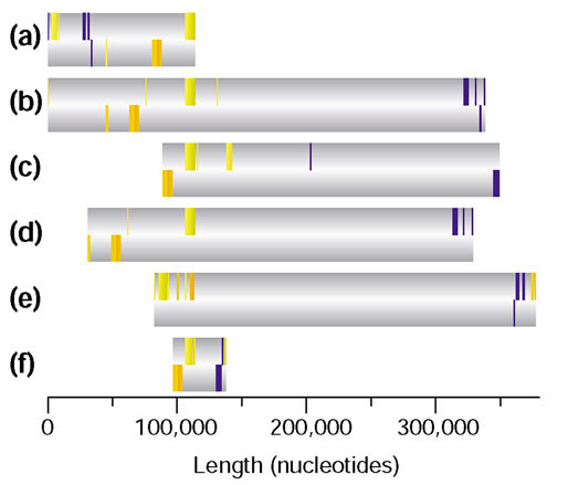
Background: Symbiotic bacteria known as rhizobia interact with the roots of legumes and induce the formation of nitrogen-fixing nodules. In rhizobia, essential genes for symbiosis are compartmentalized either in symbiotic plasmids or in chromosomal symbiotic islands. To understand the structure and evolution of the symbiotic genome compartments (SGCs), it is necessary to analyze their common genetic content and organization as well as to study their differences. To date, five SGCs belonging to distinct species of rhizobia have been entirely sequenced. We report the complete sequence of the symbiotic plasmid of Rhizobium etli CFN42, a microsymbiont of beans, and a comparison with other SGC sequences available.

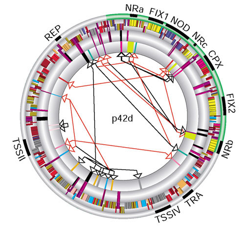
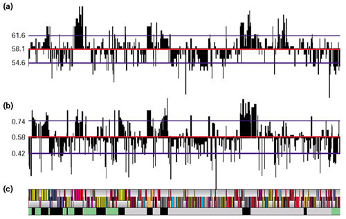
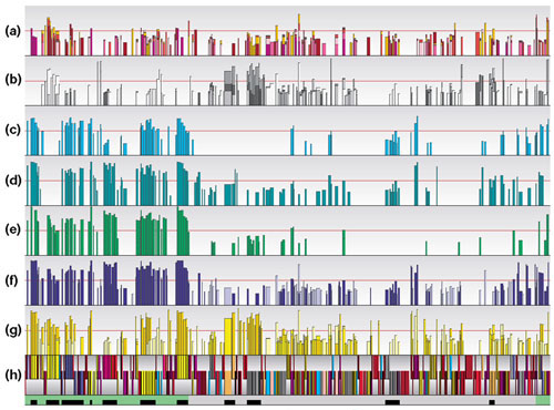
Results: The symbiotic plasmid is a circular molecule of 371,255 base-pairs containing 359 coding sequences. Nodulation and nitrogen-fixation genes common to other rhizobia are clustered in a region of 125 kilobases. Numerous sequences related to mobile elements are scattered throughout. In some cases the mobile elements flank blocks of functionally related sequences, thereby suggesting a role in transposition. The plasmid contains 12 reiterated DNA families that are likely to participate in genomic rearrangements. Comparisons between this plasmid and complete rhizobial genomes and symbiotic compartments already sequenced show a general lack of synteny and colinearity, with the exception of some transcriptional units. There are only 20 symbiotic genes that are shared by all SGCs.

Conclusions: Our data support the notion that the symbiotic compartments of rhizobia genomes are mosaic structures that have been frequently tailored by recombination, horizontal transfer and transposition.


Figures

Figure 1
Figure 1
Structure of the symbiotic plasmid p42d of R. etli CNF42. The structure of p42d is represented in five concentric circles. Outermost circle, relevant regions referred to in the text: NRa, b and c, regions containing nitrogenase structural genes; FIX1 and FIX2, clusters containing nitrogen-fixation genes; NOD, major cluster of nodulation genes; CPX, cluster for cytochrome P450; TRA, cluster for tra genes; REP, replicator region; TSSIII and IV, clusters for transport secretion system genes. The 125 kb region that contains most of the symbiotic genes, described in the text as a putative mobile element, is shown in green. Second circle, organization of predicted CDSs located according to the direction of transcription color-coded as below; those transcribed on the plus strand are shown in the outer half of the circle. For each class, the number of CDSs and the percentage of the total are: hypothetical (70) 19.5% (dark red); hypothetical conserved (62) 17.3% (red); integration recombination (55) 15.3% (purple); various enzymatic functions (45) 12.3% (khaki); transport secretion systems (37) 10.3% (gray); nitrogen fixation (35) 9.8% (yellow); nodulation (18) 5% (dark blue); transcriptional regulation (15) 4.2% (light blue); plasmid maintenance (10) 2.8% (orange); electron transfer (7) 2.1% (magenta); chemotaxis (3) 0.8% (pink); and polysaccharide synthesis (2) 0.6% (green). Third circle, elements related to insertion sequences (ERIS). Putative partial ISs (purple), and putative complete ISs (black). Fourth circle, reiterated DNA families. The major reiterated families (see text) are shown in different colors. Innermost circle, potential genomic rearrangements. Arrowheads indicate the sites for homologous recombination leading to genomic rearrangements. Black lines connect sites for amplification or deletion events; red lines connect sites for inversion.
Figure 2
Figure 2
Compositional features of the coding sequences (CDS) of p42d. (a) GC content, and (b) CU of the 359 CDS of p42d. Red lines indicate the average in GC (58.1%) and CU (0.58). Blue lines indicate 1 standard deviation of GC ± 3.5% and CU ± 0.16. Highest and lowest percentage values of GC are 69.4 and 45.8 respectively. The CU limit values varies from 0.11 to 1.00. (c) CDS distribution with the color codes for functional classes and the relevant regions described in Figure 1.
Figure 3
Figure 3
Comparison of predicted proteins from p42d with those from other genomes and SGCs. Bidirectional best hits (BDBHs) between p42d and other genomes are shown. The bars in all rows represent the percentage identity (number of identities/length of the alignment) of BDBHs between p42d and the indicated genome (see below for color code). The horizontal red line in each row indicates 50% of similarity. A color code is shown for each genome or compartment. (a) Different organisms: Bacillus subtilis (dark magenta); Brucella melitensis (yellow); Caulobacter crescentus (red); Escherichia coli K12 (light magenta), Methanobacterium thermoautotrophicum (dark purple), and Ralstonia solanacearum (purple). (b) A. tumefaciens C58 circular chromosome (white), linear chromosome (pale gray), pAT (gray), and pTi (dark gray). (c) B. japonicum USDA110 SGC (turquoise). (d) pNGR234a (blue green). (e) M. loti R7A SGC (green). (f) M. loti MAFF303099 SGC (dark blue), and the rest of the chromosome (light blue). (g) S. meliloti pSymA (pale yellow), pSymB (yellow), and the chromosome (dark yellow). (h) CDS distribution for p42d with the color codes for functional classes and the relevant regions as indicated in Figure 1.
Figure 4
Figure 4
Analysis of synteny among the SGCs. Pairs of orthologous proteins among different genomes or SGCs are plotted. Each protein pair is shown according to the location of the corresponding coordinate of the predicted translation start of the gene on the DNA region. The axes correspond to the total length of the respective DNA region: p42d 371,255 bp; M. loti MAFF303099 symbiotic island 610,975 bp; M. loti R7A symbiotic island 502,000 bp; S. meliloti pSymA 354,226 bp; pNGR234a 536,165 bp and B. japonicum symbiotic region 410,573 bp. For each group the first region mentioned corresponds to the x-axis. (a) p42d vs pNGR234a; (b) p42d vs pSymA; (c) p42d vs B. japonicum symbiotic region; (d) p42d vs M. loti MAFF303099 symbiotic island; (e) p42d vs M. loti R7A, symbiotic island; (f) pNGR234a vs S. meliloti pSymA; (g) pNGR234a vs B. japonicum symbiotic region; (h) pNGR234a vs M. loti 303099 symbiotic island; (i) pNGR234a vs M. loti R7A symbiotic island; (j) M. loti MAFF303099 symbiotic island vs M. loti R7A symbiotic island.
Figure 5
Figure 5
Distribution of the 20 genes common to all the SGCs analyzed. (a) p42d; (b) M. loti MAFF303099 SGC; (c) pNGR234a; (d) M. loti R7A SGC; (e) B. japonicum SGC; (f) S. meliloti pSymA. The color bars indicate the position of the genes. The nodulation genes nodABCDIJ are represented in blue, and the nitrogen-fixation genes nifHDKNEXAB, fixABCX and fdxBN are represented in yellow.

Similar articles

Cited by

References

    1. Garrity GM, Johnson KL, Bell JA, Searles DB. In Bergey's Manual of Systematic Bacteriology. New York: Springer-Verlag; 2002. Taxonomic outline of the Procaryotes. Release 3.0.
    1. Kaneko T, Nakamura Y, Sato S, Asamizu E, Kato T, Sasamoto S, Watanabe A, Idesawa K, Ishikawa A, Kawashima K, et al. Complete genome structure of the nitrogen-fixing symbiotic bacterium Mesorhizobium loti. DNA Res. 2000;7:331–338. - PubMed
    1. Barnett MJ, Fisher RF, Jones T, Komp C, Abola AP, Barloy-Hubler F, Bowser L, Capela D, Galibert F, Gouzy J, et al. Nucleotide sequence and predicted functions of the entire Sinorhizobium meliloti pSymA megaplasmid. Proc Natl Acad Sci USA. 2001;98:9883–9888. - PMC - PubMed
    1. Capela D, Barloy-Hubler F, Gouzy J, Bothe G, Ampe F, Batut J, Boistard P, Becker A, Boutry M, Cadieu E, et al. Analysis of the chromosome sequence of the legume symbiont Sinorhizobium meliloti strain 1021. Proc Natl Acad Sci USA. 2001;98:9877–9882. - PMC - PubMed
    1. Finan TM, Weidner S, Wong K, Buhrmester J, Chain P, Vorholter FJ, Hernández-Lucas I, Becker A, Cowie A, Gouzy J, et al. The complete sequence of the 1,683-kb pSymB megaplasmid from the N2-fixing endosymbiont Sinorhizobium meliloti. Proc Natl Acad Sci USA. 2001;98:9889–9894. - PMC - PubMed

Publication types

MeSH terms

LinkOut - more resources