

ATP binding cassette transporter gene expression in rat liver progenitor cells
- PMID: 12801967
- PMCID: PMC1773728
- DOI: 10.1136/gut.52.7.1060
ATP binding cassette transporter gene expression in rat liver progenitor cells
Abstract
Background and aim: Liver regeneration after severe liver damage depends in part on proliferation and differentiation of hepatic progenitor cells (HPCs). Under these conditions they must be able to withstand the toxic milieu of the damaged liver. ATP binding cassette (ABC) transporters are cytoprotective efflux pumps that may contribute to the preservation of these cells. The aim of this study was to determine the ABC transporter phenotype of HPCs.
Methods: HPC activation was studied in rats treated with 2- acetylaminofluorene (2-AAF) followed by partial hepatectomy (PHx). ABC transporter gene expression was determined by real time detection reverse transcription-polymerase chain reaction in isolated HPCs, hepatocytes, cholangiocytes, and cultured progenitor cell-like RLF phi 13 cells and by immunohistochemistry of total liver samples. ABC transporter efflux activity was studied in RLF phi 13 cells by flow cytometry.
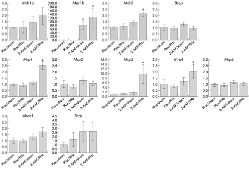
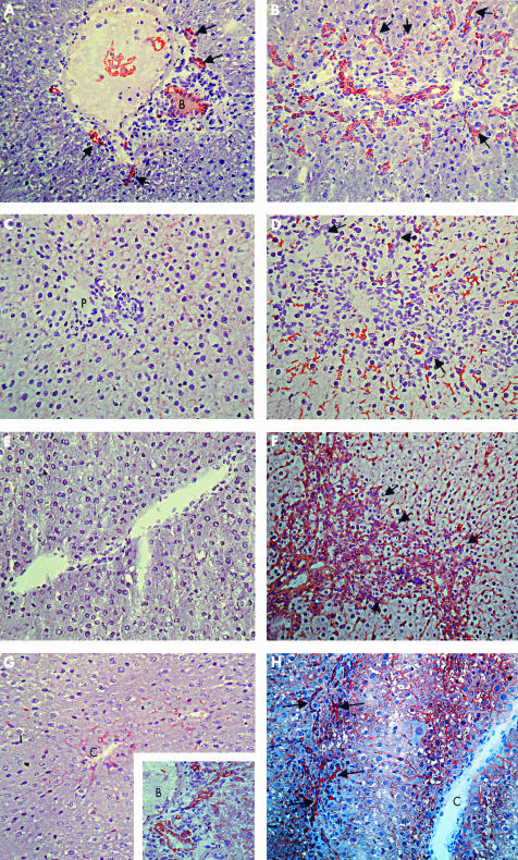
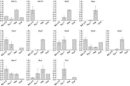
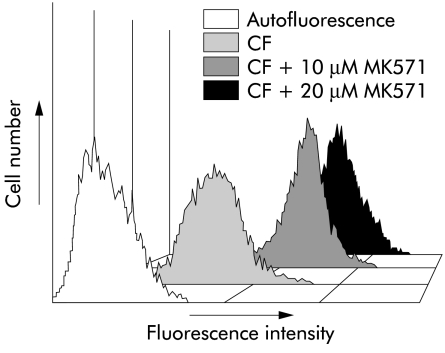
Results: 2-AAF/PHx treated animals showed increased hepatic mRNA levels of the genes encoding multidrug resistance proteins Mdr1b, Mrp1, and Mrp3. Immunohistochemistry demonstrated expression of Mrp1 and Mrp3 proteins in periportal progenitor cells and of the Mdr1b protein in periportal hepatocytes. Freshly isolated Thy-1 positive cells and cultured RLF phi 13 progenitor cells highly expressed Mrp1 and Mrp3 mRNA while the hepatocyte specific transporters Mdr2, Bsep, Mrp2, and Mrp6 were only minimally expressed. Blocking Mrp activity by MK-571 resulted in accumulation of the Mrp specific substrate carboxyfluorescein in RLF phi 13 cells.
Conclusion: HPCs express high levels of active Mrp1 and Mrp3. These may have a cytoprotective role in conditions of severe hepatotoxicity.
Figures
References
-
- Evarts RP, Nagy P, Nakatsukasa H, et al. In vivo differentiation of rat liver oval cells into hepatocytes. Cancer Res 1989;49:1541–7. - PubMed
-
- Trautwein C, Will M, Kubicka S, et al. 2-Acetaminofluorene blocks cell cycle progression after hepatectomy by p21 induction and lack of cyclin E expression. Oncogene 1999;18:6443–53. - PubMed
-
- Theise ND, Saxena R, Portmann BC, et al. The canals of Hering and hepatic stem cells in humans. Hepatology 1999;30:1425–33. - PubMed
-
- Petersen BE, Goff JP, Greenberger JS, et al. Hepatic oval cells express the hematopoietic stem cell marker Thy-1 in the rat. Hepatology 1998;27:433–45. - PubMed
-
- Petersen BE, Zajac VF, Michalopoulos GK. Hepatic oval cell activation in response to injury following chemically induced periportal or pericentral damage in rats. Hepatology 1998;27:1030–8. - PubMed
Publication types
MeSH terms
Substances
LinkOut - more resources
Full Text Sources
Medical
Research Materials
Miscellaneous