. 2003 Jun 24;100(13):7480-5.
doi: 10.1073/pnas.1332608100. Epub 2003 Jun 11.

Efficient biotinylation and single-step purification of tagged transcription factors in mammalian cells and transgenic mice

Affiliations

Efficient biotinylation and single-step purification of tagged transcription factors in mammalian cells and transgenic mice

Ernie de Boer et al. Proc Natl Acad Sci U S A. .

Abstract

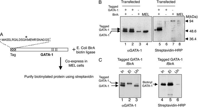
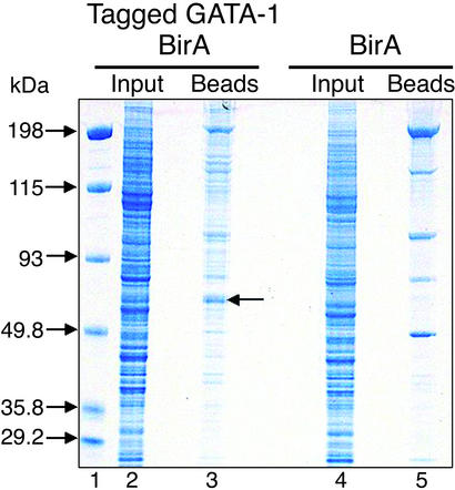
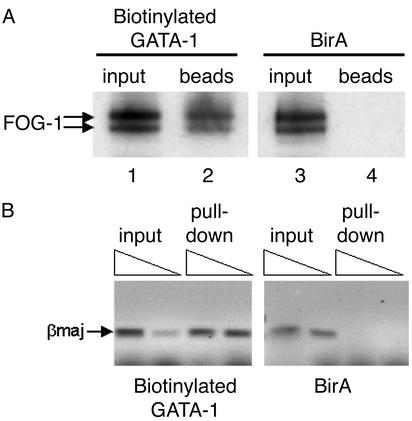
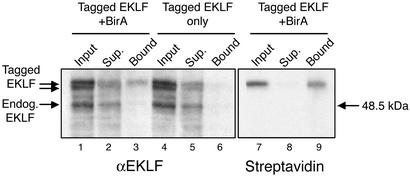
Proteomic approaches require simple and efficient protein purification methodologies that are amenable to high throughput. Biotinylation is an attractive approach for protein complex purification due to the very high affinity of avidin/streptavidin for biotinylated templates. Here, we describe an approach for the single-step purification of transcription factor complex(es) based on specific in vivo biotinylation. We expressed the bacterial BirA biotin ligase in mammalian cells and demonstrated very efficient biotinylation of a hematopoietic transcription factor bearing a small (23-aa) artificial peptide tag. Biotinylation of the tagged transcription factor altered neither the factor's protein interactions or DNA binding properties in vivo nor its subnuclear distribution. Using this approach, we isolated the biotin-tagged transcription factor and at least one other known interacting protein from crude nuclear extracts by direct binding to streptavidin beads. Finally, this method works efficiently in transgenic mice, thus raising the prospect of using biotinylation tagging in protein complex purification directly from animal tissues. Therefore, BirA-mediated biotinylation of tagged proteins provides the basis for the single-step purification of proteins from mammalian cells.


Figures

Fig. 1.
Fig. 1.
(A) Scheme for the specific biotinylation of tagged GATA-1 by BirA biotin ligase in MEL cells. The sequence of the 23-aa peptide tag fused to the N terminus of GATA-1 is shown. The asterisk indicates the lysine residue that becomes specifically biotinylated by BirA. Speckled boxes indicate the positions of the two GATA-1 Zinc-fingers. Tagged GATA-1 and BirA were cloned separately in a mammalian erythroid expression cassette and coexpressed in MEL cells. (B) Biotinylation of tagged GATA-1 in MEL cells. (Left) Western blot with an anti-GATA-1 antibody to detect endogenous and tagged GATA-1 proteins. (Right) Western blot of the same extracts with streptavidin–HRP conjugate to detect biotinylated GATA-1. Nuclear extracts (5 μg per lane) from the double transfectants (lanes 1 and 5) and single transfectants (lanes 2, 3, 6, and 7) for tagged GATA-1 and Bir A were tested. Lanes 4 and 8, nuclear extract from nontransfected MEL cells. Biotinylated GATA-1 (asterisk) is clearly visible in only in the lane of the double transfected cells. Also indicated is the low background detected by streptavidin in MEL nuclear extracts from cells expressing only BirA (Right, lane 7). (C) Efficiency of GATA-1 biotinylation and binding to streptavidin beads. (Left) Western blot using anti-GATA-1 antibody to detect binding of tagged GATA-1 to streptavidin beads (lane 2; starting material for the binding was 2.5 times the amount of nuclear extract shown in the input lane). Input and unbound material are shown in lanes 1 and 3. (Right) The same filter stripped and reprobed with streptavidin–HRP to detect the binding of biotinylated GATA-1 to streptavidin beads (lane 5). Lane 6 shows that very little tagged GATA-1 remains unbound by streptavidin. In this binding experiment, the beads were washed under stringent conditions (0.5 M NaCl/0.3% Triton X-100 in PBS). In, input (nuclear extract); El, eluted material; Un, unbound material.
Fig. 2.
Fig. 2.
Colloidal blue-stained gel of a binding experiment of crude nuclear extracts to streptavidin beads. Lane 1, marker (M). Lane 2, input nuclear extract from tagged GATA-1/BirA double transfected cells (≈12 μg). Lane 3, proteins eluted after direct binding to streptavidin beads of ≈5 mg of crude nuclear extracts from tagged GATA-1/BirA transfected cells. Lane 4, input nuclear extract from tagged GATA-1/BirA transfected cells. Lane 5, proteins eluted after binding to streptavidin beads ≈5 mg of nuclear extract from BirA transfected cells. Arrow in lane 3 indicates protein band containing purified biotinylated GATA-1, as determined by MS.
Fig. 3.
Fig. 3.
(A) Binding biotinylated GATA-1 to streptavidin beads specifically pulls down FOG-1, as detected by Western blotting by using a FOG-1 antibody. By contrast, FOG-1 cannot be pulled down by streptavidin in nuclear extracts expressing biotinylated expressing BirA only (Right). FOG-1 is detected as a doublet (26). (B) Streptavidin pull-down of βmaj globin promoter sequences from crosslinked chromatin from MEL cells expressing biotinylated GATA-1 (Left) or BirA only (Right). Triangles indicate increasing amounts of pulled-down crosslinked chromatin used as template in PCR reactions in detecting amplification of the βmaj sequences. Specific enrichment for βmaj sequences is observed in pulled-down chromatin from cells expressing biotinylated GATA-1 but not from cells expressing BirA only.
Fig. 4.
Fig. 4.
Specific biotinylation of tagged EKLF in transgenic mouse embryos. Nuclear extracts from the fetal liver of 13.5-days postcoitum embryos from a tagged EKLF/BirA double transgenic line (lanes 1–3 and 7–9) and from a tagged EKLF transgenic line (lanes 4–6) were bound to streptavidin beads. Tagged and biotinylated EKLF in input nuclear extract, unbound material (sup., supernatant), and bound material was detected by an EKLF antibody (Left) or by streptavidin–HRP (Right). EKLF biotinylation and binding to the beads is detected only in extracts from double transgenic embryos.

Similar articles

Cited by

References

    1. Lamond, A. I. & Mann, M. (1997) Trends Cell Biol. 7, 139–142. - PubMed
    1. Kumar, A. & Snyder, M. (2002) Nature 415, 123–124. - PubMed
    1. Jarvik, J. W. & Telmer, C. A. (1998) Annu. Rev. Genet. 32, 601–618. - PubMed
    1. Rigaut, G., Shevchenko, A., Rutz, B., Wilm, M., Mann, M. & Seraphin, B. (1999) Nat. Biotechnol. 17, 1030–1032. - PubMed
    1. Chapman-Smith, A. & Cronan, J. E., Jr. (1999) Trends Biochem. Sci. 24, 359–363. - PubMed

Publication types