. 2003 Jun 24;100(13):7626-31.
doi: 10.1073/pnas.0932599100. Epub 2003 Jun 11.

TSG101 interaction with HRS mediates endosomal trafficking and receptor down-regulation

Affiliations

TSG101 interaction with HRS mediates endosomal trafficking and receptor down-regulation

Quan Lu et al. Proc Natl Acad Sci U S A. .

Abstract

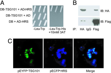
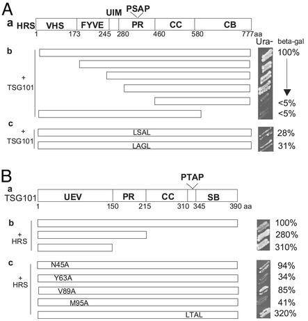
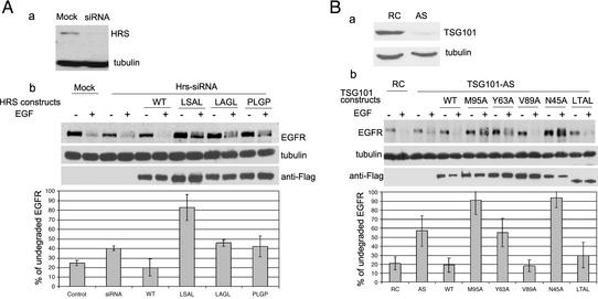
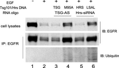
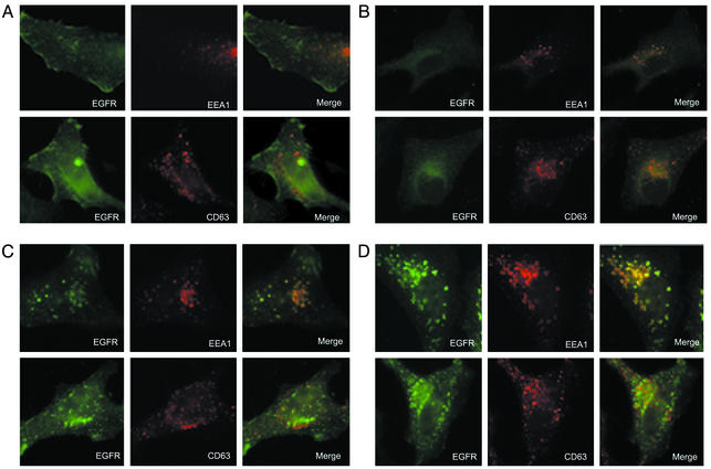
Down-regulation of mitogenic signaling in mammalian cells relies in part on endosomal trafficking of activated receptors into lysosomes, where the receptors are degraded. These events are mediated by ubiquitination of the endosomal cargo and its consequent sorting into multivesicular bodies that form at the surfaces of late endosomes. Tumor susceptibility gene 101 (tsg101) recently was found to be centrally involved in this process. Here we report that TSG101 interacts with hepatocyte growth factor-regulated tyrosine kinase substrate (HRS), an early endosomal protein, and that disruption of this interaction impedes endosomal trafficking and endocytosis-mediated degradation of mitogenic receptors. TSG101/HRS interaction occurs between a ubiquitin-binding domain of TSG101 and two distinct proline-rich regions of HRS, and is modulated by a C-terminal TSG101 sequence that resembles a motif targeted in HRS. Mutational perturbation of TSG101/HRS interaction prevented delivery of epidermal growth factor receptor (EGFR) to late endosomes, resulted in the cellular accumulation of ubiquitinated EGFR in early endosomes, and inhibited ligand-induced down-regulation of EGFR. Our results reveal the TSG101 interaction with HRS as a crucial step in endocytic down-regulation of mitogenic signaling and suggest a role for this interaction in linking the functions of early and late endosomes.


Figures

Fig. 1.
Fig. 1.
Physical interaction between TSG101 and HRS. (A) TSG101 and HRS interact in a yeast two-hybrid assay. Bait (TSG101) and prey (HRS) two-hybrid constructs were introduced by transformation into yeast strain MaV203, and three random transformants were transferred onto SC-Leu-Trp media (to demonstrate the presence of both plasmids) and onto SC-Leu-Trp-His + 10 mM 3-amino-triazole media (to examine induction of the HIS3 reporter gene). (B) TSG101 and HRS coimmunoprecipitate. 293T cells were transfected with constructs containing HA-tagged TSG101 and Flag-tagged HRS. Cell lysates were made 48 h after transfection. TSG101 was immunoprecipitated (IP) with anti-HA antibody, and HRS was immunoprecipitated with anti-Flag antibody conjugated beads. The complexes were immunoblotted with anti-HA or anti-Flag antibodies. Mouse IgG was used in the control IP. (C) Partial colocalization of TSG101 and HRS. NIH 3T3 cells were transfected with the pEYFP-TSG101 and pECFP-HRS constructs. Twenty-four hours after transfection, cells were fixed and observed under a deconvolution fluorescence microscope. The images are pseudocolored.
Fig. 2.
Fig. 2.
Identification of structural elements involved in TSG101/HRS interaction. TSG101 (fused to Gal4-activation domain) and HRS (fused to Gal4-DNA-binding domain) constructs were introduced by cotransformation into yeast Mav203 cells. Interactions were indicated by growth on uracil-minus plate and quantitated by liquid β-galactosidase assay. Averages from three independent β-gal assays were used, and the interaction between wild-type TSG101 and wild-type HRS was designated as 100%. (A) Interaction between TSG101 and HRS with deletions (b) or mutations (c). (a) The schematic domain structure of Hrs. VHS, Vps27-Hrs-STAM; FYVE, lipid interaction domain; UIM, ubiquitin interaction motif; PR, proline-rich; CC, coiled-coil; CB, clathrin binding. (B) Interaction between HRS and TSG101 with deletion (b) or mutations (c). (a) The schematic domain structure of TSG101. PR, proline-rich; CC, coiled-coil; SB, steadiness box.
Fig. 3.
Fig. 3.
Assay of ligand-induced EGF receptor degradation. (A) Effects of HRS mutations on EGFR degradation. HeLa cells (in a six-well plate) were made deficient in HRS by transfection of 50 nM Hrs-siRNA (a) for 24 h. The cells were then transfected with Flag-tagged HRS expression plasmids (siRNA-resistant) (50 ng per well) or control DNA (pBi-Luc) along with pCDNA3-EGFR (200 ng per well). Twenty-four hours after plasmid transfection the cells was starved in Opti-MEM medium for 1 h and then either mock-treated or induced with 150 ng/ml EGF for 90 min. Proteins from cell lysates were separated on SDS-8% polyacrylamide gel, electrotransferred, and immunoblotted with anti-EGFR, antitubulin, and anti-Flag antibodies. EGFR signals were quantitated by x-ray film densitometry (normalized against tubulin signals). Data from three independent experiments were averaged, and percentages of undegraded EGFR upon EGF induction were plotted. (B) Effects of TSG101 mutations on EGFR degradation. HeLa cells were made deficient in TSG101 by transfection of 100 nM TSG101 antisense (AS) RNA oligonucleotide (a). Cells were then transfected with various TSG101 expression constructs (resistant to antisense inhibition) (200 ng per well) or control DNA along with pCDNA3-EGFR (200 ng per well). Assays were performed as in A.
Fig. 4.
Fig. 4.
Effects of TSG101/HRS interaction on accumulation of ubiquitinated EGFR. HeLa cell (in six-well plates) were first mock-treated (lanes 1 and 2), or transfected with 100 nM TSG101 antisense RNA oligonucleotide (lanes 3 and 4) or 50 nM Hrs-siRNA (lanes 5 and 6) for 24 h. The cells were then transfected with various TSG101 or HRS expression constructs (resistant to antisense inhibition) along with pCDNA3-EGFR (200 ng per well). Twenty-four hours after plasmid transfection the cells were starved and induced with EGF (150 ng/ml) for 90 min. A small portion of cell lysates was analyzed by anti-EGFR Western blot, and the remaining lysates were immunoprecipitated (IP) for EGFR. IP samples were Western-blotted with anti-EGFR and antiubiquitin antibodies.
Fig. 5.
Fig. 5.
EGFR colocalization with early and late endosomal markers. (A and B) Control naïve HeLa cells. (C) HeLa cells with mutant TSG101 were made by first transfection of TSG101-antisensed RNA oligo and subsequent transfection of a mutant TSG101 construct (M95A). (D) HeLa cells with mutant HRS were made by transfection of HRS siRNA and subsequent transfection of a mutant HRS construct (HRS-LSAL). Cells were either mock-treated (A) or induced (B–D) with EGF (150 ng/ml) for 60 min. Cells were then fixed, permeabilized, and immunostained for EGFR and EEA1 (or CD63), and imaged under a deconvolution fluorescence microscope as described in Materials and Methods.

Similar articles

Cited by

References

    1. Hunter, T. (2000) Cell 100, 113–127. - PubMed
    1. Schlessinger, J. (2000) Cell 103, 211–225. - PubMed
    1. Blume-Jensen, P. & Hunter, T. (2001) Nature 411, 355–365. - PubMed
    1. Di Fiore, P. P. & De Camilli, P. (2001) Cell 106, 1–4. - PubMed
    1. Sorkin, A. & Von Zastrow, M. (2002) Nat. Rev. Mol. Cell Biol. 3, 600–614. - PubMed

Publication types

MeSH terms