. 2003 May;14(5):1780-9.
doi: 10.1091/mbc.e02-12-0794. Epub 2003 Jan 26.

Human PIG-U and yeast Cdc91p are the fifth subunit of GPI transamidase that attaches GPI-anchors to proteins

Affiliations

Human PIG-U and yeast Cdc91p are the fifth subunit of GPI transamidase that attaches GPI-anchors to proteins

Yeongjin Hong et al. Mol Biol Cell. 2003 May.

Abstract

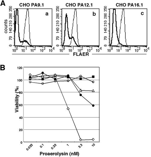
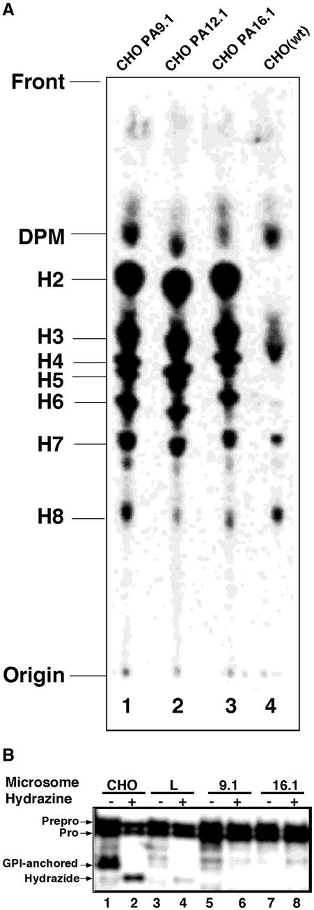
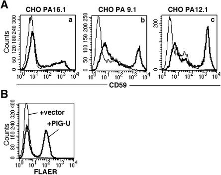
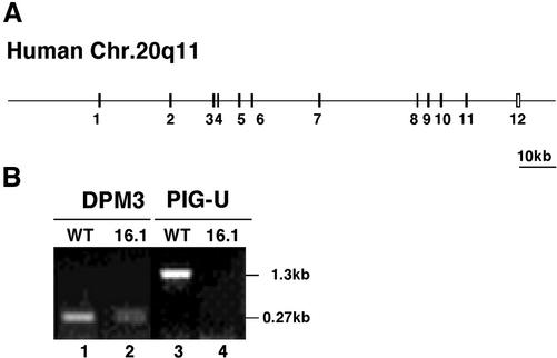
Many eukaryotic proteins are anchored to the cell surface via glycosylphosphatidylinositol (GPI), which is posttranslationally attached to the carboxyl-terminus by GPI transamidase. The mammalian GPI transamidase is a complex of at least four subunits, GPI8, GAA1, PIG-S, and PIG-T. Here, we report Chinese hamster ovary cells representing a new complementation group of GPI-anchored protein-deficient mutants, class U. The class U cells accumulated mature and immature GPI and did not have in vitro GPI transamidase activity. We cloned the gene responsible, termed PIG-U, that encoded a 435-amino-acid hydrophobic protein. The GPI transamidase complex affinity-purified from cells expressing epitope-tagged-GPI8 contained PIG-U and four other known components. Cells lacking PIG-U formed complexes of the four other components normally but had no ability to cleave the GPI attachment signal peptide. Saccharomyces cerevisiae Cdc91p, with 28% amino acid identity to PIG-U, partially restored GPI-anchored proteins on the surface of class U cells. PIG-U and Cdc91p have a functionally important short region with similarity to a region conserved in long-chain fatty acid elongases. Taken together, PIG-U and the yeast orthologue Cdc91p are the fifth component of GPI transamidase that may be involved in the recognition of either the GPI attachment signal or the lipid portion of GPI.


Figures

Figure 1.
Figure 1.
Expression of GPI-anchored proteins on three class U mutant CHO cells. Cells were stained for CD59 and DAF and analyzed by flow cytometry. (A) Control staining of the parental cell, CHO(wt) with nonrelevant first antibodies; (B) CHO(wt); (C) CHOPA9.1; (D) CHOPA12.1; (E) CHOPA16.1 cells.
Figure 2.
Figure 2.
Aerolysin-binding and -sensitivity of class U mutant CHO cells. (A) Binding of fluorescence-tagged proaerolysin (FLAER) on class U cells. Thick lines, CHOPA9.1 (a); CHOPA12.1 (b); CHOPA16.1 (c) cells. Thin lines, CHO(wt) cells. (B) Sensitivity to aerolysin. The cell viability after a 3-h incubation with increasing concentrations of proaerolysin was measured by an MTT assay. Diamonds, CHO(wt); ▪, GPI(-). O cells defective in the PIG-O gene; triangles, CHOPA9.1; •, CHOPA12.1; ○, CHOPA16.1.
Figure 3.
Figure 3.
(A) GPI biosynthesis in class U cells. Lipids from cells labeled with [3H]mannose were separated by TLC in a solvent, CHCl3:MeOH:H2O = 10:10:3. Lane 1, CHOPA9.1; lane 2, CHOPA12.1; lane 3, CHOPA16.1; lane 4, CHO(wt) cells. The origin, front, and identities of the mannolipids are on the left. DPM, dolichol-phosphate-mannose; H2–H6, GPI intermediates; H7 and H8, mature GPI (Hirose et al., 1992). (B) Class U cells do not have GPI transamidase activity in a cell-free system. Mini-PLAP mRNA was translated using rabbit reticulocyte lysates and microsomes from the indicated cells in the absence (-) or presence (+) of hydrazine. Lanes 1 and 2, CHO(wt); lanes 3 and 4, class L CHO cells defective in the PIG-L gene; lanes 5 and 6, CHOPA9.1; lanes 7 and 8, CHOPA16.1. The identities of the forms of mini-PLAP according to Kodukula et al. (1991) are shown on the left.
Figure 4.
Figure 4.
PIG-U restores the surface expresssion of GPI-anchored proteins and aerolysin binding to class U cells. Class U cells were transfected with PIG-U cDNA or a mock vector, and 2 days later were stained with an anti-CD59 antibody or incubated with 5 nM FLAER. Thick lines, PIG-U-transfected; thin lines, mock vectortransfected. (A) CD59 expression on CHOPA16.1 (a), 9.1 (b), and 12.1 (c) cells. (B) Binding of FLAER to CHOPA16.1 cells.
Figure 5.
Figure 5.
Characterization of PIG-U proteins. (A) Amino acid sequences of rat, Chinese hamster, and human PIG-U and S. cerevisiae Cdc91p. Two highly conserved regions are underlined. Black and gray squares are identical and similar amino acids, respectively. (B) Hydropathy plot (Kyte and Doolittle, 1982) of human PIG-U.
Figure 6.
Figure 6.
Genomic structure of human PIG-U and RT-PCR analysis of PIG-U mRNA. (A) Exon-intron organization of human PIG-U. (B) RT-PCR analysis of CHO(wt) and CHOPA16.1 cells for PIG-U and DPM3 mRNAs. Lanes 1 and 2, DPM3; lanes 3 and 4, PIG-U; lanes 1 and 3, CHO(wt); lanes 2 and 4, CHOPA16.1.
Figure 7.
Figure 7.
PIG-U is the fifth subunit of the GPI transamidase complex. (A) Two-step purification of the GPI transamidase complex. Class K cells stably expressing GST-FLAG-tagged GPI8 (lane 2) were solubilized in a buffer with 1% NP40. GST-FLAG-tagged GPI8 was purified with anti-FLAG beads followed by glutathione beads. The identities of the bands and the determined N-terminal sequence of PIG-U protein (AAPLVLV) are shown on the right. Lane 1, size markers. (B) Formation of a stable complex of PIG-S, PIG-T, GAA1, and GPI8 in the absence of PIG-U. A mixture of HA-PIG-S, Myc-PIG-T, HSV-GAA1, and GST-GPI8 was transfected into CHOPA16.1 cells with or without FLAG-PIG-U cDNA. Complexes were precipitated with anti-HA to collect HA-PIG-S, and the other coprecipitated components were assessed by Western blotting using antitag antibodies. Lanes 1, 3, 5, 7, and 9, cells with PIG-U; lanes 2, 4, 6, 8, and 10, cells without PIG-U; lanes 1 and 2, anti-HA; lanes 3 and 4, anti-Myc; lanes 5 and 6, anti-HSV; lanes 7 and 8, anti-GST; lanes 9 and 10, anti-FLAG.
Figure 8.
Figure 8.
S. cerevisiae Cdc91p partially restores the expression of GPI-anchored proteins on class U cells. CHOPA16.1 cells were stably transfected with PIG-U, CDC91, or a mock vector and stained with anti-CD59 and anti-DAF antibodies. Left panel, PIG-U-transfected; middle panel, CDC91-transfected; right panel, mock vectortransfected cells.
Figure 9.
Figure 9.
Functionally important short sequence in PIG-U. (A) Alignment of similar short sequences of PIG-U (residues 273–289), Cdc91p (residues 239–255) and long-chain fatty acid elongases. (B) Flow-cytometric analysis of a function of the F274L/W275L mutant of hamster PIG-U. CHOPA16.1 cells were transfected with wild-type PIG-U (thin line), the mutant PIG-U (thick line) and a mock vector (broken line), and 2 days later, stained for CD59. (C and D) Normal expression and incorporation into the GPI transamidase complex of the F274L/W275L mutant of PIG-U. The HSV-tagged mutant and wild-type PIG-U cDNAs were transfected into CHOPA16.1 cells together with HA-PIG-S, Myc-PIG-T, FLAG-GAA1, GST-GPI8, and HSV-ALDH (as a control to assess transfection efficiency). To measure the expression level, the mutant and wild-type PIG-U, and ALDH were immunoprecipitated by anti-HSV beads and analyzed by Western blotting with anti-HSV antibody (C). To assess incorporation of the mutant PIG-U into the GPI transamidase complex, HA-PIG-S was immunoprecipitated with anti-HA antibody and coprecipitated four other proteins were determined by Western blotting with antitag antibodies (D). Lane 1, wild-type PIG-U; lane 2, F274L/W275L PIG-U.

References

    1. Abrami, L., Fivaz, M., Kobayashi, T., Kinoshita, T., Parton, R.G., and van der Goot, F.G. (2001). Cross-talk between caveolae and glycosylphosphatidylinositol-rich domains. J. Biol. Chem. 276, 30729–30736. - PubMed
    1. Abrami, L., Fivaz, M., and van der Goot, F.G. (2000). Adventures of a pore-forming toxin at the target cell surface. Trends Microbiol 8, 168–172. - PubMed
    1. Benghezal, M., Benachour, A., Rusconi, S., Aebi, M., and Conzelmann, A. (1996). Yeast Gpi8p is essential for GPI anchor attachment onto proteins. EMBO J. 15, 6575–6583. - PMC - PubMed
    1. Buckley, J.T. (1999). The channel-forming toxin aerolysin. In: The Comprehensive Sourcebook of Bacterial Protein Toxins, ed. J.E. Alouf and J.H. Freer, London: Academic Press, 362–372.
    1. Fraering, P., Imhof, I., Meyer, U., Strub, J.M., van Dorsselaer, A., Vionnet, C., and Conzelmann, A. (2001). The GPI transamidase complex of Saccharomyces cerevisiae contains Gaa1p, Gpi8p, and Gpi16p. Mol. Biol. Cell 12, 3295–3306. - PMC - PubMed

Publication types