. 2003 May;14(5):1790-800.
doi: 10.1091/mbc.e02-09-0583. Epub 2003 Jan 26.

Tumor necrosis factor-alpha stimulates the epithelial-to-mesenchymal transition of human colonic organoids

Affiliations

Tumor necrosis factor-alpha stimulates the epithelial-to-mesenchymal transition of human colonic organoids

Richard C Bates et al. Mol Biol Cell. 2003 May.

Abstract

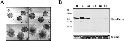
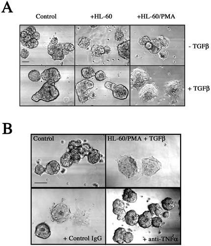
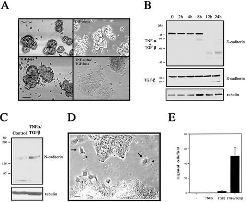
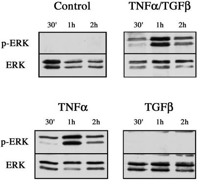
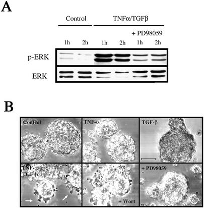
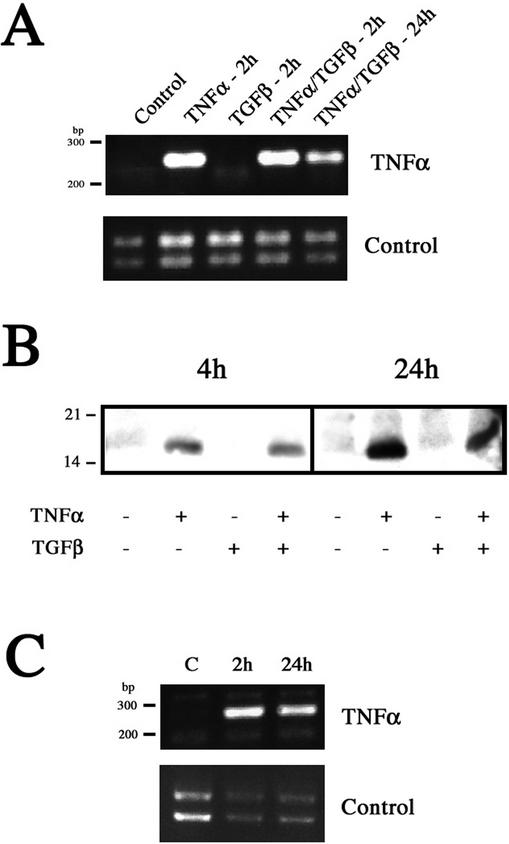
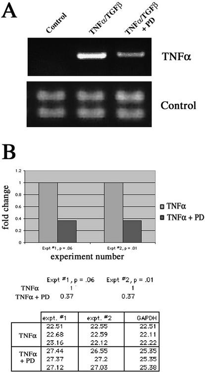
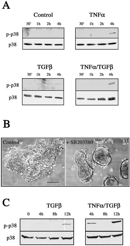
An epithelial-mesenchymal transition (EMT) characterizes the progression of many carcinomas and it is linked to the acquisition of an invasive phenotype. Given that the tumor microenvironment is an active participant in tumor progression, an important issue is whether a reactive stroma can modulate this process. Using a novel EMT model of colon carcinoma spheroids, we demonstrate that their transforming-growth factor-beta1 (TGF-beta)-induced EMT is accelerated dramatically by the presence of activated macrophages, and we identify tumor necrosis factor-alpha (TNF-alpha) as the critical factor produced by macrophages that accelerates the EMT. A synergy of TNF-alpha and TGF-beta signaling promotes a rapid morphological conversion of the highly organized colonic epithelium to dispersed cells with a mesenchymal phenotype, and this process is dependent on enhanced p38 MAPK activity. Moreover, exposure to TNF-alpha stimulates a rapid burst of ERK activation that results in the autocrine production of this cytokine by the tumor cells themselves. These results establish a novel role for the stroma in influencing EMT in colon carcinoma, and they identify a selective advantage to the stromal presence of infiltrating leukocytes in regulating malignant tumor progression.


Figures

Figure 1.
Figure 1.
TGF-β induces EMT in LIM 1863 organoids. (A) TGF-β induces EMT. LIM 1863 organoids were seeded in the presence of TGF-β (2 ng/ml) and allowed to undergo transition in culture. Morphological changes were documented by light microscopy using phase contrast optics at 1, 3, 5, and 7 d after exposure to the cytokine. Bar, 150 μm. (B) E-cadherin loss characterizes EMT. Cell extracts were prepared over the time course shown after addition of TGF-β and immunoblotted with an E-cadherin–specific antibody. Relative molecular masses are shown to the left in kDa. Equal protein loading was confirmed by tubulin immunoblotting (bottom panel).
Figure 2.
Figure 2.
Augmentation of LIM 1863 organoid EMT by a stromal factor. (A) Activated HL-60 cells secrete a factor that accelerates the EMT. LIM 1863 organoids were seeded in a coculture assay as described in MATERIALS AND METHODS. LIM 1863 cells were seeded in the absence (Control) or presence of HL-60 cells (+HL-60), or activated HL-60 cells that had been pretreated with PMA (+HL-60/PMA) as described in MATERIALS AND METHODS. Cells were cultured for 24 h in the absence or presence of TGF-β (top and bottom panels, respectively). Bar, 150 μm. (B) Anti–TNF-α antibody inhibits the stromally accelerated EMT. LIM 1863 cells were cultured in the presence of TGF-β for 24 h alone (Control), or in coculture with activated HL-60 cells (HL60/PMA + TGF-β). Either a neutralizing TNF-α antibody (1 μg/ml) or an isotype matched IgG control antibody was added to the cocultured cells as indicated. The degree of phenotypic transition was assessed by light microscopy. Bar, 150 μm.
Figure 3.
Figure 3.
TNF-α synergizes with TGF-β to promote EMT. (A) Recombinant human TNF-α recapitulates stromally derived TNF-α effect. LIM 1863 organoids were unstimulated (Control), treated with TNF-α (10 ng/ml), TGF-β (2 ng/ml) or the combination of cytokines for 24 h. The extent of morphological transformation was photographed under light microscopy. Bar, 150 μm. (B) Rapid and complete loss of E-cadherin during the TNF-α/TGF-β–induced EMT. Cell extracts were prepared over the time course shown after addition of TNF-α/TGF-β (top panel) or TGF-β alone (middle panel) and immunoblotted with an E-cadherin–specific antibody. Relative molecular masses are shown to the left in kDa. Equal protein loading was confirmed by tubulin immunoblotting (bottom panel). (C) Upregulation of the mesenchymal marker N-cadherin after EMT. Cell extracts from untreated organoids (Control) or cells treated with TNF-α/TGF-β for 24 h and immunoblotted with a N-cadherin–specific antibody. Relative molecular masses are shown to the left in kDa. Equal protein loading was confirmed by tubulin immunoblotting (bottom panel). (D) The EMT promotes a migratory phenotype in LIM 1863 cells. Photomicrograph of LIM 1863 cells after EMT induced by combination TNF-α/TGF-β treatment for 24 h. Individual migrating cells (arrows) exhibit broad lamellae (arrowheads). Bar, 10 μm. (E) The EMT induces chemotaxis. Chemotactic migration assay of LIM 1863 cells, treated with cytokines as indicated, for 3 d on laminin-coated Transwells toward conditioned NIH-3T3 medium. Data are expressed as the means and SDs of five individual fields randomly selected for each well.
Figure 4.
Figure 4.
ERK activation in response to cytokine stimulation. Cell extracts were prepared from untreated LIM 1863 organoids (Control), or cells treated with TNF-α, TGF-β, or the combination of cytokines, for the times indicated. ERK 1/2 activity was determined by immunoblotting with a phospho-specific ERK antibody (top panels). ERK protein expression was confirmed using an ERK antibody (bottom panels).
Figure 5.
Figure 5.
The ERK inhibitor PD98059 prevents the accelerated EMT induced by TNF-α. (A) PD98059 inhibits the 1-h peak of ERK activity induced by TNF-α treatment. Extracts were prepared from control LIM 1863 cells, or TNF-α/TGF-β–treated cells cultured in the absence or presence of the ERK inhibitor PD98059 for the times indicated. ERK 1/2 activation was determined using the phospho-specific ERK antibody (top panel). ERK protein expression was confirmed using an ERK antibody (bottom panel). (B) PD98059 prevents the accelerated EMT phenotype. Photomicrographs of LIM 1863 cells seeded in the cytokine assay for 6 h. Cells were untreated (Control) or stimulated with either TNF-α or TGF-β, or the combination of the two cytokines, as indicated. Individual cells can be seen emerging from the periphery of the organoids (arrows). Cells were pretreated with PD98059 for 20 min before stimulation with both cytokines and seeding in the assay (bottom left panel). The PI3K inhibitor wortmannin (Wort) was also used, after a 2-h pretreatment. Bar, 100 μm.
Figure 6.
Figure 6.
TNF-α treatment induces its own expression in colon carcinoma cells. (A) RT-PCR for TNF-α was performed on RNA purified from untreated LIM 1863 cells (Control), cells treated with TNF-α or TGF-β for 2 h, and cells treated with both cytokines for 2 and 24 h, as described in MATERIALS AND METHODS. The predicted size of the PCR product is 254 base pairs. Control reactions were performed using integrin α6 primers (bottom panel). (B) TNF-α protein expression. Cell extracts were prepared from untreated, TNF-α–, TGF-β–, or TNF-α/TGF-β–treated LIM 1863 organoids harvested after 4 and 24 h. Extracts were analyzed by SDS-PAGE and immunoblotting with an anti–TNF-α antibody. Equal protein loading was confirmed by reprobing with tubulin (unpublished data). Relative molecular masses are shown to the left in kDa. (C) RT-PCR was performed as described above using RNA from Clone A colon carcinoma cells treated with TNF-α for 2 and 24 h. Control reactions were performed using integrin α6 primers (bottom panel).
Figure 7.
Figure 7.
Inhibition of ERK activity reduces autocrine induction of TNF-α. (A) RT-PCR of untreated LIM 1863 cells (Control), or TNF-α/TGF-β–treated cells for 2 h in either the absence or presence (+PD) of the ERK inhibitor PD98059. Control reactions shown in bottom panel using integrin α6 primers. (B) Real time quantitative PCR (RQ-PCR) of cytokine-treated cells in the absence or presence of PD98059 for 2 h. Increases in fluorescence signal (ΔRn) from each PCR reaction were monitored in real time by the ABI Prism 7700 Sequence Detector. The fold change between treatments (a consistent 2.7-fold reduction in the presence of PD98059) in two separate experiments is represented graphically (top panel). Ct values, the PCR cycle at which a statistically significant difference in the ΔRn is first detected, are shown in the table. Ct values, when normalized to the internal reference gene (GAPDH), are inversely related to the magnitude of mRNA expression.
Figure 8.
Figure 8.
p38 MAPK is activated in response to TNF-α and is required for the accelerated EMT. (A) p38 MAPK activation in response to cytokine stimulation. Cell extracts were prepared from untreated LIM 1863 organoids (Control) or cells treated with TNF-α, TGF-β, or the combination of cytokines, for the times indicated. p38 MAPK activity was determined by immunoblotting with a phospho-specific p38 MAPK antibody (top panels). p38 MAPK protein expression was confirmed using a p38 antibody (bottom panels). (B) SB203580 prevents the accelerated EMT phenotype. Photomicrographs of LIM 1863 cells seeded in the cytokine assay for 24 h. Control cells were treated with DMSO (left panel) or SB203580 (right panel) and stimulated with TNF-α/TGF-β. Bar, 150 μm. (C) TNF-α and TGF-β cause a synergistic activation of p38 MAPK. Cell extracts were prepared over the time course shown from cells treated with TGF-β alone or the combination of cytokines, as indicated. p38 MAPK activity was determined by immunoblotting as described above.

References

    1. Arias, A.M. (2001). Epithelial mesenchymal interactions in cancer and development. Cell 105, 425–431. - PubMed
    1. Bakin, A.V., Rinehart, C., Tomlinson, A.K., and Arteaga, C.L. (2002). p38 mitogen-activated kinase is required for TGF beta-mediated fibroblastic transdifferentiation, and cell migration. J. Cell Sci. 115, 3193–3206. - PubMed
    1. Barth, R.J., Jr., Camp, B.J., Martuscello, T.A., Dain, B.J., and Memoli, V.A. (1996). The cytokine microenvironment of human colon carcinoma. Lymphocyte expression of tumor necrosis factor-alpha and interleukin-4 predicts improved survival. Cancer 78, 1168–1178. - PubMed
    1. Bates, R.C., Buret, A., van Helden, D.F., Horton, M.A., and Burns, G.F. (1994). Apoptosis induced by inhibition of intercellular contact. J. Cell Biol. 125, 403–415. - PMC - PubMed
    1. Bates, R.C., Edwards, N.S., and Yates, J.D. (2000). Spheroids and cell survival. Crit. Rev. Oncol. Hematol. 36, 61–74. - PubMed

Publication types

MeSH terms

Substances

LinkOut - more resources