. 2003 May;14(5):1953-63.
doi: 10.1091/mbc.e02-10-0657. Epub 2003 Feb 6.

Constriction and Dnm1p recruitment are distinct processes in mitochondrial fission

Affiliations

Constriction and Dnm1p recruitment are distinct processes in mitochondrial fission

Aster Legesse-Miller et al. Mol Biol Cell. 2003 May.

Abstract

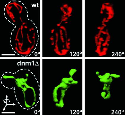
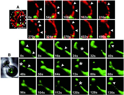
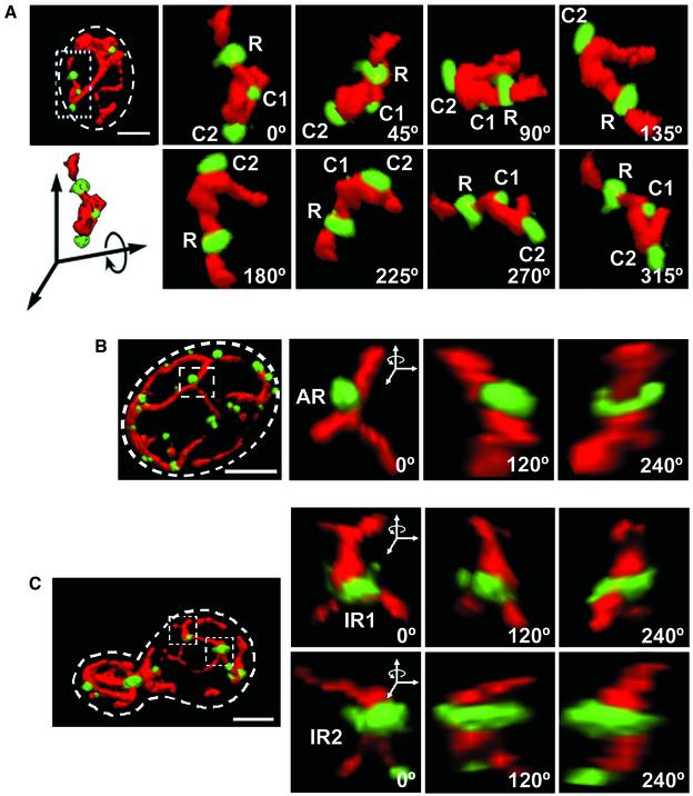
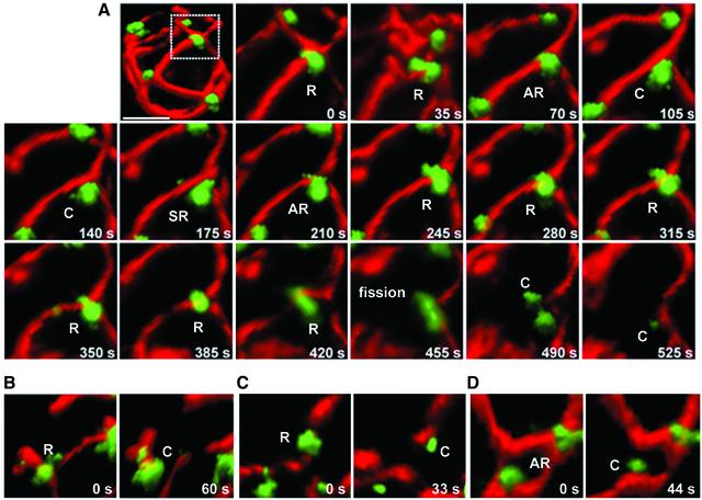
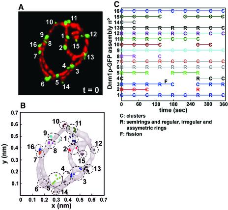
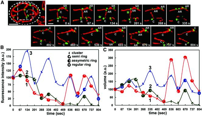
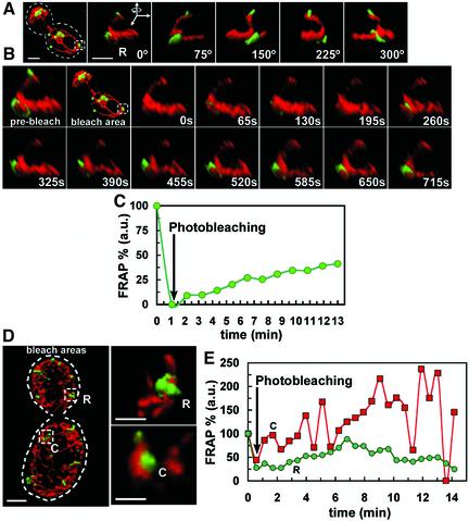
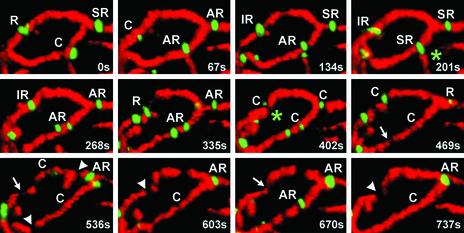
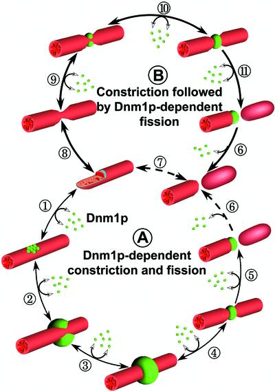
Mitochondria undergo cycles of fusion and fission crucial for organelle homeostasis. Fission is regulated partially by recruitment of the large GTPase Dnm1p to the outer mitochondrial membrane. Using three-dimensional time-lapse fluorescence imaging of Saccharomyces cerevisiae cells, we found that Dnm1p-EGFP appears and disappears at "hot spots" along mitochondrial tubes. It forms patches that convert rapidly into different shapes regardless of whether mitochondrial fission ensues or not. Moreover, the thickness of the mitochondrial matrix displays frequent temporal fluctuations apparently unrelated to fission or to recruitment of Dnm1p-EGFP. These results suggest that mitochondrial fission requires coordination of at least two distinct processes.


Figures

Figure 1.
Figure 1.
Three-dimensional imaging of mitochondria in living yeast cells. The mitochondrial matrix of S. cerevisae cells expressing (wt, top) or lacking (dnm1Δ, bottom) Dnm1p were labeled with mito-RFP or mito-EGFP, respectively. 3D image stacks were obtained by collecting 15 consecutive optical sections (step size 0.15 μm, 63×, 1.4 numerical aperture objective lens) followed by image restoration with eight cycles of constrained iterative deconvolution. The views correspond to 3D surface renderings of the image stacks rotated 120° around the y-axis. The cell contour (stippled line) is shown. Bar, 2 μm.
Figure 2.
Figure 2.
Thickness of the mitochondria matrix fluctuates as a function of time. Time-lapse combined with 3D surface rendering acquired from selected areas of yeast cells (stippled rectangles) expressing mito-RFP and Dnm1p-EGFP (A) or mito-EGFP (B) in the presence or absence of Dnm1p, respectively. Transient constrictions of the mitochondria matrix (arrowheads) were observed regardless of Dnm1p expression. Example of extensive mitochondrial matrix constriction or fission occurring in the absence of Dnm1p is shown (stars). The area selected for visualization in B comprises the mitochondria compartment in the budding daughter cell and the connecting mitochondrial tubule in the mother cell. Each of the dual color image stacks for A required 24 s of acquisition, whereas the single color stacks for B required 3 s. Bar, 2 μm.
Figure 3.
Figure 3.
Dnm1p-EGFP assemblies adopt different shapes. 3D renditions of yeast cells expressing Dnm1p-EGFP and mito-RFP visualized from different viewpoints rotated along the indicated axis. Selected areas (stippled rectangles and squares) were selected to highlight Dnm1p-EGFP assemblies of different shapes position on different locations along the mitochondria. (A) Dnm1p-EGFP arranged in the form of a regular ring surrounding a mitochondria tubule (R) or as a cluster on one side of the mitochondria tubule (C1 and C2). (B) Dnm1p-EGFP arranged as an asymmetric ring (AR) with the majority of the molecules located toward one side of the ring. (C) Dnm1p-EGFP arranged as an irregular ring around a mitochondrial branch (IR1, top) or surrounding two juxtaposed mitochondrial tubules (IR2, bottom). Bar, 2 μm.
Figure 7.
Figure 7.
Relation between mitochondrial fission and Dnm1p assemblies. (A) Time-lapse 3D renditions were acquired every 35 s from different yeast cells to follow the coupling between mitochondria fission events and the appearance of Dnm1p-EGFP assemblies adjacent to the fission event (monitored with mito-RFP). (B–D) These images illustrate several examples acquired from different cells before and after fission occurring next to Dnm1p-EGFP rings. After fission and separation of the free mitochondrial ends, Dnm1p-EGFP remained associated with one but not the other end. Bar, 2 μm.
Figure 4.
Figure 4.
Assembly and disassembly of Dnm1p-EGFP occurs on mitochondria hot spots. Consecutive nine timelapse 3D renditions from a yeast cell expressing Dnm1p-EGFP and mito-RFP were acquired every 44 s to monitor the temporal and spatial characteristics of Dnm1p-EGFP. (A) The image depicts the 3D rendition for the first time point (t = 0) and highlights the location of 16 different Dnm1p-EGFP assemblies whose presence, relative positions, and shape were monitored as a function of time. (B) Plots of the centers of mass presented as an orthogonal projection along the z-axis corresponding to the 16 Dnm1p-EGFP selected in A. The absolute position of the mass centers was determined in each time frame and is represented by a colored dot; each cluster (stippled circle) corresponds to a single Dnm1pEGFP assembly. (C) Schematic representation of the Dnm1p-EGFP assemblies high lighting variations in their shape and presence during the imaging period. Scoring of the assemblies as clusters (C) or rings (R) was done by visual inspection of the 3D renditions. For simplicity, all forms of rings were grouped together. About one-half of the Dnm1p-EGFP assemblies disappeared during the imaging period and subsequently reappeared at approximately the same position, irrespective of the original shape and of a mitochondria fission event (F).
Figure 5.
Figure 5.
Shape transitions of Dnm1p-EGFP assemblies. Fourteen sequential 3D renditions were obtained every 67 s from a single yeast cell expressing Dnm1p-EGFP and mito-RFP, and three Dnm1p-EGFP spots (labeled 1–3) located within the broken square area were selected for analysis. (A) Images correspond to the time-lapse series of the selected area visualized from the same viewpoint. The shapes of the Dnm1p-EGFP assemblies, scored by inspection from this and other viewpoints (our unpublished data), indicated frequent changes in shape between clusters (C) and rings of variable appearance (R, AR, or SR); these changes occurred at sites unrelated to a mitochondria fission event. Mitochondria fissions (*) occurred adjacent to Dnm1p-EGFP rings (R, SR, and AR) but never clusters. (B and C) Time-dependent plots corresponding to the fluorescence intensities (proportional to the number of Dnm1p-EGFP molecules) and volumes (proportional to the spatial density of Dnm1p-EGFP) determined for the Dnm1p-EGFP assemblies 1–3; they were normalized to the values obtained at the beginning of the time series. The plots highlight the apparent lack of correlation between shape transitions, fluorescence intensity and relative volume. Bar, 2 μm.
Figure 6.
Figure 6.
Exchange between the cytosolic and mitochondria-bound pools of Dnm1p-EGFP. FRAP experiments were carried out on single Dnm1p-EGFP assemblies imaged in yeast cells expressing Dnm1p-EGFP and mito-RFP. Photobleaching restricted to the EGFP but not the RFP signal was achieved in <1 s of illumination of the selected Dnm1p-EGFP assemblies by using a diffraction-limited laser whose wavelength was tuned using a coumarin-based fluorescent dye optimized for GFP photobleaching. 3D renditions were obtained at the indicated times. (A) General and close-up views of a Dnm1p-EGFP ring (R) selected for photobleaching visualized immediately before FRAP (see Movie 9 in Supplementary Materials). (B) Recovery of the fluorescent signal of the Dnm1p-EGFP assembly imaged in A after photobleaching. (C) Plot as a function of time corresponding to the recovery of fluorescent signal of the Dnm1p-EGFP assembly depicted in B normalized to its fluorescence intensity before photobleaching; the recovery reflects the incomplete exchange between the cytosolic and mitochondria-bound pools of Dnm1p-EGFP when organized as a ring. (D and E) Comparison between the relative recoveries of fluorescent signals determined for a Dnm1p-EGFP ring (R) and a cluster (C) after photobleaching. Fluorescence recovery for a ring was slower and less complete than for a cluster. Bar, 2 μm.
Figure 8.
Figure 8.
Mitochondrial constriction and fission in the absence of Dnm1p-EGFP assemblies. 3D image stacks of a wt cell coexpressing Dnm1p-EGFP and mito-RFP were captured every 67 s and surface rendered as mentioned previously. Examination of an area of this cell reveals that mitochondria matrix constriction (arrows) and fissions (arrowheads) do not necessarily require the presence of Dnm1p. During the time lapse it is also possible to observe Dnm1p-dependent fission events (see green stars). Rapid oscillations in the shape of several Dnm1p assemblies and formation of new ones occurred during the time lapse, without necessarily concluding in mitochondria fission.
Figure 9.
Figure 9.
Schematic representation for two models of mitochondrial fission. (A) Cytosolic Dnm1p (green spheres) is actively recruited to specific sites (hot spots) on the surface of the outer mitochondrial membrane where it first organizes as clusters (1) and continues growing to become a semi-ring (SR, 2) to end as a complete ring (3). These steps are reversible. Eventually, the Dnm1p ring induces deformation of the outer membrane coupled with inner membrane constriction (4) followed by scission (5). Dnm1p remains attached to one mitochondrial free end although full disassembly back to the cytosol is not required (6). (B) Inner and outer mitochondrial membranes can fluctuate in diameter irrespective of Dnm1p presence (7). If recruitment of cytosolic Dnm1p coincides with a constricted region that is followed by the rapid assembly of Dnm1p to form a ring (8) then scission (9) and dissociation occurs (10). Although with significantly less efficiency, mitochondria scission might also occur without Dnm1p (11).

References

    1. Agard, D.A., Hiaroka, Y., Shaw, P., and Sedat, J.W. (1989). Fluorescence microscopy in three dimensions. Methods Cell Biol. 30, 353–377. - PubMed
    1. Bleazard, W., McCaffery, J.M., King, E.J., Bale, S., Mozdy, A., Tieu, Q., Nunnari, J., and Shaw, J.M. (1999). The dynamin-related GTPase Dnm1 regulates mitochondrial fission in yeast. Nat. Cell Biol. 1, 298–304. - PMC - PubMed
    1. Cerveny, K.L., McCaffery, J.M., and Jensen, R.E. (2001). Division of mitochondria requires a novel DMN1-interacting protein, Net2p. Mol. Biol. Cell 12, 309–321. - PMC - PubMed
    1. Gammie, A.E., Kurihara, L.J., Vallee, R.B., and Rose, M.D. (1995). DNM1, a dynamin-related gene, participates in endosomal trafficking in yeast. J. Cell Biol. 130, 553–566. - PMC - PubMed
    1. Hoffmann, H.P., and Avers, C.J. (1973). Mitochondrion of yeast: ultrastructural evidence for one giant, branched organelle per cell. Science 181, 749–751. - PubMed

Publication types

MeSH terms

Substances

LinkOut - more resources