. 2003 May;14(5):1978-92.
doi: 10.1091/mbc.e02-08-0548. Epub 2003 Jan 26.

Interaction of the bullous pemphigoid antigen 1 (BP230) and desmoplakin with intermediate filaments is mediated by distinct sequences within their COOH terminus

Affiliations

Interaction of the bullous pemphigoid antigen 1 (BP230) and desmoplakin with intermediate filaments is mediated by distinct sequences within their COOH terminus

Lionel Fontao et al. Mol Biol Cell. 2003 May.

Abstract

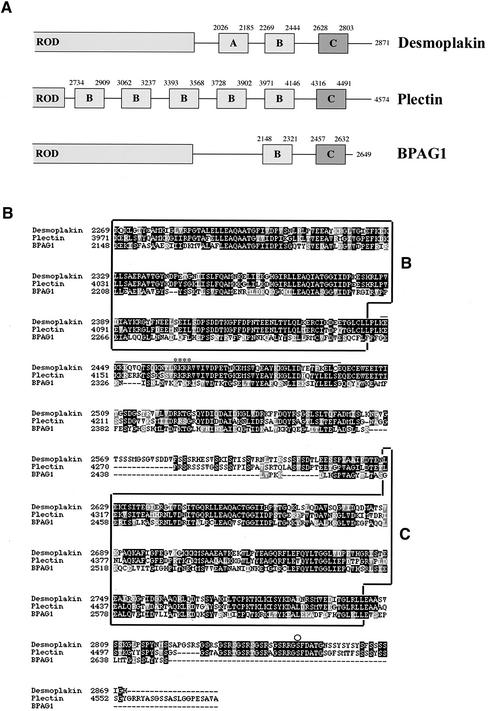
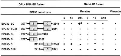
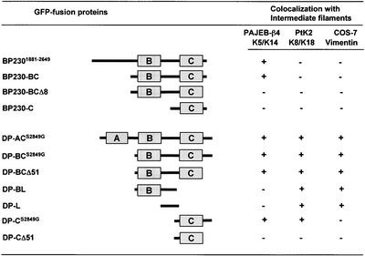
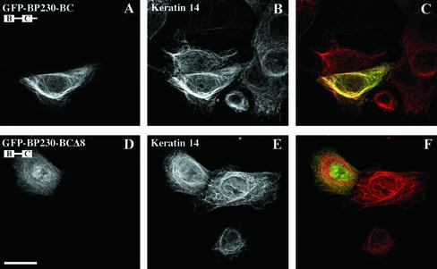
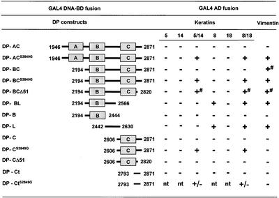
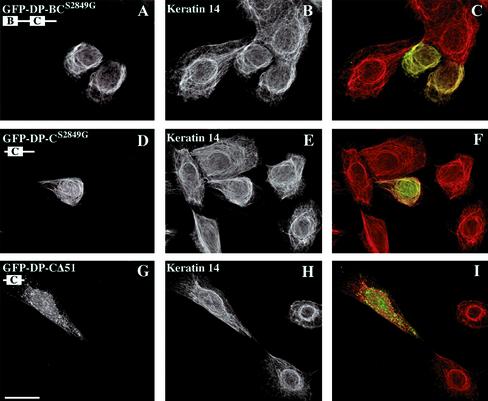
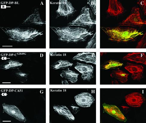
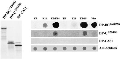
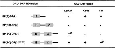
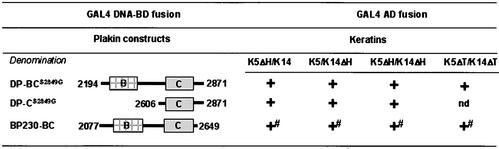
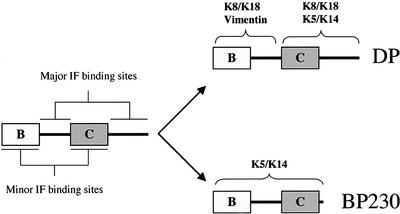
The bullous pemphigoid antigen 1 (BP230) and desmoplakin (DP) are members of the plakin protein family of cytolinkers. Despite their homology, their COOH termini selectively bind distinct intermediate filaments (IFs). We studied sequences within their COOH termini required for their interaction with the epidermal keratins K5/K14, the simple epithelial keratins K8/K18, and type III IF vimentin by yeast three-hybrid, cell transfection, and overlay assays. The results indicate that BP230 interacts with K5/K14 but not with K8/K18 or vimentin via a region encompassing both the B and C subdomains and the COOH extremity, including a COOH-terminal eight-amino-acid stretch. In contrast, the C subdomain with the COOH-terminal extremity of DP interacts with K5/K14 and K8/K18, and its linker region is able to associate with K8/K18 and vimentin. Furthermore, the potential of DP to interact with IF proteins in yeast seems to be regulated by phosphorylation of Ser 2849 within its COOH terminus. Strikingly, BP230 and DP interacted with cytokeratins only when both type I and type II keratins were present. The head and tail domains of K5/K14 keratins were dispensable for their interaction with BP230 or DP. On the basis of our findings, we postulate that (1) the binding specificity of plakins for various IF proteins depends on their linker region between the highly homologous B and C subdomains and their COOH extremity and (2) the association of DP and BP230 with both epidermal and simple keratins is critically affected by the tertiary structure induced by heterodimerization and involves recognition sites located primarily in the rod domain of these keratins.


Figures

Figure 1.
Figure 1.
(A) Schematic of the COOH-terminal half of DP, PL, and BP230 showing the predicted subdomains within their COOH-terminal tail. (B) Sequence alignment of the COOH termini of human DP I, epithelial PL, and BP230 (GenBank accession numbers P15924, G02520, and Q03001, respectively). Alignment was performed with the CLUSTAL-W program. Black boxes indicate identical amino acids; gray boxes denote amino acid similarity. The bar indicates the stretch of amino acid residues of PL involved in binding to vimentin (Nikolic et al., 1996). The asterisks denote residues crucial for the interaction between PL and vimentin. The circle denotes Ser 2849 in DP. The subdomains B and C are de-lineated by boxes.
Figure 2.
Figure 2.
Survey of the sites of interaction between the COOH-terminal domain of BP230 fused to GAL4-BD and IF proteins fused to GAL4-AD: + and - indicate growth or no growth, respectively, on selective media. The PJ69–4A yeast strain was cotransformed (triple transformation) with pACT2, pACT2-URA plasmids encoding IF proteins in fusion with GAL4-AD and pAS2–1 plasmids encoding BP230 fused at its NH2-terminus with GAL4-DNA-BD. To test the interaction with either vimentin or monomeric keratins, triple transformations were performed with either pACT2 or pACT2-URA without insert. After selection on SC-LWUra agar plates, eight colonies of each transformation were arrayed in 96-well microtiter plates and then transferred onto agar SC-LWUra (positive control), SC-LWUra without adenine, and SC-LWUra without histidine and supplemented with 2 mM 3-amino 1,2,3-triazole. Growth was estimated after 5 d of incubation at 30°C. Transactivation controls were performed systematically for each construct with the opposite vectors without insert. # indicates no growth on medium lacking adenine. Each experiment was repeated at least twice.
Figure 3.
Figure 3.
Summary of the data obtained in cell transfection experiments in PA-JEB/β4, PtK2, and COS-7 cell lines: + and - indicate colocalization or not between GFP-tagged BP230 or DP fusion proteins and the network of IFs consisting of K5/K14 in PAJEB-β4 cells, K8/K18 in PtK 2 cells, and vimentin in COS-7 cells.
Figure 4.
Figure 4.
The codistribution of BP230 with the epidermal keratin network is mediated by sequences contained within the B and C subdomains and the most COOH-terminal extension. Ec-topic expression of the COOH-terminal domain of BP230 fused to GFP in PA-JEB/β4 keratinocytes. Cells were transiently transfected with either GFP-BP230-BC (A–C) or GFP-BP230-BCΔ8 cDNA (D–F). After 48 h, cells were stained with anti-GFP (A and D) and anti-keratin 14 antibodies (B and E). Overlays are shown in C and F. The deletion of the eight COOH-terminal amino acid residues impairs the ability of the BP230 tail to codistribute with epidermal keratins. Bar, 25 μm
Figure 5.
Figure 5.
Yeast three-hybrid analysis of the interaction between various subdomains of the COOH-terminal tail of DP fused to GAL4-BD and IF proteins fused to GAL4-AD: +, +/- and - indicate growth, slow growth, or no growth, respectively, on selective media, tested as described under MATERIALS AND METHODS and in Figure 2. # indicates slow growth on medium lacking adenine; nt, not tested.
Figure 6.
Figure 6.
Ser 2849 in the DP tail is phosphorylated in yeast. GAL4-recombinant proteins DP-CS2849G (lanes 1 and 2) and DP-C (lanes 3 and 4) were immunoprecipitated from yeast extracts and subjected (lanes 2 and 4) or not (lanes 1 and 3) to dephosphorylation by CIAP and immunoblotted with mouse anti-GAL4 antibody. DP-CS2849G and DP-C produce three distinct bands presumably as a result of a proteolytic activity in yeast cells and extracts. DP-C has a lower electrophoretic mobility than DP-CS2849G. Treatment with CIAP has no impact on the electrophoretic mobility of recombinant protein DP-CS2849G (lanes 1 and 2), whereas it affects recombinant protein DP-C (lane 3), the electrophoretic mobility of which (lane 4) becomes indistinguishable from that of recombinant protein DP-CS2849G (lanes 1 and 2).
Figure 7.
Figure 7.
Distinct subdomains affect the coalignment potential of DP tail with the epidermal K5/K14 keratin network. Representatives of double immunofluorescence microscopy analyses of PA-JEB/β4 keratinocytes transiently transfected with cDNAs encoding deletion mutants of the DP tail fused to GFP. Cells were transfected with the cDNA constructs GFP-DP-BCS2849G (A–C), GFP-DP-CS2849G (D–F), and GFP-DP-CΔ51 (G–I), respectively, and stained with anti-GFP (A, D, and G) and anti-keratin 14 antibodies (B, E, and H). Overlays are shown in the right panel. Bar, 25 μm
Figure 8.
Figure 8.
The ability of the COOH-terminal tail of DP to codistribute with the K8/K18 IF network is affected by sequences contained within either the B subdomain and the linker or the C subdomain and the COOH-terminal extension. PtK2 cells were transiently transfected with the cDNA construct GFP-DP-BL (A–C), GFP-DP-CS2849G (D–F), or GFP-DP-CΔ51 (G–I), respectively. After 48 h, cells were double-stained with anti-GFP (A, D, and G) and anti-keratin 18 antibodies (B, E, and H). Overlays are shown in the right panel. Bar, 25 μm
Figure 9.
Figure 9.
Dot-blot overlay assays of recombinant purified monomeric or polymerized IF proteins with radiolabeled fragments of the tail of DP. Left, 35[S]-radiolabeled c-myc–tagged recombinant forms of DP, DP-BCS2849G, DP-CS2849G, or DP-CΔ51 were generated by coupled in vitro transcription/translation and analyzed by SDS-PAGE and autoradiography. Right, identical amounts (1 μg) of monomeric or polymerized IF proteins were immobilized on a nitrocellulose membrane by dot blotting and incubated with 35[S]-radiolabeled DP-BCS2849G, DP-CS2849G, or DP-CΔ51. Protein loading was verified by amino-black staining of the spotted proteins. Overlay assays using recombinant protein DP-BC yielded results identical to those with DP-BCS2849G (not shown).
Figure 10.
Figure 10.
Yeast three-hybrid analysis of the interaction between IF proteins and chimeric proteins BP230 and DP. + and - indicate growth or no growth, respectively, on selective media, tested as described under MATERIALS AND METHODS and in Figure 2. # indicates no growth on medium lacking adenine.
Figure 11.
Figure 11.
Yeast three-hybrid analysis of the interaction between the COOH terminus of either DP or BP230 and K5/K14 keratins, from which the head or tail domains were removed. + and - indicate growth or no growth, respectively, on selective media, tested as described under MATERIALS AND METHODS and in Figure 2. # indicates no growth on medium lacking adenine.
Figure 12.
Figure 12.
Binding sites for IF proteins on plakin family members. The linker region between the B and C subdomains and the COOH extremity contain the major IF binding sites and ensure binding specificity. The B and C subdomains contribute to the association with IF proteins by providing additional binding sites and/or by affecting the proper folding of the linker region and the COOH extremity. DP binds to vimentin and K8/K18 via the B subdomain and the linker, whereas the C subdomain and the COOH extremity mediate the association with K8/K18 and K5/K14. In contrast, the ability of BP230 to associate with K5/K14 depends on a region encompassing the B and C subdomains and the COOH extremity. Although there is no direct evidence that they contain a recognition site, the linker region and the COOH extremity are essential for IF binding.

References

    1. Andrä, K., Lassmann, H., Bittner, R., Shorny, S., Fässler, R., Propst, F., and Wiche, G. (1997). Targeted inactivation of plectin reveals essential function in maintaining the integrity of skin, muscle, and heart cytoarchitecture. Genes Dev. 11, 3143–3156. - PMC - PubMed
    1. Angst, B.D., Nilles, L.A., and Green, K.J. (1990). Desmoplakin II expression is not restricted to stratified epithelia. J. Cell Sci. 97, 247–257. - PubMed
    1. Arnemann, J., Sullivan, K.H., Magee, A.I., King, I.A., and Buxton, R.S. (1993). Stratification-related expression of isoforms of the desmosomal cadherins in human epidermis. J. Cell Sci. 104, 741–750. - PubMed
    1. Borradori, L., and Sonnenberg, A. (1999). Structure and function of hemidesmosomes: more than simple adhesion complexes. J. Invest. Dermatol. 112, 411–418. - PubMed
    1. Brown, A., Bernier, G., Mathieu, M., Rossant, J., and Kothary, R. (1995). The mouse dystonia musculorum gene is a neural isoform of bullous pemphigoid antigen 1. Nat. Genet. 10, 301–306. - PubMed

Publication types

MeSH terms

LinkOut - more resources