. 2003 May;14(5):2071-87.
doi: 10.1091/mbc.e02-10-0653. Epub 2003 Jan 26.

p38 Mitogen-activated protein kinase mediates cell death and p21-activated kinase mediates cell survival during chemotherapeutic drug-induced mitotic arrest

Affiliations

p38 Mitogen-activated protein kinase mediates cell death and p21-activated kinase mediates cell survival during chemotherapeutic drug-induced mitotic arrest

Karl Deacon et al. Mol Biol Cell. 2003 May.

Abstract

Activation of the mitotic checkpoint by chemotherapeutic drugs such as taxol causes mammalian cells to arrest in mitosis and then undergo apoptosis. However, the biochemical basis of chemotherapeutic drug-induced cell death is unclear. Herein, we provide new evidence that both cell survival and cell death-signaling pathways are concomitantly activated during mitotic arrest by microtubule-interfering drugs. Treatment of HeLa cells with chemotherapeutic drugs activated both p38 mitogen-activated protein kinase (MAPK) and p21-activated kinase (PAK). p38 MAPK was necessary for chemotherapeutic drug-induced cell death because the p38 MAPK inhibitors SB203580 or SB202190 suppressed cell death. Dominant-active MKK6, a direct activator of p38 MAPK, also induced cell death by stimulating translocation of Bax from the cytosol to the mitochondria in a p38 MAPK-dependent manner. Dominant active PAK suppressed this MKK6-induced cell death. PAK seems to mediate cell survival by phosphorylating Bad, and inhibition of PAK in mitotically arrested cells reduced Bad phosphorylation and increased apoptosis. Our results suggest that therapeutic strategies that suppress PAK-mediated survival signals may improve the efficacy of current cancer chemotherapies by enhancing p38 MAPK-mediated cell death.


Figures
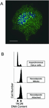
Figure 1.
Figure 1.
Nocodazole activates the mitotic checkpoint and causes mitotic cell cycle arrest. (A) HeLa cells were transfected with HA-MAD2 as described in MATERIALS AND METHODS. After 24 h, the cell were treated with nocodazole (3 μM) for 12 h and the mitotic cells collected by “shake-off.” The mitotic cells were attached to poly-d-lysine–coated coverslips and the cells processed for immunofluorescence microscopy as described in MATERIALS AND METHODS. HA-MAD2 (green) localized to the condensed chromosomes (blue) in the nocodazole-arrested mitotic cells. Bar, 10 μm. (B) Flow cytometry DNA profiles of exponentially growing HeLa cells (top), nocodazole-arrested mitotic cells (middle), and nocodazole-treated attached cells (bottom).
Figure 2.
Figure 2.
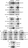
Activation of p38 MAPK, but not JNK or Erk, occurs during mitotic arrest but not during normal mitosis. (A) Exponentially growing HeLa cells were treated with either dimethyl sulfoxide alone (asynchronous population [Asyn]) or with nocodazole (3 μM) for 12 h, and the cells separated into a mitotic (Mit) and an attached (Att) population. Specific antibodies were used to immunoprecipitate (IP) p38 MAPK, JNK, and cdk1 from the cell lysates for immunocomplex kinase assays and Western blotting (WB) with anti-p38 MAPK, anti-JNK, and anti-cdk1. A sample of the mitotic cell lysate was also incubated with protein A-Sepharose beads alone (C). Protein extracts prepared from HeLa cells treated with anisomycin (100 ng/ml for 0.5 h) were also analyzed in parallel as a positive control for the activation of both p38 MAPK and JNK. (B) Activation of Erk1, Erk2, and cdk1 in nocodazole-arrested mitotic cells was determined as described above. Protein extracts from phorbol 12-myristate 13-acetate–treated (100 nM for 0.5 h) HeLa cells were also analyzed in parallel as a positive control for the activation of both Erk1 and Erk2. (C) Activation of p38 MAPK, JNK, Erk1, Erk2, and cdk1 after treatment of exponentially growing HeLa cells with either nocodazole (3 μM), taxol, vincristine, or vinblastine (all 1 μM) for 12 h. (D) HeLa cells were synchronized using the aphidicolin-thymidine double block method as described in MATERIALS AND METHODS. Eight to 10 h after release from the cell cycle block, mitotic cells were collected by shake-off and the activation of p38 MAPK, JNK, and cdk1 in this mitotic cell population (Mit) determined as described in A. Protein extracts from nocodazole-treated (3 μM for 12 h) HeLa cells were also analyzed in parallel as a positive control for the activation of p38 MAPK.
Figure 3.
Figure 3.
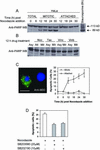
p38 MAPK activation and inactivation correlates with cdk1 activation and inactivation respectively. (A) Exponentially growing HeLa cells were treated with nocodazole (3 μM) and cell extracts prepared at the indicated time points after nocodazole addition. The activation of p38 MAPK, JNK, and cdk1 was assessed using immunocomplex kinase assays as described in MATERIALS AND METHODS. (B) Quantitation of the data shown in A. (C) Exponentially growing HeLa cells were treated with nocodazole (3 μM) for 12 h and the mitotic cells collected by shake-off. After the removal of nocodazole cell extracts were prepared at the indicated time points to assess the activation of both p38 MAPK and cdk1 using immunocomplex kinase assays as described in MATERIALS AND METHODS. (D) Quantitation of the data shown in C.
Figure 4.
Figure 4.
PAK activation correlates with the activation of p38 MAPK in mitotically arrested HeLa cells but PAK does not activate p38 MAPK. (A) Exponentially growing HeLa cells were treated with nocodazole (3 μM) and cell extracts prepared, after separation of the attached (Att) and the mitotic cell populations (Mit), at the indicated time points. Activation of p38 MAPK and PAK was assessed using immunocomplex kinase assays as described in MATERIALS AND METHODS. The cell extracts were also immunoblotted (WB) with specific antibodies for p38α and PAKα. (B) Activation of p38 MAPK and PAK after treatment of exponentially growing HeLa cells were treated with nocodazole (3 μM), taxol, vincristine, or vinblastine (all 1 μM) for 12 h. The cell extracts were also immunoblotted (WB) with anti-p38α and PAKα. (C) Activation of p38 MAPK and PAKα at the indicated times after treatment of exponentially growing HeLa cells were treated with nocodazole (3 μM) for 12 h. (D) HeLa cells were transfected with FLAG epitope-tagged p38α alone or together with either HA epitope-tagged wild-type (wt) PAKα or dominant active (L107F) HA-PAKα. Twenty-four hours after transfection, cells transfected with p38α alone were treated with anisomycin (100 ng/ml for 0.5h) or an equivalent volume of dimethyl sulfoxide. The activation of PAKα and p38 MAPK was determined using immunocomplex kinase assays and cell extracts immunoblotted with either an anti-HA or anti-FLAG antibody to verify transfection efficiency.
Figure 5.
Figure 5.
Nocodazole-mediated induction of apoptosis in HeLa cells requires p38 MAPK activity. (A) Immunoblot analysis (WB) of nocodazole-induced apoptosis, assessed by PARP cleavage, after treatment of exponentially growing HeLa cells with nocodazole (3 μM) for the indicated times. At the 6-h time point a mitotic shake-off was not performed as few mitotic cells were present (<10%). Protein extracts prepared from HeLa cells treated with staurosporine (1 μM for 6 h) were analyzed in parallel as a positive control for PARP cleavage. Arrowheads indicate the position of the immunoreactive bands corresponding to intact PARP (113 kDa) and the caspase-cleaved PARP (89 kDa). (B) Immunoblot analysis of PARP cleavage after treatment of exponentially growing HeLa cells with nocodazole (3 μM), taxol, vincristine, or vinblastine (all 1 μM) for 12 h. (C) HeLa cells were plated onto sterile glass coverslips. Twenty-four hours later, the cells were treated with nocodazole (3 μM) for the indicated times. The mitotic cells were recovered by shake-off, and the cells were reattached to poly-d-lysine–coated glass coverslips. Both the mitotic and attached cells were fixed and stained with the M30 antibody as described IN MATERIALS AND METHODS. Left, indirect immunofluorescence staining of nocodazole-arrested mitotic cells (24h treatment) with the M30 antibody (green) and the DNA stain Hoechst 33342 (blue). The cell on the right does not show M30 immunoreactivity and is not apoptotic. Bar, 10 μm. Right, quantitation of the number of M30 immunoreactive cells in the mitotic and attached cell populations, at the indicated time points, after treatment with nocodazole. At least 100 cells were counted in randomly selected fields for each time point, and the data shown represents the mean ± SEM of three independent experiments. (D) Effect of SB203580 (20 μM) and SB202190 (10 μM) on nocodazole (3 μM for 24 h)-induced apoptosis. Control cells were treated with an equivalent vol-ume of dimethyl sulfoxide. After 24 h, the mitotic cells were collected by shake-off and reattached to poly-l-lysine–coated coverslips. The cells were fixed and immunostained with the M30 antibody as described in MATERIALS AND METHODS. At least 100 cells were counted in randomly selected fields for each condition and the bars represent the mean ± SEM of three independent experiments.
Figure 6.
Figure 6.
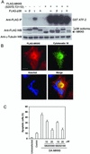
DAMKK6 induces apoptosis in HeLa cells by activating p38 MAPK, whereas DAPAK inhibits daMKK6-induced apoptosis. (A) HeLa cells were transfected with either the FLAG epitope-tagged p38 isotypes or FLAG-DAMKK6 (S207D, T211D) alone or in combination with each other. Twenty-four hours after transfection, the activation of p38 MAPK isotypes was determined using an immunocomplex kinase assay as described in MATERIALS AND METHODS. The cell extracts were also immunoblotted with either an anti-FLAG antibody or an anti-γ-tubulin antibody. The arrowhead indicates the position of FLAG-MKK6. (B) Exponentially growing HeLa cells were transfected with FLAG-DAMKK6. Twenty-four hours after transfection, the cells were fixed and immunostained sequentially with the M30 antibody and an anti-FLAG antibody as described in MATERIALS AND METHODS. Indirect immunofluorescence of an MKK6 transfected cell (top left) showing cleaved cytokeratin 18 (top right) and DNA fragmentation (bottom left). The merged image is also shown (bottom right). Bar, 10 μm. (C) HeLa cells were transfected with either FLAG-DAMKK6, empty vector alone (control) or left untransfected. Eight hours after transfection the cells were treated with either SB203580 (10–20 μM), SB202190 (10–20 μM), or with an equivalent volume of dimethyl sulfoxide alone. Twenty-four hours after transfection, the cells were fixed and immunostained with the M30 antibody as described in MATERIALS AND METHODS. At least 100 transfected cells were counted in randomly selected fields for each condition, and the bars represent the mean ± SEM of three independent experiments. (D) Exponentially growing HeLa cells were transfected with either FLAG-DAMKK6, FLAG-kinase dead (KD)MKK6, HA-DAPAK, HA-KDPAK, empty vector alone (control), or cotransfected with FLAG-DAMKK6 and HA-DAPAK or FLAG-DAMKK6 and HA-KDPAK. Twenty-four hours after transfection cell extracts were prepared and immunoblotted with either with a mixture of anti-FLAG and anti-HA antibodies or with an anti-γ-tubulin antibody. (E) Exponentially growing HeLa cells were transfected with the indicated constructs. Twenty-four hours after transfection, the cells were fixed and immunostained with an anti-FLAG antibody to identify the transfected cells and with the M30 antibody to identify the apoptotic cells as described in MATERIALS AND METHODS. At least 100 MKK6-transfected cells were counted in randomly selected fields for each condition and the bars represent the mean ± SEM of three independent experiments.
Figure 6.
Figure 6.
DAMKK6 induces apoptosis in HeLa cells by activating p38 MAPK, whereas DAPAK inhibits daMKK6-induced apoptosis. (A) HeLa cells were transfected with either the FLAG epitope-tagged p38 isotypes or FLAG-DAMKK6 (S207D, T211D) alone or in combination with each other. Twenty-four hours after transfection, the activation of p38 MAPK isotypes was determined using an immunocomplex kinase assay as described in MATERIALS AND METHODS. The cell extracts were also immunoblotted with either an anti-FLAG antibody or an anti-γ-tubulin antibody. The arrowhead indicates the position of FLAG-MKK6. (B) Exponentially growing HeLa cells were transfected with FLAG-DAMKK6. Twenty-four hours after transfection, the cells were fixed and immunostained sequentially with the M30 antibody and an anti-FLAG antibody as described in MATERIALS AND METHODS. Indirect immunofluorescence of an MKK6 transfected cell (top left) showing cleaved cytokeratin 18 (top right) and DNA fragmentation (bottom left). The merged image is also shown (bottom right). Bar, 10 μm. (C) HeLa cells were transfected with either FLAG-DAMKK6, empty vector alone (control) or left untransfected. Eight hours after transfection the cells were treated with either SB203580 (10–20 μM), SB202190 (10–20 μM), or with an equivalent volume of dimethyl sulfoxide alone. Twenty-four hours after transfection, the cells were fixed and immunostained with the M30 antibody as described in MATERIALS AND METHODS. At least 100 transfected cells were counted in randomly selected fields for each condition, and the bars represent the mean ± SEM of three independent experiments. (D) Exponentially growing HeLa cells were transfected with either FLAG-DAMKK6, FLAG-kinase dead (KD)MKK6, HA-DAPAK, HA-KDPAK, empty vector alone (control), or cotransfected with FLAG-DAMKK6 and HA-DAPAK or FLAG-DAMKK6 and HA-KDPAK. Twenty-four hours after transfection cell extracts were prepared and immunoblotted with either with a mixture of anti-FLAG and anti-HA antibodies or with an anti-γ-tubulin antibody. (E) Exponentially growing HeLa cells were transfected with the indicated constructs. Twenty-four hours after transfection, the cells were fixed and immunostained with an anti-FLAG antibody to identify the transfected cells and with the M30 antibody to identify the apoptotic cells as described in MATERIALS AND METHODS. At least 100 MKK6-transfected cells were counted in randomly selected fields for each condition and the bars represent the mean ± SEM of three independent experiments.
Figure 7.
Figure 7.
DAMKK6 induces p38 MAPK-dependent redistribution of the proapoptotic protein Bax in HeLa cells. HeLa cells were transfected with either GFP-Bax or FLAG-DAMKK6 alone or cotransfected with either GFP-Bax and FLAG-DAMKK6, GFP-Bax, and FLAG-KDMKK6 or EGFP and FLAG-DAMKK6. Sixteen hours (A–C, E, and F) or 36 h after transfection (D), the cells were fixed and immunostained with an anti-FLAG antibody to detect the MKK6 (red) and the DNA was stained with Hoechst 33342 (blue) as described in MATERIALS AND METHODS. Indirect immunofluorescence showing the intracellular distribution at 16 h after transfection of GFP-Bax (A), FLAG-DAMKK6 (B), GFP-Bax in a cell overexpressing DAMKK6 (C), GFP-bax distribution in a cell overexpressing DAMKK6 at 36 h after transfection (D), GFP-Bax intracellular localization in the presence of KDMKK6 (E), and the intracellular distribution of GFP in the presence of DAMKK6 (F). Bar, 10 μm A–C, E and F); 5 μm (D). (G) Exponentially growing HeLa cells were transfected with either GFP-Bax alone or cotransfected with GFP-Bax and FLAG-DAMKK6. Six hours after transfection the cells cotransfected with GFP-Bax and FLAG-DAMKK6 were treated with either SB203580 (20 μM) or SB202190 (10 μM). Twenty-four hours after transfection, the cells were fixed and immunostained with an anti-FLAG antibody to detect the MKK6 as described in MATERIALS AND METHODS. At least 100 MKK6-transfected cells were counted in randomly selected fields for each condition and the bars represent the mean ± SEM of three independent experiments.
Figure 7.
Figure 7.
DAMKK6 induces p38 MAPK-dependent redistribution of the proapoptotic protein Bax in HeLa cells. HeLa cells were transfected with either GFP-Bax or FLAG-DAMKK6 alone or cotransfected with either GFP-Bax and FLAG-DAMKK6, GFP-Bax, and FLAG-KDMKK6 or EGFP and FLAG-DAMKK6. Sixteen hours (A–C, E, and F) or 36 h after transfection (D), the cells were fixed and immunostained with an anti-FLAG antibody to detect the MKK6 (red) and the DNA was stained with Hoechst 33342 (blue) as described in MATERIALS AND METHODS. Indirect immunofluorescence showing the intracellular distribution at 16 h after transfection of GFP-Bax (A), FLAG-DAMKK6 (B), GFP-Bax in a cell overexpressing DAMKK6 (C), GFP-bax distribution in a cell overexpressing DAMKK6 at 36 h after transfection (D), GFP-Bax intracellular localization in the presence of KDMKK6 (E), and the intracellular distribution of GFP in the presence of DAMKK6 (F). Bar, 10 μm A–C, E and F); 5 μm (D). (G) Exponentially growing HeLa cells were transfected with either GFP-Bax alone or cotransfected with GFP-Bax and FLAG-DAMKK6. Six hours after transfection the cells cotransfected with GFP-Bax and FLAG-DAMKK6 were treated with either SB203580 (20 μM) or SB202190 (10 μM). Twenty-four hours after transfection, the cells were fixed and immunostained with an anti-FLAG antibody to detect the MKK6 as described in MATERIALS AND METHODS. At least 100 MKK6-transfected cells were counted in randomly selected fields for each condition and the bars represent the mean ± SEM of three independent experiments.
Figure 8.
Figure 8.
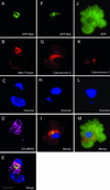
Both DAMKK6 and nocodazole induce Bax translocation to the mitochondria. Exponentially growing HeLa cells were cotransfected with GFP-Bax and FLAG-DAMKK6 (A–E) or with either GFP-Bax (F–I) or EGFP (J–M) alone. Sixteen hours after transfection, the cells cotransfected with GFP-Bax and FLAG-DAMKK6 were stained with MitoTracker red to detect the mitochondria (red), fixed, and immunostained with anti-FLAG antibody to detect the MKK6 (pseudocolored magenta) as described in MATERIALS AND METHODS. The DNA was stained with Hoechst 33342 (blue). Sixteen hours after transfection, the cells transfected with either EGFP or GFP-Bax alone were treated with nocodazole (3 μM) for 12 h and then stained with MitoTracker red for 0.5 h at 37°C. The mitotic cells were collected by shake-off and reattached to poly-d-lysine–coated glass coverslips, fixed, and the DNA stained with Hoechst 33342 as described in MATERIALS AND METHODS. Indirect immunofluorescence showing the intracellular localization of GFP-Bax (A), MitoTracker red (B), Hoechst (C), DAMKK6 (D), and a merged image indicating overlap of the GFP-Bax and MitoTracker red fluorescence signals (E). Indirect immunofluorescence of nocodazole-arrested mitotic cells showing the intracellular distribution of GFP-Bax (F), MitoTracker red (G), Hoechst (H), and a merged image (I) indicating the localization of Bax to a mitochondrial-rich region of the cell. Indirect immunofluorescence of a nocodazole-arrested mitotic cell showing the intracellular distribution of EGFP (J), MitoTracker red (K), Hoechst (L), and a merged image (M). Bar, 10 μm.
Figure 9.
Figure 9.
PAK phosphorylates the proapoptotic protein Bad. (A) Immunoblot analysis of native Bad after treatment of HeLa cells with nocodazole (Noc, 3 μM), taxol (Tax), vincristine (Vinc), or vinblastine (Vinb) (all 1 μM) for 12 h. The cell extracts were also immunoblotted with an anti-γ-tubulin antibody. The upper and lower arrowheads indicate the positions of the slower and faster migrating form of Bad, respectively. Asynchronous, untreated cells (Asynch), mitotic (Mit), and attached (Att). (B) Immunoblot analysis of GST-mBad phosphorylation after treatment of HeLa cells with the indicated drugs as described in (A). HeLa cells were transfected with pEBGmBAD and 24 h later treated with the indicated drugs. Cell extracts were immunoblotted with the indicated phospho-specific Bad antibodies and an anti-Bad antibody. (C) HeLa cells were transfected with pEBGmBAD wild-type, pEBGmBAD (S155A), HA-DAPAK (L107F), or HA-KDPAK (K298A) alone or in the indicated combinations. Twenty-four hours after transfection cell extracts were prepared and immunoblotted with either anti-HA, anti-Bad antibody, phospho-specific Bad antibodies, or an anti-β-actin antibody. The arrowheads indicate the positions of the Bad and PAK proteins. (D) HeLa cells were transfected with pEBGmBAD wild-type alone or in combination with pEBG-PAK (83–149). Twenty-four hours after transfection, cells were treated with nocodazole (3 μM) for 12 h. Cell extracts of the mitotic and attached populations were immunoblotted with either phospho-specific Bad antibodies or a GST antibody. The arrowheads indicate the positions of the Bad and PAK proteins. (E) HeLa cells were transfected with pEBG-PAK (83–149) or vector alone (pEBG). Twenty-four hours after transfection the cell were treated with nocodazole (3 μM) for 12 h. The mitotic cells were fixed and immunostained with an anti-GST antibody to identify the transfected cells and with the M30 antibody to identify the apoptotic cells as described in MATERIALS AND METHODS. At least 100 GST-positive cells were counted in randomly selected fields and the bars represent the mean ± SEM of three independent experiments.
Figure 9.
Figure 9.
PAK phosphorylates the proapoptotic protein Bad. (A) Immunoblot analysis of native Bad after treatment of HeLa cells with nocodazole (Noc, 3 μM), taxol (Tax), vincristine (Vinc), or vinblastine (Vinb) (all 1 μM) for 12 h. The cell extracts were also immunoblotted with an anti-γ-tubulin antibody. The upper and lower arrowheads indicate the positions of the slower and faster migrating form of Bad, respectively. Asynchronous, untreated cells (Asynch), mitotic (Mit), and attached (Att). (B) Immunoblot analysis of GST-mBad phosphorylation after treatment of HeLa cells with the indicated drugs as described in (A). HeLa cells were transfected with pEBGmBAD and 24 h later treated with the indicated drugs. Cell extracts were immunoblotted with the indicated phospho-specific Bad antibodies and an anti-Bad antibody. (C) HeLa cells were transfected with pEBGmBAD wild-type, pEBGmBAD (S155A), HA-DAPAK (L107F), or HA-KDPAK (K298A) alone or in the indicated combinations. Twenty-four hours after transfection cell extracts were prepared and immunoblotted with either anti-HA, anti-Bad antibody, phospho-specific Bad antibodies, or an anti-β-actin antibody. The arrowheads indicate the positions of the Bad and PAK proteins. (D) HeLa cells were transfected with pEBGmBAD wild-type alone or in combination with pEBG-PAK (83–149). Twenty-four hours after transfection, cells were treated with nocodazole (3 μM) for 12 h. Cell extracts of the mitotic and attached populations were immunoblotted with either phospho-specific Bad antibodies or a GST antibody. The arrowheads indicate the positions of the Bad and PAK proteins. (E) HeLa cells were transfected with pEBG-PAK (83–149) or vector alone (pEBG). Twenty-four hours after transfection the cell were treated with nocodazole (3 μM) for 12 h. The mitotic cells were fixed and immunostained with an anti-GST antibody to identify the transfected cells and with the M30 antibody to identify the apoptotic cells as described in MATERIALS AND METHODS. At least 100 GST-positive cells were counted in randomly selected fields and the bars represent the mean ± SEM of three independent experiments.

Similar articles

Cited by

References

    1. Aoshiba, K., Yasui, S., Hayashi, M., Tamaoki, J., and Nagai, A. (1999). Role of p38-mitogen-activated protein kinase in spontaneous apoptosis of human neutrophils. J. Immunol. 162, 1692–1700. - PubMed
    1. Bagrodia, S., and Cerione, R.A. (1999). PAK to the future. Trends Cell Biol. 9, 350–355. - PubMed
    1. Bagrodia, S., Derijard, B., Davis, R.J., and Cerione, R.A. (1995). Cdc42 and PAK-mediated signaling leads to Jun kinase and p38 mitogen-activated protein kinase activation. J. Biol. Chem. 270, 27995–27998. - PubMed
    1. Bueno, O.F., et al. (2000). The MEK1-ERK1/2 signaling pathway promotes compensated cardiac hypertrophy in transgenic mice. Cancer Res. 60, 5887–5894. - PMC - PubMed
    1. Burke, D. (2000). Complexity in the spindle checkpoint. Curr. Opin. Gen. Dev. 10, 26–31. - PubMed

Publication types

MeSH terms