. 2003 Jun 16;22(12):2959-69.
doi: 10.1093/emboj/cdg290.

The mechanism of action of the Pseudomonas aeruginosa-encoded type III cytotoxin, ExoU

Affiliations

The mechanism of action of the Pseudomonas aeruginosa-encoded type III cytotoxin, ExoU

Hiromi Sato et al. EMBO J. .

Abstract

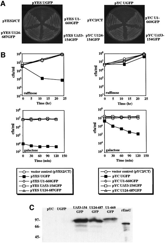
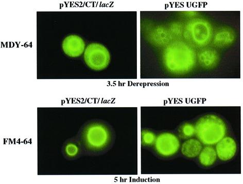
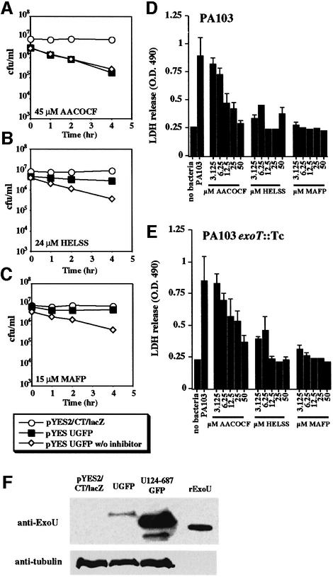
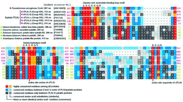
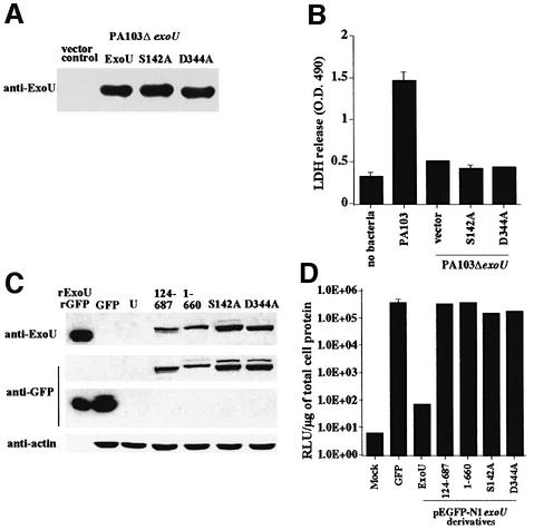
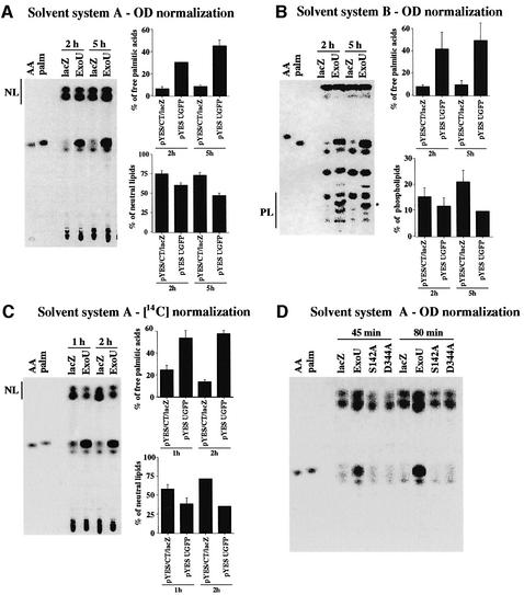
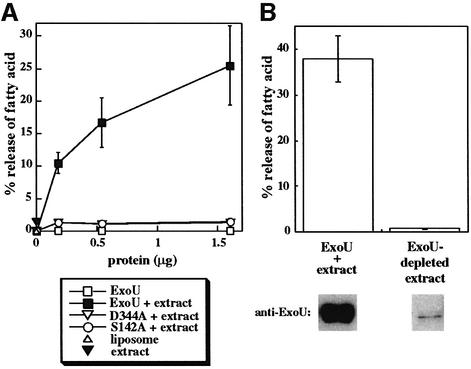
Pseudomonas aeruginosa delivers the toxin ExoU to eukaryotic cells via a type III secretion system. Intoxication with ExoU is associated with lung injury, bacterial dissemination and sepsis in animal model and human infections. To search for ExoU targets in a genetically tractable system, we used controlled expression of the toxin in Saccharomyces cerevisiae. ExoU was cytotoxic for yeast and caused a vacuolar fragmentation phenotype. Inhibitors of human calcium-independent (iPLA(2)) and cytosolic phospholipase A(2) (cPLA(2)) lipase activity reduce the cytotoxicity of ExoU. The catalytic domains of patatin, iPLA(2) and cPLA(2) align or are similar to ExoU sequences. Site-specific mutagenesis of predicted catalytic residues (ExoUS142A or ExoUD344A) eliminated toxicity. ExoU expression in yeast resulted in an accumulation of free palmitic acid, changes in the phospholipid profiles and reduction of radiolabeled neutral lipids. ExoUS142A and ExoUD344A expressed in yeast failed to release palmitic acid. Recombinant ExoU demonstrated lipase activity in vitro, but only in the presence of a yeast extract. From these data we conclude that ExoU is a lipase that requires activation or modification by eukaryotic factors.


Figures

None
Fig. 1. (A) Saccharomyces cerevisiae strain K699 containing high copy number pYES2/CT or low copy number pYC2/CT with ExoUGFP or derivatives of ExoUGFP (ExoU124-687GFP, ExoU1-660GFP or ExoUΔ53-154GFP). Both plates shown contain galactose. (B) Quantitation of the number of colony forming units after the induction of ExoU expression in pYES2/CT (left column) or pYC2/CT (right column). Open symbols represent strains containing vector controls or non-toxic expression constructs of ExoUGFP [as demonstrated in (A)] and filled squares represent strains expressing ExoUGFP under the control of the GAL1 promoter. (C) Western blot analysis of ExoU expression in the low copy number vector pYC2/CT. Proteins present in yeast lysates were separated by SDS-PAGE and transferred to nitrocellulose for western blot analysis with 5 ng of recombinant ExoU (rExoU) as a positive control.
None
Fig. 2. Fluorescence microscopy of yeast stained with a vacuolar membrane marker. Yeast cells with vector controls (left panels) or strains expressing an ExoUGFP fusion protein (right panels) were stained with the vacuole membrane marker MDY-64 after 3.5 h (top) of growth in raffinose, or stained with FM4-64 overnight and induced for 5 h (bottom). Yeast cells were not fixed during these experiments.
None
Fig. 3. Inhibitors of human cytosolic (cPLA2) and Ca2+-independent (iPLA2) lipases reduce the cytotoxic effects of ExoU expression in yeast (AC) and during infection of BEAS-2B human bronchial cells with P.aeruginosa (D and E). Yeast transformants or BEAS-2B cells were treated with inhibitors at the indicated concentrations. In (A–C), yeast viability was measured over time after a shift to induce ExoU (by growth in galactose) in the presence or absence of the inhibitor. In (D), BEAS-2B cells were infected with a strain of P.aeruginosa producing both ExoU and ExoT, and the level of cytotoxicity was measured using the release of lactate dehydrogenase. In (E), BEAS-2B cells were co-cultivated with a P.aeruginosa strain delivering only ExoU. (F) Western blot of yeast extracts derived from cultures grown with 15 µM MAFP. pYES2/CT/lacZ is a vector control lane and UGFP contains an extract from yeast cells induced to express a toxic fusion protein (ExoUGFP). U124-687GFP, an extract from a yeast strain producing a non-toxic N-terminal truncation of ExoU, and 5 ng of His-tagged rExoU were used as markers in this experiment. Reactivity with an antibody to yeast tubulin was used as a loading control (anti-tubulin).
None
Fig. 4. The alignment of the primary sequences of P.aeruginosa ExoU (amino acids 107–154 and 316–352), and patatin-like phospholipase A2 domains of human iPLA2 (A and B), human cPLA2 (α, β and γ) and plant patatins using the NCBI Conserved Domain Database. A color index for conserved amino acid residues among listed proteins was based on the PAM250 substitution score matrix (Dayhoff et al., 1978).
None
Fig. 5. Expression of point mutants during infection (A and B) and transfection (C and D). (A) Point mutations in the predicted catalytic dyad were constructed in exoU cloned in vectors compatible for replication in P.aeruginosa strain PA103ΔexoU. Vector control strains and transformants with plasmids encoding wild-type ExoU or point mutations in ExoU (S142A or D344A) were induced for type III secretion. Supernatant material was concentrated 20-fold. Each lane was loaded with an equivalent quantity of supernatant proteins based on the optical density of the culture at the time of harvest. Monoclonal antibody was used to detect ExoU after transfer to nitrocellulose. (B) Release of LDH after infection of BEAS-2B cells with strains of PA103ΔexoU (1 × 108 c.f.u./ml) expressing ExoUS142A or D344A. No bacteria or vector containing strains served as the negative controls and strain PA103 served as a positive ExoU-expressing strain. (C) Expression of ExoU during transfection analysis of CHO cells. Cells were transfected as described and analyzed for protein expression using western blots. The amount of sample loaded per lane was normalized to actin content in the cellular extract. Recombinant ExoU (rExoU) or GFP (rGFP) were loaded as positive controls. Cells transfected with GFP expression vectors, non-toxic ExoUGFP derivatives (amino acids 124–687GFP and 1–660GFP) and luciferase were processed as negative controls for ExoU-mediated cytotoxicity. (D) Cytotoxicity of ExoU or ExoU derivatives as measured by luciferase activity. CHO cells were co-transfected with a luciferase expression plasmid and constructs producing either GFP or ExoU, or derivatives of ExoU. All constructs with ExoU were made in pEGFP-N1 as GFP fusions. Cells in the ‘Mock’ lane were exposed to transfection reagents alone (no DNA). Luciferase activity is measured as relative light units (RLU) and normalized to micrograms of total cellular protein.
None
Fig. 6. TLC of lipid populations after ExoU induction. Yeast cells were labeled with 1 µCi [14C]palmitic acid. The change of lipid composition after ExoU induction was analyzed by TLC. (A) Samples were loaded onto silica gel TLC plates, based on cell number (OD600), and resolved using solvent system A. Percentages of free palmitic acids and neutral lipids of the total radioactivity were analyzed after induction times of 2 and 5 h. Arachidonic acid (AA) and palmitate (palm) were used as markers. The migration of neutral lipids is marked as NL. (B) TLC with OD600 normalization using solvent system B to resolve phospholipids. The migration of phospholipids is marked as PL. A unique radiolabeled phospholipid product (*) appears only in cultures after induction of ExoU. The radioactivity in this spot was not included in the calculation of the percentage of radioactivity in the phospholipid fraction. (C) Samples were normalized using 14C instead of cell numbers. Percentages of free palmitic acids and neutral lipids of the total radioactivity were analyzed after 1 and 2 h induction using solvent system A. (D) TLC with optical density normalization using solvent system A. TLC was performed on vector controls pYES/CT/lacZ (lacZ), strains expressing ExoUGFP (ExoU) and strains containing an alanine substitution for either of the catalytic residues (S142A and D344A) after 45 and 80 min of induction.
None
Fig. 7. (A) Lipase activity of rExoU. Purified His-tagged proteins at the indicated concentrations were added to an in vitro assay with 14C-labeled liposomes. In some cases purified proteins were preincubated with a yeast extract (+ extract) before addition to the labeled liposomes. The release of free fatty acids was measured using TLC. (B) The activity of an rExoU-replete (ExoU + extract) and an rExoU-depleted yeast extract was quantified by fatty acid release. Removal of rExoU from the treated yeast extract was accomplished using cobalt beads (97.6% reduction in protein as assessed by western blot analysis). Lipase activity was reduced by 98.1% upon removal of His-tagged rExoU.

Similar articles

Cited by

References

    1. Ackermann E.J., Conde-Frieboes,K. and Dennis,E.A. (1995) Inhibition of macrophage Ca2+-independent phospholipase A2 by bromoenol lactone and trifluoromethyl ketones. J. Biol. Chem., 270, 445–450. - PubMed
    1. Allewelt M., Coleman,F.T., Grout,M., Priebe,G.P. and Pier,G.B. (2000) Acquisition of expression of the Pseudomonas aeruginosa ExoU cytotoxin leads to increased bacterial virulence in a murine model of acute pneumonia and systemic spread. Infect. Immun., 68, 3998–4004. - PMC - PubMed
    1. Altschul S.F., Madden,T.L., Schåffer,A.A., Zhang,J., Zhang,Z., Miller,W. and Lipman,D.J. (1997) Gapped BLAST and PSI-BLAST: a new generation of protein database search programs. Nucleic Acids Res., 25, 3389–3402. - PMC - PubMed
    1. Andrews D.L., Beames,B., Summers,M.D. and Park,W.D. (1988) Characterization of the lipid acyl hydrolase activity of the major potato (Solanum tuberosum) tuber protein, patatin, by cloning and abundant expression in a baculovirus vector. Biochem. J., 252, 199–206. - PMC - PubMed
    1. Balsinde J. and Dennis,E.A. (1996) Distinct roles in signal transduction for each of the phospholipase A2 enzymes present in P388D1 macrophages. J. Biol. Chem., 271, 6758–6765. - PubMed

Publication types

MeSH terms