. 2003 Jun 16;22(12):2981-91.
doi: 10.1093/emboj/cdg302.

Phospholipase Cdelta1 is required for skin stem cell lineage commitment

Affiliations

Phospholipase Cdelta1 is required for skin stem cell lineage commitment

Yoshikazu Nakamura et al. EMBO J. .

Abstract

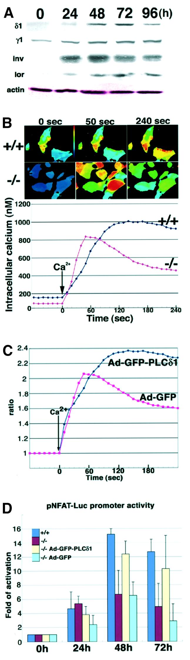
Phosphoinositide-specific phospholipase C (PLC) is a key enzyme in phosphoinositide turnover and is involved in a variety of physiological functions. Here we report that PLCdelta(1)-deficient mice undergo progressive hair loss in the first postnatal hair cycle. Epidermal hyperplasia was observed, and many hairs in the skin of PLCdelta(1)-deficient mice failed to penetrate the epidermis and became zigzagged owing to occlusion of the hair canal. Two major downstream signals of PLC, calcium elevation and protein kinase C activation, were impaired in the keratinocytes and skin of PLCdelta(1)-deficient mice. In addition, many cysts that had remarkable similarities to interfollicular epidermis, as well as hyperplasia of sebaceous glands, were observed. Furthermore, PLCdelta(1)-deficient mice developed spontaneous skin tumors that had characteristics of both interfollicular epidermis and sebaceous glands. From these results, we conclude that PLCdelta(1) is required for skin stem cell lineage commitment.


Figures

None
Fig. 1. Targeted disruption of the PLCδ1 gene in mice. (A) Gene targeting strategy. Exons encoding the X and Y domains were replaced with a neomycin-resistance cassette (neo). gpt, xanthine/guanine phosphoribosyl transferase gene; DTA, diphtheria toxin A gene; PH, pleckstrin homology domain; X, X domain; Y, Y domain; D, DraI. (B) Southern blot analysis of mouse genomic DNA. DraI-digested genomic DNAs isolated from the tails of wild-type (+/+), heterozygous (+/–) and PLCδ1-deficient (–/–) mice were hybridized with the 3′ probe. DraI digestion yields a 12.5 kb fragment for the wild-type allele and a 6.5 kb fragment for the mutant allele. (C) Western blotting of protein extracts from the testes of wild-type (+/+), heterozygous (+/–) and PLCδ1- deficient (–/–) mice. (D) Dorsal and (E) ventral views of 3.5-month-old wild-type (+/+) and PLCδ1-deficient (–/–) mice.
None
Fig. 2. Histological abnormalities in the skin of PLCδ1-deficient mice at 8 days of age. HE staining of dorsal skin sections from (A and C) wild-type (+/+) and (B and D) PLCδ1-deficient (–/–) mice at 8 days of age. The arrow in (D) indicates hair canal occlusion by differentiated keratinocytes. The length of both heads and arrows (C and D) indicates the thickness of the epidermis except for the cornified layers. Analysis of keratinocyte proliferation in dorsal skin sections from (E) wild-type and (F) PLCδ1-deficient mice at 8 days of age by BrdU incorporation. Sections were counterstained with hematoxylin. (G) Number of BrdU-positive cells per field. Values represent the average number of BrdU-positive cells in 30 fields from three sections from different mice. The statistical significance was determined by Student’s t-test (p < 0.05). (H–J) Immunohistochemical detection of PLCδ1 in dorsal skin sections. Immunohistochemical staining of (H) wild-type and (I) PLCδ1-deficient mice at 8 days of age with a polyclonal anti-PLCδ1 antibody. (J) Immunohistochemical staining with antigen-absorbed antibody on wild-type skin was also carried out. Bars: (A and B), shown in (A), 100 µm; (C and D), shown in (C), 50 µm; (E and F), shown in (E), 50 µm; (H–J), shown in (H), 50 µm.
None
Fig. 3. Abnormal differentiation of keratinocytes in the epidermis and hair follicles of PLCδ1-deficient mice at 8 days of age. Green corresponds to antibody staining, and red corresponds to PI (BO-PRO-3) staining. Dorsal skin sections from wild-type (left, +/+) and PLCδ1-deficient (right, –/–) mice at 8 days of age were stained with antibody against (A and B) cytokeratin 1 (K1), (C and D) cytokeratin 5 (K5), (E and F) cytokeratin 6 (K6) and (G and H) loricrin (lor). Arrows indicate aberrant K1 (B) and loricrin (H) expression in the upper portion of mutant hair follicles. Bars: (A–H), shown in (A), 100 µm.
None
Fig. 4. Changes in calcium signaling in PLCδ1-deficient primary cells. (A) PLCδ1 and PLC γ1 contents during calcium-induced keratinocyte differentiation. Primary keratinocytes were treated with 1 mM CaCl2 for 0, 24, 48, 72 and 96 h, and 15 µg of total protein were subjected to SDS–PAGE and western blot analysis was performed. Actin was used as the loading control. δ1, PLCδ1; γ1, PLCγ1; inv, involucrin; lor, loricrin. (B) Keratinocytes from wild-type and PLCδ1-deficient mice differentiated for 72 h were stimulated with 1.6 mM CaCl2. Fluorescence ratio (F340/F380) imagings of wild-type (+/+) and PLCδ1-deficient (–/–) keratinocytes at the indicated times are shown in the upper panels. Time courses of [Ca2+]i mobilization in wild-type (+/+) and PLCδ1-deficient (–/–) keratinocytes are shown in the lower panel. Data were collected in independent experiments (n = 40). (C) Adenoviral infection of PLCδ1 or GFP was performed in PLCδ1-deficient keratinocytes. PLCδ1- and GFP-infected keratinocytes from PLCδ1-deficient mice differentiated for 72 h were stimulated with 1.6 mM CaCl2. Time courses of [Ca2+]i mobilization in PLCδ1-infected (Ad-GFP-PLCδ1) and GFP-infected (Ad-GFP) keratinocytes are shown. Values were calculated by comparison with the 340/380 ratio of cells without Ca2+stimulation. (D) The change in NFAT transcriptional activity in keratinocytes. Adenoviral infection of PLCδ1 or GFP was performed in PLCδ1-deficient keratinocytes. Luciferase activity was measured 76 h after transfection of pNFAT-Luc in wild-type (+/+), PLCδ1-deficient (–/–), PLCδ1-infected PLCδ1-deficient (–/– Ad-GFP-PLCδ1) and GFP-infected PLCδ1-deficient(–/– Ad-GFP) cells untreated or treated with high Ca2+(1.0 mM) for the last 24, 48 and 72 h of the experiment (0 h, 24 h, 48 h, 72 h). Values were calculated by comparison with the activity of cells untreated with high Ca2+ (0 h). Statistical significance between +/+ and –/– at 48 and 72 h was determined by Student’s t-test (p < 0.05).
None
Fig. 5. Downstream effectors of PLCδ1. Green corresponds to antibody staining (A, B, D, E), and red corresponds to PI (BO-PRO-3) staining (A and B). Immunohistochemical detection of active PKC in dorsal skin sections from (A) wild-type (+/+) and (B) PLCδ1-deficient (–/–) mice at 8 days of age. (C) Protein levels of phosphorylated PKCs and PKC isozymes. Western blot analysis of protein extracts from the epidermis of wild-type (+/+) and PLCδ1-deficient (–/–) mice was carried out. Arrowheads indicate the molecular size of full-length phosphorylated PKCs and PKC isozymes. Arrows indicate possible degraded PKC isozymes. Actin was used as the loading control. Localization of NF-κB (p50) in dorsal skin sections from (D) wild-type (+/+) and (E) PLCδ1-deficient (–/–) mice at 8 days of age. Arrows in (D) indicate nuclear localization of p50 in normal skin. Back skins of (F) wild-type (+/+) and (G) PLCδ1-deficient mice (–/–) were treated with 1 µg/ml PMA in acetone for 2 h and harvested. Immuno histochemical detection of NF-κB p65 subunit was performed on frozen sections. Bars: (A and B), shown in (A), 50 µm; (D and E), shown in (D), 50 µm; (F and G), shown in (F), 50 µm.
None
Fig. 6. Abnormal cyst formation and sebaceous gland hyperplasia in PLCδ1-deficient mice. HE staining of dorsal skin sections from (AC and E) wild-type (+/+) and (BD and F) PLCδ1-deficient (–/–) mice at 15 days of age. Arrows in (C) and (D) indicate sebaceous glands. Arrow in (E) indicates granular layers of wild-type IFE. Arrow in (F) indicates cells of abnormal cysts, which resemble those of the granular layers of wild-type IFE. HE staining of dorsal skin sections from (G) wild-type and (H) PLCδ1-deficient mice at 25 days of age. Comparison of ultrastructure of (I) normal epidermis and (J) hair follicle-derived cysts. Arrows in (I) and (J) indicate desmosomes. Arrowheads in (I) and (J) indicate keratohyalin granules. Bars: (A, B, G and H), shown in (A), 100 µm; (C, D and F), shown in (C), 50 µm; (E), 50 µm; (I and J), shown in (I), 2 µm.
None
Fig. 7. Similarities in expression of molecular markers between cysts and IFE. In (A–H), green corresponds to antibody staining, and red corresponds to PI (BO-PRO-3) staining. Dorsal skin sections from wild-type (left, +/+) and PLCδ1-deficient (right, –/–) mice at 15 days of age were stained with antibody against (A and B) cytokeratin 1 (K1), (C and D) cytokeratin 5 (K5), (E and F) cytokeratin 6 (K6) and (G and H) loricrin (lor). Arrows indicate aberrant K1 (B) and loricrin (H) expression in hair follicle-derived cysts. In (I–L), green corresponds to antibody staining, and red corresponds to K5 staining. Dorsal skin sections from wild-type (left, +/+) and PLCδ1-deficient (right, –/–) mice at 15 days of age were stained with antibody against (I and J) AE13 and (K and L) AE15. (M and N) The similarity between hair follicle-derived cyst in (M) PLCδ1-deficient mice and (N) normal epidermis. Bars: (A–H), shown in (A), 100 µm; (I–L), shown in (I), 50 µm.
None
Fig. 8. Development of spontaneous skin tumors in the skin of PLCδ1-deficient mice. (A and B) Skin tumors developed in PLCδ1-deficient mice. Arrows indicate tumors. (C and D) HE staining of tumor sections from PLCδ1-deficient mice at 8 days of age. Arrows in (C) indicate large cysts and the arrow in (D) indicates sebocyte-like cells observed in these tumors. Sections were stained with (E and F) Oil Red O, (G and H) cytokeratin 1 and (I and J) cytokeratin 5. (K) Summary of disturbed lineage commitment in PLCδ1-deficient mice. Bars: (C), 200 µm; (D and H), shown in (D), 25 µm; (E),100 µm; (F and J), shown in (F), 50 µm; (G and I), shown in (G), 50 µm.

Similar articles

Cited by

References

    1. Allen V., Swigart,P., Cheung,R., Cockcroft,S. and Katan,M. (1997) Regulation of inositol lipid-specific phospholipase cδ by changes in Ca2+ ion concentrations. Biochem. J., 327, 545–552. - PMC - PubMed
    1. Beg A.A. and Baltimore,D. (1996) An essential role for NF-κB in preventing TNF-α-induced cell death. Science, 274, 782–784. - PubMed
    1. Berridge M.J. and Irvine,R.F. (1984) Inositol trisphosphate, a novel second messenger in cellular signal transduction. Nature, 312, 315–321. - PubMed
    1. Chattopadhyay N., Mithal,A. and Brown,E.M. (1996) The calcium-sensing receptor: a window into the physiology and pathophysiology of mineral ion metabolism. Endocr. Rev., 17, 289–307. - PubMed
    1. Dolmetsch R.E., Lewis,R.S., Goodnow,C.C. and Healy,J.I. (1997) Differential activation of transcription factors induced by Ca2+ response amplitude and duration. Nature, 386, 855–858. - PubMed

MeSH terms