. 2003 Jun 16;22(12):2992-3003.
doi: 10.1093/emboj/cdg291.

Noggin overexpression inhibits eyelid opening by altering epidermal apoptosis and differentiation

Affiliations

Noggin overexpression inhibits eyelid opening by altering epidermal apoptosis and differentiation

Andrei A Sharov et al. EMBO J. .

Abstract

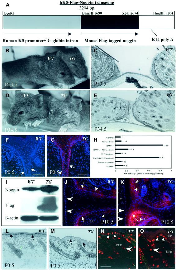
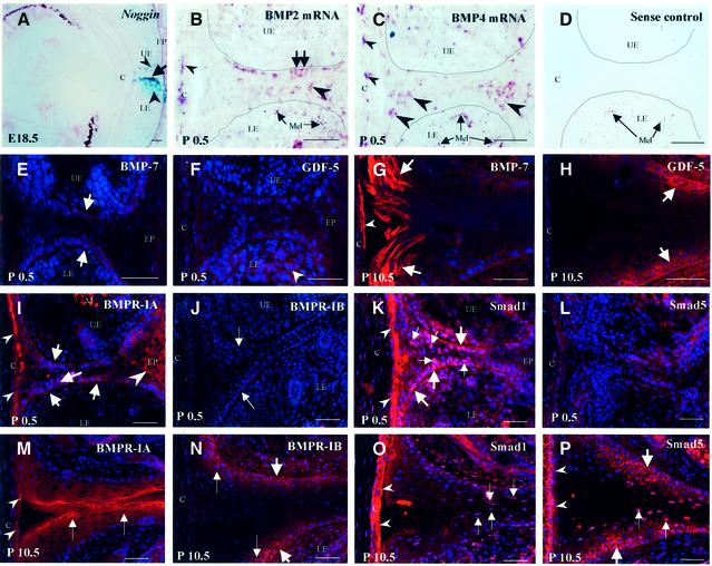
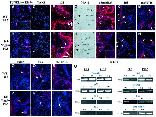
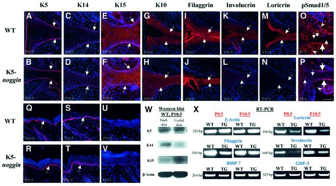
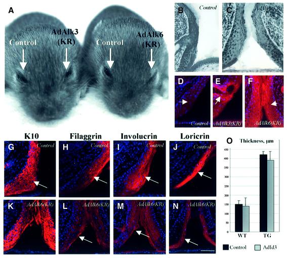
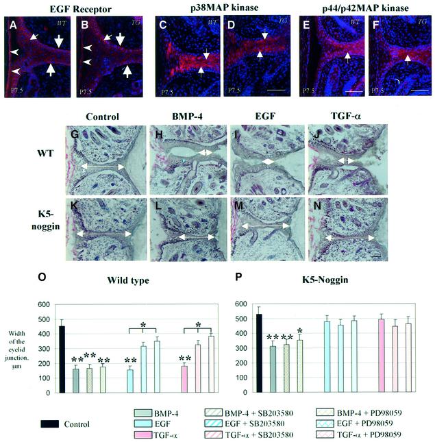
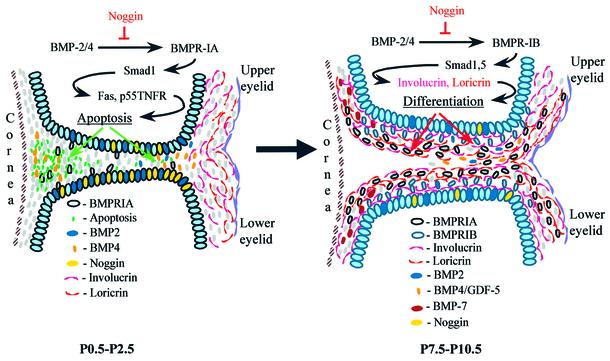
Contact of developing sensory organs with the external environment is established via the formation of openings in the skin. During eye development, eyelids first grow, fuse and finally reopen, thus providing access for visual information to the retina. Here, we show that eyelid opening is strongly inhibited in transgenic mice overexpressing the bone morphogenetic protein (BMP) antagonist noggin from the keratin 5 (K5) promoter in the epidermis. In wild-type mice, enhanced expression of the kinase-inactive form of BMPR-IB mediated by an adenovirus vector also inhibits eyelid opening. Noggin overexpression leads to reduction of apoptosis and retardation of cell differentiation in the eyelid epithelium, which is associated with downregulation of expression of the apoptotic receptors (Fas, p55 kDa TNFR), Id3 protein and keratinocyte differentiation markers (loricrin, involucrin). BMP-4, but not EGF or TGF-alpha, accelerates opening of the eyelid explants isolated from K5-Noggin transgenic mice when cultured ex vivo. These data suggest that the BMP signaling pathway plays an important role in regulation of genetic programs of eyelid opening and skin remodeling during the final steps of eye morphogenesis.


Figures

None
Fig. 1. Creation of transgenic mice overexpressing noggin in the epidermis. (A) Scheme of the transgene construct. (B and D) Inhibition of eyelid opening observed in transgenic (TG) mice at P13.5 and P34.5. (C and E) Microscopy of eyelids in WT and TG mice. (F and G) Expression of Flag in the EJE of TG mice (G, arrows) using antiserum against Flag protein. Lack of expression in WT mice (F). (H) Alkaline phosphatase activity in murine osteoblasts induced by BMP-4 after incubation with media isolated from cultured TG or WT keratinocytes (KCs), or with purified noggin protein. (I) Western blot analysis of 64 kDa noggin protein detected by antisera to full-length mouse noggin or to the Flag protein tagged to the uncleaved noggin in lysates of WT and TG eyelid skin at P0.5. (J and K) Immunofluorescence with antiserum against noggin shows expression in the EJE, the dermis and the corneal epithelium of TG mice (K, arrows, small and large arrowheads, respectively), and in WT mice (J) at P10.5. (L and M) Histological images of dorsal skin of newborn WT and TG mice. Lack of granular layer in the TG epidermis (M, arrows), compared with the WT epidermis (L, arrows). (N and O) Immuno-visualization of Ki67 (red fluorescence) and TUNEL (green fluorescence) in dorsal skin of WT and TG at P7.5. Proliferating cells in the epidermis and dermis are indicated by arrows and arrowheads, respectively. Epidermal/dermal and dermal/subcutaneous borders are indicated by dotted lines. C, cornea; LE and UE, lower and upper eyelids, respectively. Scale bars: 50 µm.
None
Fig. 2. Expression patterns for noggin, BMP-2 and BMP-4 mRNAs, BMP-7, GDF-5, BMP receptors and Smad1/5 proteins during eyelid opening. Cryo-sections of E18.5 murine embryos and P0.5–P10.5 postnatal mice were processed for microscopic visualization of noggin, BMP-2/4 mRNAs, BMP-7, GDF-5, BMPR-IA/IB and Smad1/5. (ANoggin. LacZ activity in the epithelium of the lower eyelid (arrow) and in the mesenchyme of the upper and lower eyelids (arrowheads) of noggin knockout (+/–) mice. (BDBMP-2/4 mRNAs. In situ hybridization signals in basal and suprabasal cells of the eyelid junction (arrows and large arrowheads, respectively), and in corneocytes (small arrowheads) at P0.5. Sense control for BMP-4 mRNA is shown in (D). Melanin granules in (C) and (D) are shown by arrows. (E and G) BMP-7. Expression in basal (E, arrows) and differentiating cells (G, arrows) of the EJE at P0.5 (E) and P10.5 (G). (F and H) GDF-5. Expression in the eyelid mesenchyme (arrowheads) and in basal cells of the EJE (arrows) at P0.5 (F) and P10.5 (H). (I and M) BMPR-IA. Expression in basal and differentiating cells of the EJE, and in corneocytes (arrows, large and small arrowheads, respectively) at P0.5 (I) and P10.5 (M). (J and N) BMPR-IB. Lack of expression in the eyelids at P0.5 (J, arrows). Expression in basal and suprabasal cells of the EJE at P10.5 (N, large and small arrows, respectively). (K and O) Smad1. Nuclear expression in basal and suprabasal cells of the EJE and in corneocytes (large and small arrows and arrowheads, respectively) at P0.5 (K) and P10.5 (O). (L and P) Smad5. Lack of nuclear expression in the eyelids at P0.5 (L). Expression in the basal and suprabasal cells of the EJE and in corneocytes at P10.5 (P, large and small arrows and arrowheads, respectively). C, cornea; LE and UE, lower and upper eyelids, respectively; Mel, melanin. In (B–D), a border between the EJE and the dermis is indicated by dotted line. Scale bars: 50 µm.
None
Fig. 3. Alterations in expression of the apoptotic markers in K5-Noggin mice. Eyelid skin was isolated from newborn WT or transgenic (TG) mice and processed for immunofluorescence (A–F, I–T), in situ hybridization (G, H) or semi-quantitative RT–PCR (U) protocols for analyses of the expression of apoptotic markers. (A and B) TUNEL (green) plus Ki67 (red). Marked decrease of TUNEL+ cells in TG mice (B, labeled area) compared with WT mice (A, labeled area). (C and D) TAK1. Lack of expression in the EJE of both WT and TG mice. (E and F) p21. Strong expression in basal and suprabasal eyelid keratinocytes (KCs) (large and small arrows, respectively). (G and H) Msx-2. Lack of in situ hybridization signal in the EJE and weak expression in the corneal epithelium (arrowheads). The border between eyelid epithelium and mesenchyme is indicated by the dotted lines. (I and J) pSmad1/5. Decrease of nuclear pSmad1/5 expression in the EJE (J, labeled area) and mesenchyme (J, arrowheads) of TG mice compared with the same structures in WT mice (I). (K and L) Id3. Decrease of expression in the EJE of TG mice (L, arrows) compared with WT mice (K). (M and N) p75NTR. Expression in mesenchyme around developing hair follicles (arrows). (O and P) Edar. Expression in the epithelium of developing hair follicles (arrows). (Q and T) Fas and p55TNFR. Marked decrease of expression in the eyelids of TG mice (R, T, arrows), compared with WT mice (Q, S, arrows). (U) Semi-quantitative RT–PCR. Eyelid skin dissected from WT and K5-Noggin TG mice was processed for semi- quantitative RT–PCR of β-actin, p21, Id1, Id2, Id3, p75NTR, Fas and p55TNFR. Representative gels from one of three experiments each are shown. C, cornea; LE and UE, lower and upper eyelids, respectively. Scale bars: (A–D) and (I–T), 50 µm; (E–F), 25 µm.
None
Fig. 4. Differences in the expression of keratinocyte differentiation markers in eyelid epithelium between WT and K5-Noggin mice. Eyelid and back skin was isolated from WT or TG mice at P7.5–P10.5 and processed for analyses of the expression of keratinocyte (KC) markers, BMP-7, GDF-5 and pSmad1/5 using immunofluorescence (A–V), western blot analysis (W) or semi-quantitative RT–PCR (X). (A and B) and (Q and R): keratin 5. Expression in basal KCs (arrows) of the EJE (A and B) and in back skin (Q and R). (C and D) and (S and T): keratin 14. Decrease of expression (arrows) in the EJE (C and D) compared with back skin (S and T). (E and F) and (U and V): keratin 15. Marked increase of expression (arrows) in the EJE (E and F) compared with back skin (U and V). (G–J) Keratin 10 (G and H) and filaggrin (I and J). Expression in suprabasal KCs of the EJE (arrows). (K–N) Involucrin (K and L) and loricrin (M and N). Marked decrease of expression in the EJE of TG mice compared with WT mice (arrows). (O and P) pSmad1/5. Decrease of pSmad1/5 expression in the basal and suprabasal KCs and in eyelid mesenchyme (P, large arrows, small arrows and arrowheads, respectively) of TG mice compared with the same structures of WT mice (O). Scale bars for (A–L) are 50 µm. (W) Western blot analysis of the K5 (60 kDa), K14 (55 kDa) and K15 (49 kDa) proteins in lysates of the WT eyelid and back skin at P10.5. (X) Semi-quantitative RT–PCR. Eyelid skin dissected from WT and TG mice at P0.5 and P10.5 was processed for semi-quantitative RT–PCR with primers specific for β-actin, filaggrin, involucrin, loricirn, BMP-7 and GDF-5. Representative gels from one of three experiments each are shown.
None
Fig. 5. Administration of an adenoviral vector carrying the kinase-inactive form of BMPR-IB inhibits eyelid opening and keratinocyte differentiation in WT mice. Adenoviral vectors carrying kinase-inactive forms of BMPR-IA [AdAlk3(KR)], BMPR-IB [AdAlk6(KR)], Id3 cDNA (AdId3) or β-galactosidase (control) were administered into the conjunctival sac of WT C3H/HeJ mice or K5-Noggin mice at P1.5, P3.5 and P5.5. Eyelid skin was harvested at P13.5 and processed for hematoxylin staining (B, C and O), or immunohistological detection of hemagglutinin, K10, filaggrin, involucrin and loricrin (D–N). (A) Inhibition of eyelid opening after administration of AdAlk6(KR). (B and C) Histological images of eyes treated with control vector (B) or AdAlk6(KR) (C). (DF) Hemagglutinin immunoreactivity in the EJE (arrows) after AdAlk3KR or AdAlk6(KR) treatment (E and F, respectively). Lack of hemagglutinin expression in the EJE (arrow) after treatment with control vector (D). (G and K) K10 expression in eyelids after control and AdAlk6(KR) treatments (arrows). (H–N) Filaggrin (L), involucrin (M) and loricrin expression (N) after AdAlk6(KR) administration. Corresponding controls are shown in (H), (I) and (J). Scale bars: 50 µm. (O) Width of the eyelid junction in WT and TG mice after treatment with AdId3 (values are means ± SEM).
None
Fig. 6. Effects of BMP-4, EGF and TGF-α on the dynamics of eyelid opening in eyelid skin explants of K5-Noggin and WT mice. Eyelid skin was isolated from WT or transgenic (TG) mice at P7.5 and processed for immunofluorescent detection of EGFR, p38MAPK or p44/p42 MAPK (A–F) or cultured ex vivo in presence of BMP-4, EGF, TGF-α, or their combination with MAPK inhibitors (G–P). (A and B) EGF receptor. Expression in basal and suprabasal cells of the EJE (large and small arrows, respectively) and in corneal epithelium (arrowheads). (C–F) p38MAPK (C and D) and p44/p42MAPK (E and F). Expression in differentiating cells of the EJE (arrows). (G–P) Organ culture. Eyelid skin explants isolated from WT or TG mice at P7.5 were cultured ex vivo for 96 h with mouse normal serum as a control (G and K), BMP-4 (H and L), EGF (I and M), TGF-α (J and N) or with their combinations with p38MAPK inhibitor SB203580 or p44/p42MAPK inhibitor PD98059. The width of eyelid junction was evaluated and summarized in (O) and (P) (values are means ± SEM; *P < 0.05; **P < 0.01). In (G–N), white arrows indicate the width of the eyelid junction. Scale bars: 50 µm.
None
Fig. 7. Model illustrating the involvement of BMPs and noggin in the regulation of eyelid opening. At the beginning of eyelid opening (P0.5–P2.5), BMP-2/4 interact with BMPR-IA and promote apoptosis in EJE via stimulation of the expression of Fas, p55TNFR and Id3 protein. Later (P7.5–P10.5), BMP-2/4/7 and GDF-5, via an interaction with BMPR-IB, regulate keratinocyte differentiation and expression of involucrin and loricrin in the eyelid epithelium. Both BMP-mediated apoptosis and cellular differentiation are significantly modulated by noggin, which is expressed in the eyelid epithelium and regulates the amount of biologically active BMPs available for binding to BMP receptors during eyelid opening.

Similar articles

Cited by

References

    1. Aoki H., Fujii,M., Imamura,T., Yagi,K., Takehara,K., Kato,M. and Miyazono K. (2001) Synergistic effects of different bone morphogenetic protein type I receptors on alkaline phosphatase induction. J. Cell Sci., 114, 1483–1489. - PubMed
    1. Blessing M., Nanney,L., King,L., Jones,C. and Hogan,B.L. (1993) Transgenic mice as a model to study the role of TGF-β-related molecules in hair follicles. Genes Dev., 7, 204–215. - PubMed
    1. Blessing M., Schirmacher,P. and Kaiser,S. (1996) Overexpression of bone morphogenetic protein-6 in the epidermis of transgenic mice: inhibition or stimulation of proliferation depending on the pattern of transgene expression and formation of psoriatic lesions. J. Cell Biol., 135, 227–239. - PMC - PubMed
    1. Botchkarev V.A. (2003) Bone morphogenetic proteins and their antagonists in skin and hair follicle biology. J. Invest. Dermatol., 120, 36–47. - PubMed
    1. Botchkarev V., Botchkareva,N., Roth,W., Nakamura,M., McMahon,J.A., McMahon,A. and Paus,R. (1999) Noggin is a mesenchymally-derived stimulator of hair follicle induction. Nat. Cell Biol., 1, 158–164. - PubMed

Publication types

MeSH terms

Substances