. 2003 Jun 16;22(12):3004-14.
doi: 10.1093/emboj/cdg294.

GAP43 stimulates inositol trisphosphate-mediated calcium release in response to hypotonicity

Affiliations

GAP43 stimulates inositol trisphosphate-mediated calcium release in response to hypotonicity

Marco Caprini et al. EMBO J. .

Abstract

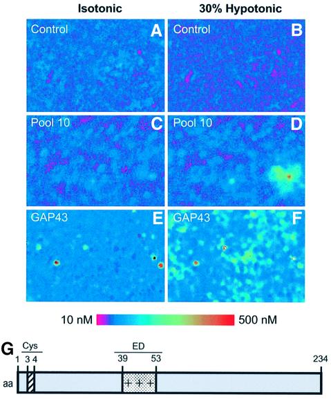
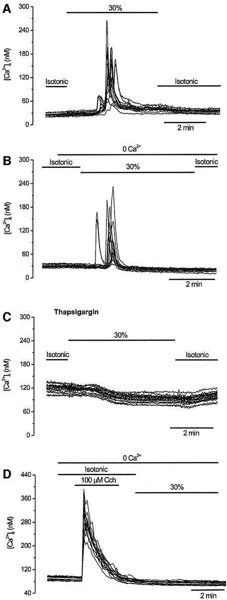
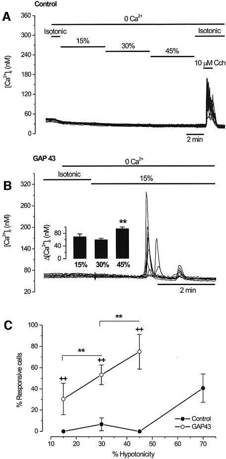
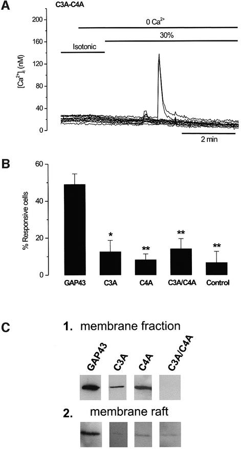
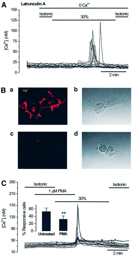
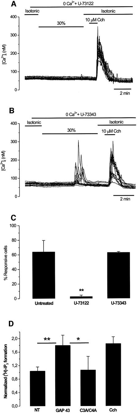
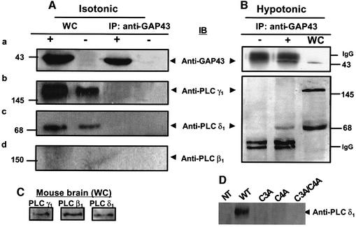
The identification of osmo/mechanosensory proteins in mammalian sensory neurons is still elusive. We have used an expression cloning approach to screen a human dorsal root ganglion cDNA library to look for proteins that respond to hypotonicity by raising the intracellular Ca(2+) concentration ([Ca(2+)](i)). We report the unexpected identification of GAP43 (also known as neuromodulin or B50), a membrane-anchored neuronal protein implicated in axonal growth and synaptic plasticity, as an osmosensory protein that augments [Ca(2+)](i) in response to hypotonicity. Palmitoylation of GAP43 plays an important role in the protein osmosensitivity. Depletion of intracellular stores or inhibition of phospholipase C (PLC) activity abrogates hypotonicity-evoked, GAP43-mediated [Ca(2+)](i) elevations. Notably, hypotonicity promoted the selective association of GAP43 with the PLC-delta(1) isoform, and a concomitant increase in inositol-1,4,5-trisphosphate (IP(3)) formation. Collectively, these findings indicate that hypo-osmotic activation of GAP43 induces Ca(2+) release from IP(3)-sensitive intracellular stores. The osmosensitivity of GAP43 furnishes a mechanistic framework that links axon elongation with phospho inositide metabolism, spontaneous triggering of cytosolic Ca(2+) transients and the regulation of actin dynamics and motility at the growth cone in response to temporal and local mechanical forces.


Figures

None
Fig. 1. Expression cloning of GAP43 protein using Ca2+ imaging. HEK293 cells transiently transfected with clones from a human DRG library were subjected to microscopic fluorescent calcium imaging in isotonic (left) and 30% hypotonic conditions (right). Non-transfected cells exhibited no response to the osmotic–mechanical stimulus (A and B). Cells transfected with pool 10 show a marked increase in cytoplasmic calcium (C and D). This pool was subdivided and re-assayed iteratively until a single positive clone (GAP43) was isolated (E and F). Elevated relative Ca2+ concentrations are indicated by an increased ratio of Fura-2 emission at 340 versus 380 nm excitation wavelenght (see calibrated colored bar). (G) Modular organization of GAP43. Palmitoylation occurs at Cys3 and Cys4. The protein displays a positively charged segment (amino acids 39–53) known as the ED domain.
None
Fig. 2. Swelling-activated Ca2+ signals involve release from intracellular stores. GAP43+ cells were loaded with Fura2-AM to record intracellular calcium signals and activated as indicated. Each panel shows superimposed traces of 15 individual cells recorded in the same experiment. (A) Extracellular Ca2+ (2.4 mM) was present throughout the experiment. After 1 min in standard isotonic solution, the tonicity of the solution was reduced to 70% for 5 min. (B) The external Ca2+ was removed from the medium (and 0.1 mM EGTA was added) 1 min before the start of the 30% hypotonic solution. (C) Responses evoked in GAP43+ cells incubated for 60 min in 2 µM thapsigargin to empty intracellular Ca2+ stores. Identical conditions to those in (A) were used. (D) Pre-treatment of GAP43+ cells with 100 µM carbachol in Ca2+-free solution abolished the subsequent response to 30% hypotonicity.
None
Fig. 3. Swelling-activated intracellular Ca2+ release is dose dependent. HEK293 cells were loaded with Fura2-AM to record intracellular calcium signals, and activated as indicated. (A) Untransfected HEK293 cells (control) were placed in a Ca2+-free (0.1 mM EGTA) isotonic solution and stimulated with solutions of increasing hypotonicity (15, 30 and 45%) for 5 min. Note the absence of response to the osmotic stimulus and the robust response to muscarinic stimulation with 10 µM carbachol. (B) In GAP43+ cells, a mild hypotonic stimulus (15%) in a Ca2+-free solution evokes robust [Ca2+]i signals. The inset shows the mean amplitude of [Ca2+]i elevations for different degrees of hypotonicity. Cells were tested with a single level of hypotonicity. The mean amplitude of the [Ca2+]i signals was larger for the strongest hypotonic stimulus. **P < 0.05 Mann–Whitney rank sum test with N (number of cells) ≥150, and n (number of experiments) ≥3. (C) The percentage of swelling-responsive cells as a function of extracellular hypotonicity. For each experiment, 50 cells in the field were marked at random and scored for a response if the [Ca2+]i was >15 nM. Data are given as mean ± SEM with N ≥ 150 cells, and n ≥ 3. (++) and (**) denote the P < 0.001 significance (Z-test) for the hypotonic responses of GAP43-transfected versus untransfected cells, and for the different hypo- osmolarities tested in GAP43+ cells, respectively.
None
Fig. 4. The palmitoylation sites of GAP43 are important for the protein osmosensitivity. (A) [Ca2+]i response in GAP43 C3A/C4A mutant-transfected cells during a 30% hypotonic stimulation in a Ca2+-free solution. (B) Bar histogram showing the percentage of cells hypotonic-responsive to a stimulus protocol similar to that in (A) in wild-type GAP43+ cells and after single and double mutations of the palmitoyl ation sites. Control denotes untransfected HEK293 cells. For each experiment, 50 cells in the field were marked at random and scored for a response if the [Ca2+]i was >15 nM. Data are given as mean ± SEM with N ≥ 150 cells, and n ≥ 3. **P < 0.001 and *P < 0.005 significance (Z-test) of GAP43 mutants versus wild type. (C) Membrane fractions (1) and membrane rafts (2) from a cell expressing the wild-type GAP43 protein and mutant species. Membrane fractions denote crude plasma membranes. Lipid rafts were obtained from detergent-insoluble complexes in Triton X-100 flotation gradients. The figure depicts the top layer of the Optiprep discontinuous gradient. Crude membranes and rafts were separated by SDS–PAGE and analyzed by western immunoblot using an anti-GAP43 monoclonal antibody.
None
Fig. 5. GAP43-dependent, hypotonicity-activated Ca2+ release is not mediated by alterations in the actin cytoskeleton but is inhibited by PKC activation. (A) [Ca2+]i response in GAP43+ cells during a 30% hypotonic stimulation in a Ca2+-free solution, after incubating the cells with 1 µM latrunculin A for 35 min. (B) Latrunculin A disrupts the cortical cytoskeleton. Panels a and c display confocal images of phalloidin–rhodamine staining of untreated and latrunculin A-treated (1 µM, 35 min) HEK293 cells. Panels b and c depict light-transmitted images of the cells exhibited in panels a and c. The calibration bar for all images is shown in panel a. (C) Pre-incubation with 1 µM PMA for 2 min reduces the number of GAP43+ cells responding to hypotonicity in Ca2+-free external medium. Inset: the percentage of untreated and PMA-treated GAP43+ cells that responded to hypotonicity. For each experiment, 50 cells in the field were marked at random and scored for a response if the [Ca2+]i was >15 nM. Data are given as mean ± SEM with N ≥ 150 cells, and n ≥3. **P < 0.001 significance (Z-test).
None
Fig. 6. GAP43-dependent, hypotonicity-activated Ca2+ release involves IP3 signaling. (A) Pre-incubation for 60 min with the PLC inhibitor U-73122 (3 µM) suppressed the hypotonicity-activated [Ca2+]i response. Note that the response to 10 µM carbacol was unaffected. (B) Pre-incubation for 60 min with the inactive control compound U-73343 (3 µM) did not prevent the hypotonicity-activated [Ca2+]i response. (C) Analysis of the effect of aminosteroid on the percentage of hypotonic-responsive (30% hypotonic stimulus) GAP43+ cells. For each experiment, 50 cells in the field were marked at random and scored for a response if the [Ca2+]i was >15 nM. Data are given as mean ± SEM with N ≥ 150 cells, and n ≥ 3. **P < 0.001 significance (Z-test) for non-treated versus U-73122-treated cells. (D) Hypotonicity induces the synthesis of IP3 in GAP43+ cells. The amount of [3H]IP3 was measured in cells labeled with [2-3H]myo-inositol in isotonic and hypotonic external conditions. Normalized values refer to the ratio of [3H]IP3 after the stimuli (hypotonicity or carbachol) with respect to that before (isotonic). NT denotes non-transfected. Values are given as mean ± SD, with n ≥ 3. **P < 0.01 and *P < 0.05 significance (Student’s t-test) for GAP43 versus NT and GAP43 versus C3A/C4A double mutant, respectively.
None
Fig. 7. Hypotonicity induces the selective association between GAP43 and PLC-δ1. Whole-cell (WC) extracts from GAP43+ cells (+) and untransfected cells (–) in isotonic (A) or 30% hypotonic media (B) were immunoprobed with specific antibodies against GAP43 (panel a), PLC-γ1 (panel b), PLC-δ1 (panel c) and PLC-β1 (panel d), or immunoprecipitated with anti-GAP43 (IP) and thereafter immunoprobed with the displayed antibodies (IB). (C) Mouse brain extracts immunoblotted with anti-PLC isoform-specific antibodies. (D) Co-immunoprecipitation of PLC-δ1 with GAP43 mutant species. Proteins were separated by 10 or 12% SDS–PAGE and electrotransferred onto cellulose membranes. Immunobands were revealed with ECL plus. Numbers indicate molecular weight markers in kDa.

Similar articles

Cited by

References

    1. Aigner L. and Caroni,P. (1993) Depletion of 43-kDa growth-associated protein in primary sensory neurons leads to diminished formation and spreading of growth cones. J. Cell Biol., 123, 417–429. - PMC - PubMed
    1. Aigner L. and Caroni,P. (1995) Absence of persistent spreading, branching and adhesion in GAP-43-depleted growth cones. J. Cell Biol., 128, 647–660. - PMC - PubMed
    1. Arni S., Keilbaugh,S.A., Ostermeyer,A.G. and Brown,D.A. (1998) Association of GAP-43 with detergent-resistant membranes requires two palmitoylated cysteine residues. J. Biol. Chem., 273, 28478–28485. - PubMed
    1. Bomze H.M., Bulsara,K.R., Iskandar,B.J., Caroni,P. and Pate Skene,J.H. (2001) Spinal axon regeneration evoked by replacing two growth cone proteins in adult neurons. Nature Neurosci., 4, 38–43. - PubMed
    1. Bourque C.W. and Oliet,S.H.R. (1997) Osmoreceptors in the central nervous system. Annu. Rev. Physiol., 59, 601–619. - PubMed

Publication types

MeSH terms