. 2003 Jun 1;23(11):4395-400.
doi: 10.1523/JNEUROSCI.23-11-04395.2003.

Math1 gene transfer generates new cochlear hair cells in mature guinea pigs in vivo

Affiliations

Math1 gene transfer generates new cochlear hair cells in mature guinea pigs in vivo

Kohei Kawamoto et al. J Neurosci. .

Abstract

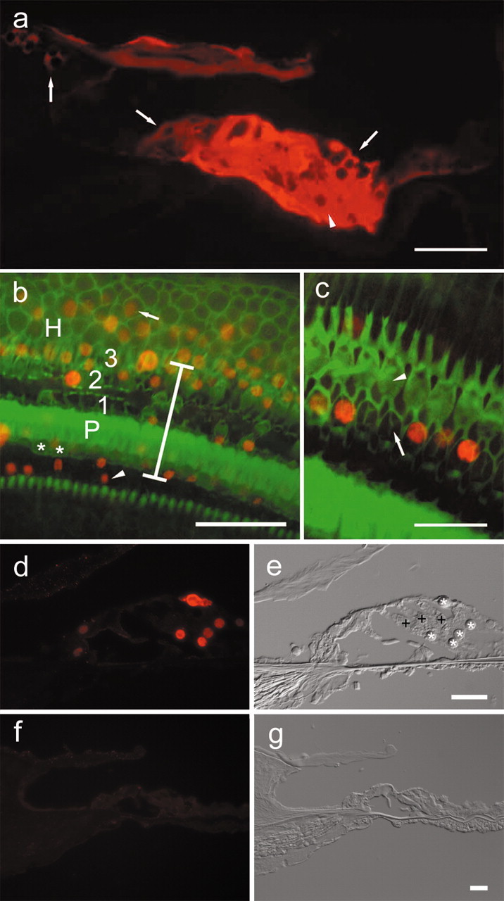
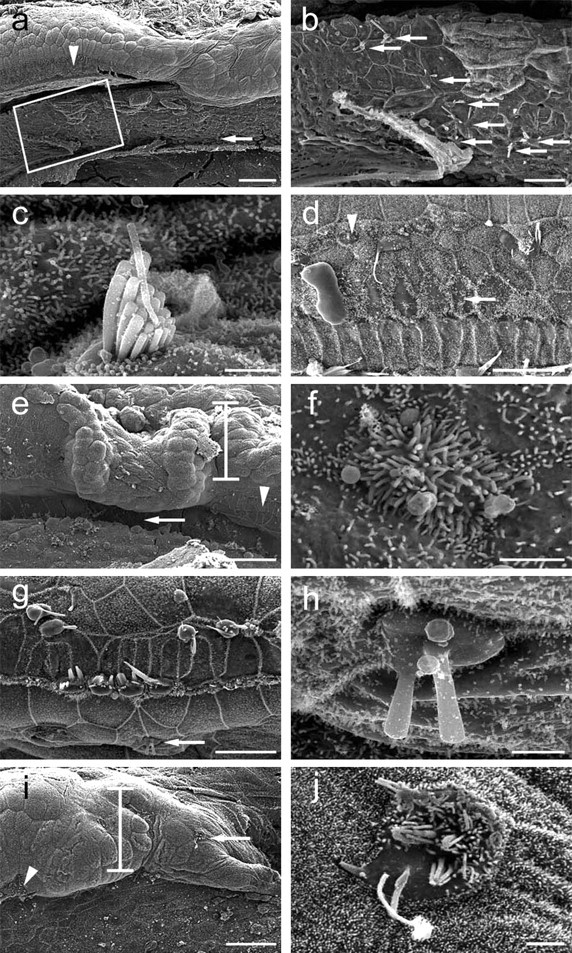
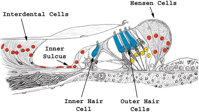
Hair cell loss in the mammalian cochlea is irreversible and results in permanent hearing loss. Math1, the basic helix-loop-helix transcription factor homolog of the Drosophila atonal gene, is a positive regulator of hair cell differentiation during cochlear development. Developing hair cells express Math1, and nonsensory cells do not. We set out to determine the outcome of overexpression of Math1 in nonsensory cells of the cochlea on the phenotype of these cells. We demonstrate that in vivo inoculation of adenovirus with the Math1 gene insert into the endolymph of the mature guinea pig cochlea results in Math1 overexpression in nonsensory cochlear cells, as evident from the presence of Math1 protein in supporting cells of the organ of Corti and in adjacent nonsensory epithelial cells. Math1 overexpression leads to the appearance of immature hair cells in the organ of Corti and new hair cells adjacent to the organ of Corti in the interdental cell, inner sulcus, and Hensen cell regions. Axons are extended from the bundle of auditory nerve toward some of the new hair cells, suggesting that the new cells attract auditory neurons. We conclude that nonsensory cells in the mature cochlea retain the competence to generate new hair cells after overexpression of Math1 in vivo and that Math1 is necessary and sufficient to direct hair cell differentiation in these mature nonsensory cells.


Figures

Figure 1.
Figure 1.
Epifluorescence ofβ-galactosidase and Math1 in the cochlear epithelium 4 d after Ad.Math1.11D inoculation. a, A cryosection showing β-galactosidase immunoreactivity in interdental(left arrow), inner sulcus(middle arrow), Hensen (right arrow), and supporting cells of the organ of Corti (arrowhead) in a cryosection of the third cochlear turn. b, A whole mount showing that Math1-positive nuclei (red) are in the inner sulcus (arrowhead), organ of Corti (vertical bar spans organ of Corti area; asterisk depicts inner hair cells; P depicts pillar cells; 1, 2, and 3 are first, second, and third row outer hair cells, respectively), and in the Hensen cell area (H) in which Math1-positive nuclei (arrow) are observed >30 μm outside the organ of Corti. Phalloidin staining (green) identifies surviving hair cells and sites of hair cell loss. c, Remaining hair cells adjacent to the inoculation site (phalloidin stain, green) are Math1 negative (arrowhead points to third row, outer hair cells). Some of the nonsensory cells that replaced lost hair cells (arrow in second row, outer hair cell area) are Math1 positive (red). d, e, Cryosection (d) of second turn of Ad.Math1.11D-inoculated organ of Corti showing Math1 immunoreactivity in nuclei of nonsensory cells (*). Outer hair cells (+) and several other cell types are negative. Nomarski optics image of same cryosection (e) identified cells shown in d. f, g, Cryosection of second-turn auditory epithelium of Ad.LacZ-inoculated cochlea. Math1 immunoreactivity is negative (f). Nomarski optics image of same cryosection (g) identifies cells. Scale bars: a, b, 50 μm; c, dg, 25 μm.
Figure 2.
Figure 2.
Myosin VIIa and neurofilaments in Ad.Math1.11D-treated and normal cochleae. a, Myosin VIIa-positive ectopic hair cells (arrows) among interdental cells. Inner hair cells (arrowheads) mark the medial border of the organ of Corti. b, A myosin VIIa-positive hair cell (arrow) among Hensen cells. c, An ectopic myosin VIIa-positive hair cell (red) in the interdental cell region. An axon (green) extends from the organ of Corti (arrowhead) to ectopic hair cell. d, An axon (green) extends laterally into the Hensen cell region (arrowheads, lateral border of organ of Corti). e, Myosin VIIa and axons in the organ of Corti (cochlear area similar to that shown in Fig. 1b, adjacent to the inoculation site) 60 d after artificial endolymph inoculation. Inner (arrowhead) and outer (arrows) hair cells are myosin VIIa positive. Several outer hair cells are missing. f, Neurofilament staining is restricted to the organ of Corti in normal (noninoculated) cochlea. Myosin VIIa (red) is in inner (arrowhead) and outer (arrows) hair cells. Micrographs are oriented with medial (modiolar) side down. Scale bars, 25 μm.
Figure 3.
Figure 3.
Scanning electron microscopy of cochleae after Ad.Math1.11D treatment. a, An interdental cell area (boxed) with several ectopic hair cells medial to the organ of Corti (arrowhead). b, Box in a enlarged to show ectopic hair cells (arrows). c, Higher magnification of ectopic hair cell in interdental cell area (a, arrow) with a well developed stereocilia bundle. d, Inoculation-lesioned organ of Corti exhibits cells with short stereocilia (arrow) and small hair cells (arrowhead). Micrographs are oriented with medial (modiolar) side down. e, The site of inoculation showing the injured organ of Corti (arrowhead) and Hensen cells (bar). f, An ectopic hair cell with short stereocilia in the inner sulcus (e, arrow). g, Hair cells in the organ of Corti distant from the inoculation site are missing or injured. h, An ectopic hair cell in the inner sulcus (g, arrow). i, Lateral to the organ of Corti (arrowhead), Hensen cells (bar) exhibit an ectopic hair cell (arrow). j, Higher magnification of ectopic hair cell depicted in i. Micrographs are oriented with medial (modiolar) side down. Scale bars: a, e, i, 50 μm; b, d, g, 20 μm; c, f, h, j, 2 μm.
Figure 4.
Figure 4.
Math1-positive nuclei and ectopic hair cells. The schematic of the organ of Corti is oriented similar to the mid-modiolar cross sections in Figure 1, a and dg, in which medial is on left and lateral is on right. The epithelial regions that exhibit Math1-positive nuclei include the organ of Corti (yellow) and ectopic areas adjacent to the organ of Corti (red). Ectopic new hair cells were identified in the interdental cell, inner sulcus, and Hensen cell regions.

References

    1. Adler HJ, Raphael Y ( 1996) New hair cells arise from supporting cell conversion in the acoustically damaged chick inner ear. Neurosci Lett 205: 17–20. - PubMed
    1. Bermingham NA, Hassan BA, Price SD, Vollrath MA, Ben-Arie N, Eatock RA, Bellen HJ, Lysakowski A, Zoghbi HY ( 1999) Math1: an essential gene for the generation of inner ear hair cells. Science 284: 1837–1841. - PubMed
    1. Brough DE, Lizonova A, Hsu C, Kulesa VA, Kovesdi I ( 1996) A gene transfer vector-cell line system for complete functional complementation of adenovirus early regions E1 and E4. J Virol 70: 6497–7501. - PMC - PubMed
    1. Brown NL, Kanekar S, Vetter ML, Tucker PK, Gemza DL, Glaser T ( 1998) Math5 encodes a murine basic helix-loop-helix transcription factor expressed during early stages of retinal neurogenesis. Development 125: 4821–4833. - PubMed
    1. Chen P, Johnson JE, Zoghbi HY, Segil N ( 2002) The role of Math1 in inner ear development: Uncoupling the establishment of the sensory primordium from hair cell fate determination. Development 129: 2495–2505. - PubMed

Publication types

MeSH terms

LinkOut - more resources