. 2003 Jun 1;23(11):4645-56.
doi: 10.1523/JNEUROSCI.23-11-04645.2003.

The distribution of climbing and mossy fiber collateral branches from the copula pyramidis and the paramedian lobule: congruence of climbing fiber cortical zones and the pattern of zebrin banding within the rat cerebellum

Affiliations

The distribution of climbing and mossy fiber collateral branches from the copula pyramidis and the paramedian lobule: congruence of climbing fiber cortical zones and the pattern of zebrin banding within the rat cerebellum

Jan Voogd et al. J Neurosci. .

Abstract

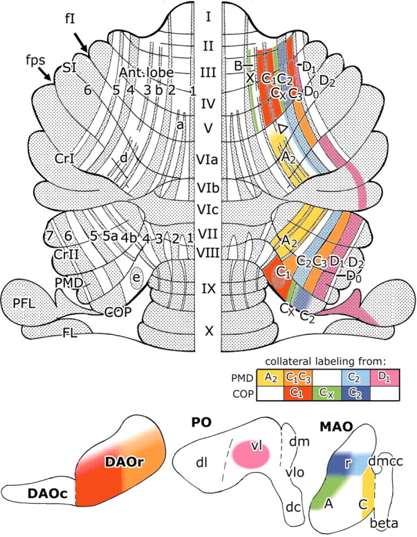
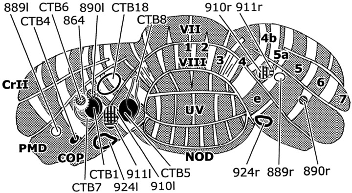
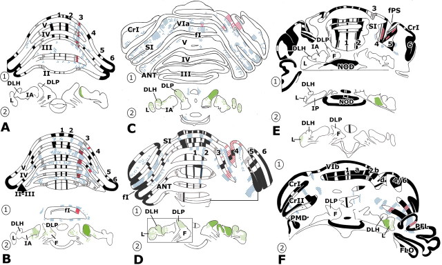
Individual cerebellar cortical zones defined by the somatotopy of climbing fiber responses and by their olivo-cortico-nuclear connections located in the paramedian lobule and the copula pyramidis of the rat cerebellum were microinjected with cholera toxin B subunit. Collateral branches of climbing and mossy fibers were mapped and related to the pattern of zebrin-positive and -negative bands of Purkinje cells. Climbing fiber collaterals from the copula distribute to the anterior lobe: from the paramedian lobule mainly to lobulus simplex and rostral crus I. Climbing fibers terminating in particular zones (X, A2, C1, CX, C2, C3, D1, and D2) in the paramedian lobule or the copula collateralize to one or two corresponding zones in lobulus simplex, crus I and II, the paraflocculus, and/or the anterior lobe. These zones can be defined by their relationship to the pattern of zebrin banding. Collaterals from mossy fibers, labeled from the same injection sites in the copula and paramedian lobule, often distribute bilaterally in a symmetrical pattern of multiple but ill-defined longitudinal strips in the anterior lobe and/or lobulus simplex. One or more of these longitudinal aggregates of mossy fiber collaterals was always found subjacent to the strip(s) of labeled climbing fiber collaterals arising from the same locus in the paramedian lobule or the copula. Corticonuclear projections focused on the target nucleus of each zone, although a bilateral plexus of thinner axons, presumably of mossy fiber collateral origin, was sometimes also present in several other regions of the cerebellar nuclei. Overall, these results suggest that climbing fiber zones and zebrin banding reflect a common organizational scheme within the cerebellar cortex.


Figures

Figure 1.
Figure 1.
Posterior view of the right side of the rat cerebellum showing the location in the PMD and COP of climbing fiber zones identified by their peripheral input, according to the key shown at the bottom (based on Atkins and Apps, 1997). A2, C1, C2, C3, Climbing fiber zones A2, C1, C2, and C3; ipsi, ipsilateral; contra, contralateral.
Figure 2.
Figure 2.
Corticonuclear and olivocerebellar projection zones of the rat cerebellum. The cortical zones (B), the subnuclei of the inferior olive that innervate particular zones (A), and their cerebellar and vestibular target nuclei (C) are indicated with the same symbols. The sections in A and C are viewed in the transverse plane, with sections 12 (A) and 8(C) being the most rostral. Redrawn from Buisseret-Delmas and Angaut (1993), with the addition of the A2 zone (asterisks)(Buisseret-Delmas, 1988a) and the X and CX zones (Buiseret-Delmas et al., 1993). A–D2, Zones A–D2; COP, copula pyramidis; CrI, crus I; CrII, crus II; DAOc, caudal part of dorsal accessory olive; DAOr, rostral part of dorsal accessoryolive; dl, dors all amina of the PO; DLH, dorsolateral hump; DLP, dorsolateral protuberance of the fastigial nucleus; DM, dorsomedial subnucleus of the PO; F, fastigial nucleus; FL, flocculus; IA, anterior interposed nucleus; IC, interstitial cell groups; IP, posterior interposed nucleus; I-X, lobules I–X; L, lateral cerebellar nucleus; LV, lateral vestibular nucleus; MAO, medial accessory olive; PFL, paraflocculus; PMD, paramedian lobule; PO, principal olive; SI, lobulus simplex; vl, ventral lamina of the PO; Y, group Y.
Figure 6.
Figure 6.
Summary of the present results and interpretation in relation to zebrin banding. Dorsal view of the flattened surface of the rat cerebellum, showing the zebrin nomenclature on the left and on the right the distribution of climbing fiber collaterals, arising from injections into different climbing fiber zones in the paramedian lobule and copula pyramidis, with their corresponding zonal identification. The block diagram contains the color code for the collateral projections for individual zones. The origin of these climbing fiber collaterals is shown in the horizontal projection diagrams of the contralateral inferior olive at the bottom. The putative CX zone in SI is indicated with an open arrowhead. Reasons for the localization of the DO and D2 zones in anterior P5- and P6+ and posterior P6- and P7+ bands, respectively, are given in Results and Discussion. 1–7, Zebrin-positive bands P1+ to P7+; A, subnucleus A of caudal MAO; A2, climbing fiber zone A2; a–e, zebrin satellite bands a, b, d, and e; ANT, anterior lobe; beta, subnucleusβ of caudal MAO; C, subnucleus C of caudal MAO; C1–C3, climbing fiber zones C1–C3; COP, copula pyramidis; II–VII, lobules VI–VII; CrI, crus I; CrII, crus II; CX, climbing fiber zone CX; D1, climbing fiber zone D1; dc, dorsal cap; dm, dorsomedial subnucleus of PO; DAOc, caudal dorsal accessory olive; DAOr, rostral dorsal accessory olive; dl, dorsal lamella of PO; dmcc, dorsomedial cell column; fI, primary fissure; fps, posterior superior fissure, MAO, medial accessory olive; PFL, paraflocculus; PMD, paramedian lobule; PO, principal olive; r, rostral; SI, lobulus simplex; vl, ventral lamella of PO; vlo, ventrolateral outgrowth; X, climbing fiber zone X.
Figure 4.
Figure 4.
A, Transverse section showing distribution of CTb-labeled mossy and climbing fiber collaterals in lobules IV and V. Note the regular and bilateral parcellation of mossy fiber terminals and two strips of labeled climbing fiber collaterals (asterisks), which convergeventrally. Case CTB8. Compare with Figure 5C. Scalebar, 500 μm.B, Transverse section showing CTb-labeled mossy and climbing fiber collaterals at higher power. Case CTB7, adjacent section to view shown in D. Scale bar, 50 μm. C, Oblique bundle of zebrin-immunoreactive axons belonging to zebrin-positive band P5+ in the lateral paramedian lobule. Case 925. Scale bar, 500 μm. D, Strips of CTb-labeled mossy and climbing fiber collaterals on facing surfaces of lobules V and VI, separated by the primary fissure, and located in zebrin-negative band P4-. The P4+ band is just visible in the top left corner. Case CTB7. Scale bar, 200 μm. E, Medial lobule of the paraflocculus containing a band of CTb-labeled mossy and climbing fiber collaterals. Case CTB4 (compare with Fig. 7F, panel 2). Scale bar, 500 μm. F, Collaterals of mossy and climbing fibers in the paraflocculus at higher power; for location, see box f in E. Case CTB4. Scale bar, 50 μm. G, Plexus of fine, CTb-labeled varicose axons in the dorsolateral protuberance of the fastigial nucleus, contralateral to the injection site. Case CTB18. Scale bar, 50 μm. H, Plexus of coarse, CTb-labeled varicose axons in the dorsolateral protuberance of the fastigial nucleus, ipsilateral to the injection site. Case CTB18. Same magnification as G. cf, Climbing fiber collaterals; m, midline; mf, mossy fiber collaterals; P4+, P5+, P5-, zebrin positive and negative bands P4+, P5+, and P5-; PFL, paraflocculus; V, VI, lobules V and VI.
Figure 3.
Figure 3.
Location of cerebellar cortical injection sites in the 18 cases used in the present study. Sites of injection projected onto the surface of a reconstruction of the caudal surface of the rat cerebellum. For additional details, see Table 1. Zebrin-positive bands are shaded and indicated with their numerals. COP, Copula pyramidis; CrII, crus II; NOD, nodulus; PMD, paramedian lobule; UV, uvula; VII, VIII, lobules VII and VIII.
Figure 5.
Figure 5.
Example injections into the copula pyramidis. In A and F, the caudal surface of the cerebellum has been reconstructed for two cases (CTB8 and 924-r, respectively). B, G and C,H are reconstructions of dorsal and rostral views of the anterior lobe. Zebrin-positive (P) bands are indicated in black and shown with their corresponding P numbers. The injection site and labeled parallel fibers are indicated in orange, and labeled mossy fiber rosettes are depicted as blue dots. Red contours indicate the position of labeled climbing fiber collaterals. For case CTB8, B′ and C′ show the right side of the cerebellum as depicted in B and C but without the mossy fiber labeling to show more clearly the relationship between the pattern of zebrin banding and the distribution of climbing fiber collateral labeling (indicated with red hatching). For both cases, the localization of retrogradely labeled neurons in the contralateral inferior olive is indicated in purple on an unfolded horizontal projection of DAO (E) and MAO (J) and on transverse sections through the olive (D, I). Scattered labeled cells are indicated with dots. The level of the transverse sections in D and I are indicated with a black horizontal line in E and J (numbers refer to levels depicted in Fig. 2 A). The broken vertical line in E indicates the junction between the dorsal and the ventral folds of DAO. 1–7, Zebrin-positive bands P1 + to P7 +; a, b, e, zebrin-positive satellite bands a, b, and e; BETA, subnucleus β of caudal MAO; COP, copulapyramidis; II–VII, lobules II–VII; CrI, crusI; CrII, crusII; DAOc, caudal part of DAO; DAOr, rostral part of DAO; DMCC, dorsomedial cell column; i, injection site; MAO, medial accessory olive; med, medial; NOD, nodulus; PMD, paramedian lobule; PO, principal olive; UV, uvula.
Figure 7.
Figure 7.
Transverse sections containing mossy fiber and climbing fiber collateral and cerebellar nuclear labeling in six cases with unilateral injections in COP or PMD. Injection sites and other particulars are documented at our website. A, CTB5; B, CTB8; C, CTB18; D, CTB7; E, 864; F, CTB4. Boxed area in D, panel 2, is taken from a more rostral section than the nuclei on the right side. Regions in the cerebellar nuclei containing labeled coarse varicose fibers are indicated with solid green, and labeled thin varicose fibers are stippled in green. Other conventions as in Figure 5. 1–7, Zebrin-positive bands P1+ to P7+; a, zebrin-positive satellite band a; ANT, anterior lobe; b, zebrin-positive satellite band b; CrI, crus I; CrII, crus II; d, zebrin-positive satellite bands d; DLH, dorsolateral hump; DLP, dorsolateral protuberance; F, fastigial nucleus; FI, primary fissure; FLO, flocculus; FPS, posterior superior fissure; IA, anterior interposed nucleus; II–VI, lobules II–VI; L, lateral cerebellar nucleus; NOD, nodulus; PFL, paraflocculus; PMD, paramedian lobule; SI, simplex lobule.

Similar articles

Cited by

References

    1. Apps R ( 2000) Rostrocaudal branching within the climbing fibre projection to forelimb-receiving areas of the cerebellar cortical C1 zone. J Comp Neurol 419: 193–204. - PubMed
    1. Apps R, Trott JR, Dietrichs E ( 1991) A study of branching in the projection from the inferior olive to the x and lateral c1 zones of the cat cerebellum using a combined electrophysiological and retrograde fluorescent double-labelling technique. Exp Brain Res 871: 141–152. - PubMed
    1. Armstrong DM, Harvey RJ, Schild RF ( 1973a) The spatial organisation of climbing fibre branching in the cat cerebellum. Exp Brain Res 18: 40–58. - PubMed
    1. Armstrong DM, Harvey RJ, Schild RF ( 1973b) Branching of inferior olivary axons to terminate in different folia, lobules or lobes of the cerebellum. Brain Res 54: 365–371. - PubMed
    1. Atkins MJ, Apps R ( 1997) Somatotopical organisation within the climbing fibre projection to paramedian lobule and copula pyramidis of the rat cerebellum. J Comp Neurol 389: 249–263. - PubMed

Publication types

MeSH terms

LinkOut - more resources