Clinical Trial
. 2003 Jun 1;23(11):4689-99.
doi: 10.1523/JNEUROSCI.23-11-04689.2003.

Functional organization of human intraparietal and frontal cortex for attending, looking, and pointing

Collaborators, Affiliations
Clinical Trial

Functional organization of human intraparietal and frontal cortex for attending, looking, and pointing

Serguei V Astafiev et al. J Neurosci. .

Abstract

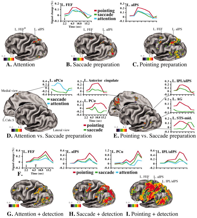
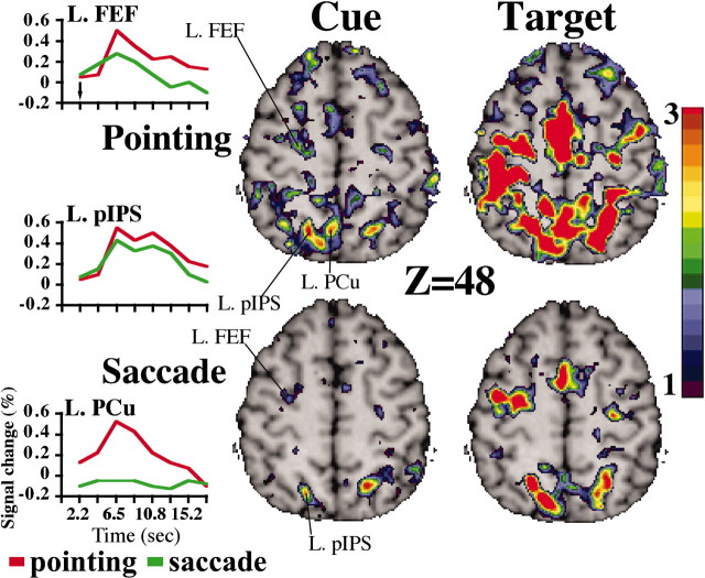
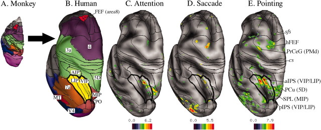
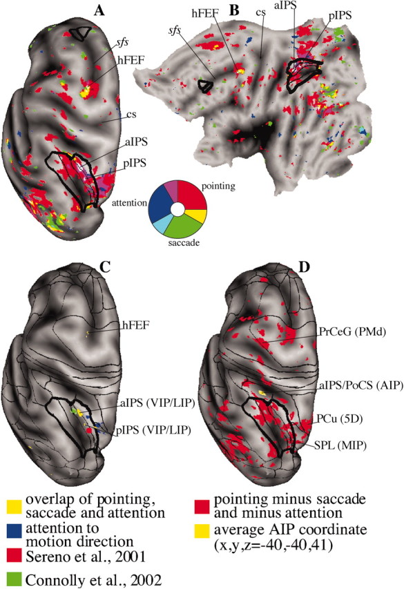
We studied the functional organization of human posterior parietal and frontal cortex using functional magnetic resonance imaging (fMRI) to map preparatory signals for attending, looking, and pointing to a peripheral visual location. The human frontal eye field and two separate regions in the intraparietal sulcus were similarly recruited in all conditions, suggesting an attentional role that generalizes across response effectors. However, the preparation of a pointing movement selectively activated a different group of regions, suggesting a stronger role in motor planning. These regions were lateralized to the left hemisphere, activated by preparation of movements of either hand, and included the inferior and superior parietal lobule, precuneus, and posterior superior temporal sulcus, plus the dorsal premotor and anterior cingulate cortex anteriorly. Surface-based registration of macaque cortical areas onto the map of fMRI responses suggests a relatively good spatial correspondence between human and macaque parietal areas. In contrast, large interspecies differences were noted in the topography of frontal areas.


Figures

Figure 1.
Figure 1.
BOLD responses on inflated surface of the left hemisphere of the Colin brain (Van Essen et al., 2001). The Front plane indicates the dorsolateral view of left hemisphere; the back plane gives the medialview. Color scale indicates z-scores. Cue period is as follows: A, Group-average ANOVA F-map, transformed to z-map and multiple-comparison corrected (F(z) mc) during covert attention. B, Preparation of saccadic eye movement. C, Preparation of pointing movement with right hand. Graphics indicate group-average BOLD time courses averaged over cue direction during attention (blue), saccade (green), and pointing (red). The y-axis indicates the percentage BOLD signal change, whereas the x-axis indicates time (in seconds). D, Differential activation during attention versus saccade preparation. E, Differential activation during saccade versus pointing preparation. Target period is as follows: F, Group-average BOLD time courses, averaged over cue and target direction, extracted over the entire trial (the black arrow shows the time of cue onset, and the black arrow with mark shows the time of target onset, indicating the temporal window of stimulus presentation) in FEF, aIPS, PCu, and IPL/aIPS. G, Group-average F(z) mc map during attention plus target detection. H, Saccade plus target detection. I, Pointing plus target detection. L, Left; Calc.S, calcarine sulcus. The asterisk indicates that there were no active voxels in the left FEF after multiple-comparison correction. The FEF response during the attention task can be seen in Figure 3B. Data sets are available at http://pulvinar.wustl.edu:8081/sums/archivelist.do?archive_id=315115.
Figure 2.
Figure 2.
Selected transverse brain slices (Z = 48) during cue and target periods for pointing and saccade tasks in a representative subject. T-maps were transformed to z-maps. BOLD time courses, averaged over cue direction, were extracted from the left FEF, PCu, and pIPS.
Figure 3.
Figure 3.
A, Macaque brain with anatomical areas (Lewis and Van Essen, 2000). B, Deformation and mapping of macaque areas onto a human atlas brain (left hemispheres, dorsal views) using surface-based registration. Red indicates the FEF (area8), purple indicates motor areas (area4), green indicates somatosensory areas (AIP, areas 5D and 3A), and yellow indicates visual parietal areas (LIP, VIP); the lines in each area define the LIP (dorsal and ventral) within the lateral intraparietal complex and VIP subdivisions (medial and lateral) within the ventral intraparietal area. Brown indicates other parietal areas (area 7A, PO), dark red indicates the MT, and blue indicates V4. C, Group-average ANOVA F(z) map, averaged over cue direction for attention. Black borders indicate deformed macaque visual areas painted in Figure 4 A. D, F(z) map during saccade preparation. E, F(z) map during pointing preparation. Labels in italic indicate the anatomical landmarks. Abbreviations include anatomical locations in the human brain and a deformed area in the monkey (in parentheses): sfs, Superior frontal sulcus; cs, central sulcus; PrCeG, precentral gyrus; PMd, premotor dorsal. Data sets are available at http://pulvinar.wustl.edu:8081/sums/archivelist.do?archive_id=315115.
Figure 4.
Figure 4.
Overlap of preparatory signals for attention (blue), saccade (green), and pointing (red). Regions of overlap are shown: White indicates all conditions, teal indicates attention/saccade, magenta indicates attention/pointing, and yellow indicates saccade/pointing. A, Left hemisphere, dorsal view. B, Flattened representation of the left hemisphere. C, Effector-independent regions (i.e., overlap of preparatory signals for attention, saccade, and pointing preparation from Fig. 4 A, B, yellow); foci of attention to motion direction from Shulman et al. (2002) are in blue. The retinotopically specific parietal area according to Sereno et al. (2001) is shown in red. The IPS area for preparation of saccadic eye movements described by Connolly et al. (2002) is shown in green. D, Hand-specific regions: responses for pointing preparation after subtraction of attention and saccade preparation from Figure 4 A (red); the coordinate of putative AIP averaged over several AIP human studies is shown in yellow (for review, see Binkofski et al., 1999; Grefkes et al., 2002). sfs, Superior frontal sulcus; cs, central sulcus; PoCS, postcentral sulcus; PrCeG, precentral gyrus; PMd, premotor dorsal.

References

    1. Andersen RA, Bracewell RM, Barash S, Gnadt JW, Fogassi L ( 1990) Eye position effects on visual, memory, and saccade-related activity in areas LIP and 7A of macaque. J Neurosci 10: 1176–1196. - PMC - PubMed
    1. Andersson JLR ( 1995) A rapid and accurate method to realign PET scans utilizing image edge information. J Nucl Med 36: 657–669. - PubMed
    1. Batista AP, Buneo CA, Snyder LH, Andersen RA ( 1999) Reach plans in eye centered coordinates. Science 285: 257–260. - PubMed
    1. Battaglia-Mayer A, Ferraina S, Genovesio A, Marconi B, Squatrito S, Molinari M, Lacquaniti F, Caminiti R ( 2001) Eye-hand coordination during reaching. II. An analysis of the relationships between visuomanual signals in parietal cortex and parieto-frontal association projections. Cereb Cortex 11: 528–544. - PubMed
    1. Binkofski F, Dohle C, Posse S, Stephan KM, Hefter H, Seitz RJ, Freund HJ ( 1998) Human anterior intraparietal area subserves prehension: a combined lesion and functional MRI activation study. Neurology 50: 1253–1259. - PubMed

Publication types

MeSH terms

LinkOut - more resources