. 2003 Jul;77(13):7383-92.
doi: 10.1128/jvi.77.13.7383-7392.2003.

Analysis of human immunodeficiency virus type 1 gene expression in latently infected resting CD4+ T lymphocytes in vivo

Affiliations

Analysis of human immunodeficiency virus type 1 gene expression in latently infected resting CD4+ T lymphocytes in vivo

Monika Hermankova et al. J Virol. 2003 Jul.

Abstract

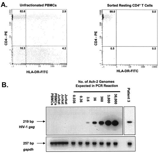
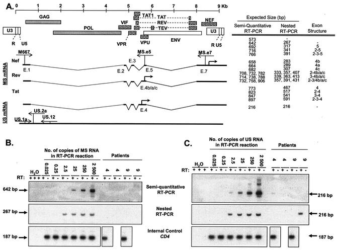
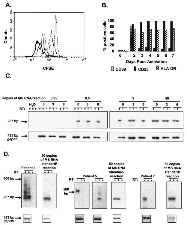
In individuals with human immunodeficiency virus type 1 (HIV-1) infection, a small reservoir of resting memory CD4(+) T lymphocytes carrying latent, integrated provirus persists even in patients treated for prolonged periods with highly active antiretroviral therapy (HAART). This reservoir greatly complicates the prospects for eradicating HIV-1 infection with antiretroviral drugs. Therefore, it is critical to understand how HIV-1 latency is established and maintained. In particular, it is important to determine whether transcriptional or posttranscriptional mechanisms are involved. Therefore, HIV-1 DNA and mRNAs were measured in highly purified populations of resting CD4(+) T lymphocytes from the peripheral blood of patients on long-term HAART. In such patients, the predominant form of persistent HIV-1 is latent integrated provirus. Typically, 100 HIV-1 DNA molecules were detected per 10(6) resting CD4(+) T cells. Only very low levels of unspliced HIV-1 RNA ( approximately 50 copies/10(6) resting CD4(+) T cells) were detected using a reverse transcriptase PCR assay capable of detecting a single molecule of RNA standard. Levels of multiply spliced HIV-1 RNA were below the limit of detection (<50 copies/10(6) cells). Only 1% of the HIV-1 DNA-positive lymphocytes in this compartment could be induced to up-regulate HIV-1 mRNAs after cellular activation, indicating that most of the proviral DNA in resting CD4(+) T cells either carries intrinsic defects precluding transcription or is subjected to transcriptional control mechanisms that preclude high-level production of multiply spliced mRNAs. Nevertheless, by inducing T-cell activation, it is possible to isolate replication-competent virus from resting CD4(+) T lymphocytes of all infected individuals, including those on prolonged HAART. Thus, a subset of integrated proviruses (1%) remains competent for high-level mRNA production after cellular activation, and a subset of these can produce infectious virus. Measurements of steady-state levels of multiply spliced and unspliced HIV-1 RNA prior to cellular activation suggest that infected resting CD4(+) T lymphocytes in blood synthesize very little viral RNA and are unlikely to be capable of producing virus. In these cells, latency appears to reflect regulation at the level of mRNA production rather than at the level of splicing or nuclear export of viral mRNAs.


Figures

FIG. 1.
FIG. 1.
Quantitative measurements of HIV-1 proviral DNA present in highly purified resting CD4+ T lymphocytes. (A) Isolation of resting CD4+ T lymphocytes from patients on HAART. The results of flow cytometric analysis of CD4 and HLA-DR expression on a gated lymphocyte population in unfractionated PBMCs (left panel) and on an ungated sorted population of highly purified resting CD4+ T lymphocytes (right panel) are shown. The numbers are the percentages of cells expressing CD4 and/or HLA-DR. (B) Quantification of HIV-1 DNA in highly purified resting CD4+ T lymphocytes from patients on HAART. To generate a standard curve, ACH-2 cells carrying a single integrated copy of the HIV-1 genome were diluted with HIV-1-negative PBMCs. Isolated DNA was amplified for HIV-1 gag as described in Materials and Methods. To control for DNA quality, the cellular gapdh gene was amplified in parallel. PCR products were confirmed by Southern hybridization using gene-specific probes.
FIG. 2.
FIG. 2.
Detection of MS and US HIV-1 mRNAs associated with resting CD4+ T lymphocytes. (A) Schematic structures of MS and US HIV-1 RNAs. The HIV-1 genome and viral proteins encoded by it are shown at the top. The proteins that are translated from MS mRNA species (dotted) and proteins translated from US mRNA species (shaded) are indicated. The exon structures of MS mRNAs for nef, rev, and tat (only two-exon tat mRNA) are shown. Exon numbers (e.g., E.1) are given below each exon (42, 44). The positions of the primers are indicated by thin arrows. The thick arrow indicates the translation initiation site. The expected sizes of PCR products with their corresponding exon structures are shown to the right of the schematic structures. Exons 1 and 7 that are present in each mRNA are not included. One spliced form that has only these two exons is represented by a hyphen. (B) Single-round (top panel) and nested (middle panel) RT-PCR assays for MS HIV-1 RNAs. Assays were calibrated by diluting radiochemically quantitated, in vitro-transcribed MS RNA into lysates of resting CD4+ T lymphocytes from healthy donors and then carrying the mixture throughout RNA isolation, DNase treatment, reverse transcription, and single-round or nested PCR. Each PCR mixture contained the indicated number of copies of standard RNA in 50,000 cell equivalents of lysate. Therefore, detection of a signal at 2.5 copies/reaction mixture indicated a sensitivity of 50 copies/106 resting CD4+ T cells. Patient samples were tested in duplicate with (+) and without (−) RT. In the experiment shown, samples from patients 4 and 9 were tested, and no MS RNA species were detected. To control for RNA isolation and cDNA synthesis, CD4 RNAs were amplified over the splice junction in each sample. (C) Single-round (top panel) and nested (middle panel) RT-PCR assays for US HIV-1 RNAs. The reactions were standardized with in vitro-transcribed US RNA standard diluted into lysates of resting CD4+ T lymphocytes from healthy donors as described above for panel B. The same lysates contained serially diluted HIV-1 MS RNA standards. US RNA species were detected only sporadically in patients' samples. To control for RNA isolation and cDNA synthesis, CD4 RNAs were amplified over the splice junction in each sample, as was done for MS RT-PCR (bottom panel).
FIG. 3.
FIG. 3.
Reversal of transcriptional silencing by T-cell activation. (A) Activation of purified resting CD4+ T lymphocytes. Prior to activation, resting CD4+ T lymphocytes were labeled with CFSE. Proliferation was measured by a twofold dilution of CFSE. After activation, lymphocytes were harvested at 16 h (- - - -), 24 h (-----), 40 h (-○-○-○), 52 h (-·-·-·), 64 h (——), and 72 h (), and CFSE fluorescence was measured. When resting CD4+ T lymphocytes were incubated in culture medium alone, no dilution of CFSE label was observed (not shown). In all experiments, >95% of CFSE-labeled lymphocytes underwent at least one cell division by day 3 postactivation. (B) Surface expression of CD69, CD25, and HLA-DR in HIV-1 DNA-positive cells measured immediately prior to and at the indicated times after activation. (C) Assay for MS HIV-1 mRNAs associated with activated CD4+ T lymphocytes. The sensitivity of the nested RT-PCR assay was assessed using in vitro-transcribed MS RNA standards. These standards were serially diluted into the lysates from activated CD4+ T lymphocytes harvested at various time points (0, 3, and 6 days) after activation. At 0.5 copy of MS RNA standard in the final nested reaction, the nested assay is positive in one of two duplicated reactions. At 0.05 copy per reaction, the PCR signals were consistently negative. (D) Reversal of HIV-1 latency in a fraction of HIV-1 DNA-positive resting CD4+ T lymphocytes after activation. Resting CD4+ T lymphocytes were seeded at approximately 10 HIV-1 DNA-positive lymphocytes per well and activated. Lymphocytes were harvested 2 to 5 days postactivation and analyzed for MS mRNA. Induced transcription of MS HIV-1 mRNA was detected in resting CD4+ T lymphocytes isolated from all patients. Representative positive wells are shown. The amplified spliced mRNA species from patients' samples had sizes ranging from 267 to 700 bp, as expected on the basis of the MS variants produced during infection (Fig. 2A).

References

    1. Adams, M., L. Sharmeen, J. Kimpton, J. M. Romeo, J. V. Garcia, B. M. Peterlin, M. Groudine, and M. Emerman. 1994. Cellular latency in human immunodeficiency virus-infected individuals with high CD4 levels can be detected by the presence of promoter-proximal transcripts. Proc. Natl. Acad. Sci. USA 91:3862-3866. - PMC - PubMed
    1. Bagnarelli, P., A. Valenza, S. Menzo, R. Sampaolesi, P. E. Varaldo, L. Butini, M. Montroni, C. F. Perno, S. Aquaro, D. Mathez, J. Leibowitch, C. Balotta, and M. Clementi. 1996. Dynamics and modulation of human immunodeficiency virus type 1 transcripts in vitro and in vivo. J. Virol. 70:7603-7613. - PMC - PubMed
    1. Bednarik, D. P., J. A. Cook, and P. M. Pitha. 1990. Inactivation of the HIV LTR by DNA CpG methylation: evidence for a role in latency. EMBO J. 9:1157-1164. - PMC - PubMed
    1. Blankson, J. N., D. Finzi, T. C. Pierson, B. P. Sabundayo, K. Chadwich, J. B. Margolick, T. C. Quinn, and R. F. Siliciano. 2000. Biphasic decay of latently infected CD4+ T cells in acute HIV-1 infection. J. Infect. Dis. 182:1636-1642. - PubMed
    1. Blankson, J. N., D. Persaud, and R. F. Siliciano. 2002. The challenge of viral reservoirs in HIV-1 infection. Annu. Rev. Med. 53:557-593. - PubMed

Publication types