. 2003 Jun 24;100(13):7785-90.
doi: 10.1073/pnas.1432908100. Epub 2003 Jun 12.

Regression of established tumors and metastases by potent vascular endothelial growth factor blockade

Affiliations

Regression of established tumors and metastases by potent vascular endothelial growth factor blockade

Jianzhong Huang et al. Proc Natl Acad Sci U S A. .

Abstract

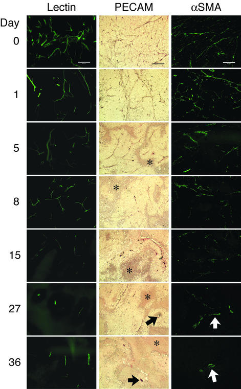
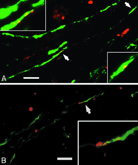
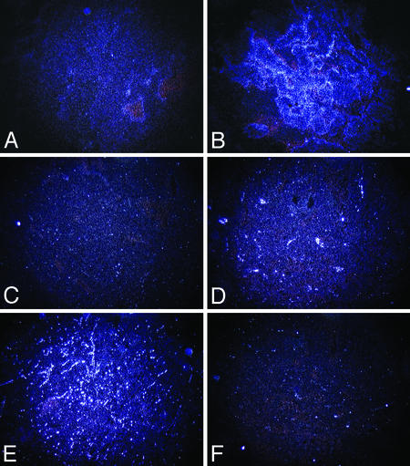
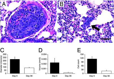
Vascular endothelial growth factor (VEGF) is a critical promoter of blood vessel growth during embryonic development and tumorigenesis. To date, studies of VEGF antagonists have primarily focused on halting progression in models of minimal residual cancer. Consistent with this focus, recent clinical trials suggest that blockade of VEGF may impede cancer progression, presumably by preventing neoangiogenesis. However, VEGF is also a key mediator of endothelial-vascular mural cell interactions, a role that may contribute to the integrity of mature vessels in advanced tumors. Here, we report that high-affinity blockade of VEGF, using the recently described VEGF-Trap, abolishes mature, preexisting vasculature in established xenografts. Eradication of vasculature is followed by marked tumor regression, including regression of lung micrometastases. Thus, the contribution of relatively low levels of VEGF to vessel integrity may be critical to maintenance of even very small tumor masses. Potent blockade of VEGF may provide a new therapeutic option for patients with bulky, metastatic cancers.


Figures

Fig. 1.
Fig. 1.
VEGF-Trap causes involution of xenograft vessels, followed by regression of tumors. Xenografts were established in NCR nude mice and were allowed to grow for 5 weeks. (A) A random cohort of mice was killed (n = 10) to provide day-0 controls (mean weight 5.8 ± 1.1 g). The remaining mice were divided into two groups and injected twice weekly with VEGF-Trap (500 μg) or an equal amount of human Fc protein. Mice were killed at days 1, 5, 8, 15, and 27 after initiation of injections (mean tumor weights ± SEM: 5.5 ± 1.02, 4.2 ± 0.66, 3.9 ± 0.87, 3.5 ± 0.91, and 2.7 ± 0.8 g, respectively). Only treated mice survived until day 36 (mean tumor weight ± SEM: 1.2 g ± 0.3 g, P < 0.0002 vs. day-0 controls). Error bars represent standard error of the mean. (B) Xenografts were initially abundantly vascular at day 0. (C) Surface vessels disappeared and tumors became pale and poorly perfused by day 15. Arrows indicate the contralateral, non-tumor-bearing kidney (approximate size: 10 × 5 mm) in B and C. (D) The native ipsilateral kidney (arrowhead) subsequently reemerged as tumors regressed (day 36).
Fig. 2.
Fig. 2.
Progressive decrease in luminal perfusion and in endothelial and vascular mural compartments of vasculature during the course of VEGF-Trap injection. Day 0 represents tumor vasculature before VEGF-Trap treatment. Before death, selected mice at each time point underwent intravascular injection of fluorescein-labeled L. esculentum lectin. Fluorescein-labeled vasculature decreased progressively, detectable 1 day after the first injection of VEGF-Trap. For immunostaining for endothelial cells (middle column), PECAM-1-stained vasculature decreased after the first dose of VEGF-Trap, with rare remaining immunopositive cells by 27 and 36 days (arrows). Areas of necrosis (asterisks) surround viable regions, which have surviving vessels centrally. For immunostaining for vascular mural cells (right column), decrease in αSMA-positive staining was observed after one dose of VEGF-Trap, and scant remaining αSMA-positive vessels were seen at 27 and 36 days with relatively large diameters (arrows). (Scale bars, 200 μm.)
Fig. 3.
Fig. 3.
Truncation of vasculature during treatment with VEGF-Trap. (A) Day-0 tumor sections during confocal microscopy of lectin-perfused sections. Colors assigned by computer algorithm indicate the relative spatial orientation of vessels. (B) Truncation of vessels is observed at day 1, with abrupt termination of branches (arrows) in midfield. A decrease in branching off large vessels is also evident. (Scale bars, 100 μm.)
Fig. 4.
Fig. 4.
Endothelial and vascular mural cell populations undergo apoptosis concurrently, detectable 1 day after initiation of VEGF-Trap injections. Double-labeling studies were performed to identify endothelial and recruited vascular mural cells (using anti-PECAM an anti-αSMA antibodies, respectively) and apoptosis (using TUNEL assays). TUNEL-positive nuclei (red) colocalize with endothelial cells (A, gray signal) and vascular mural cells (B, gray signal) in regions of intact vessels 1 day after start of VEGF-Trap treatment. (Scale bars, 25 μm.)
Fig. 5.
Fig. 5.
Increased xenograft expression of VEGF and Ang-2 over the course of VEGF-Trap-induced regression, with concurrent decrease in VEGFR-2 expression. VEGF expression is low at day 0 (A) but increases markedly by day 36 (B). Expression of Ang-2, which can cause vessel involution when VEGF is deficient, similarly increases from day 0 (C) to day 36 (D). VEGFR-2 expression decreases from day 0 (E) to day 36 (F). (Original magnification, ×4.)
Fig. 6.
Fig. 6.
Pulmonary metastases decrease in size 36 days after initiation of VEGF-Trap injections. Pulmonary micrometastases in day-0 control (A) and tumor 36 days (B) after initiation of VEGF-Trap treatment (arrow). (Scale bars, 50 μm.) The incidence of pulmonary metastasis and the pattern of adjacent lung microvessels in tumor-bearing animals did not change significantly during VEGF-Trap administration, but diameter (C), volume (D), and cell count (E) significantly decreased.

Comment in

  • Building a better Trap.
    Hood JD, Cheresh DA. Hood JD, et al. Proc Natl Acad Sci U S A. 2003 Jul 22;100(15):8624-5. doi: 10.1073/pnas.1633646100. Epub 2003 Jul 14. Proc Natl Acad Sci U S A. 2003. PMID: 12861079 Free PMC article. No abstract available.

References

    1. Kim, K. J., Li, B., Winer, J., Armanini, M., Gillett, N., Phillips, H. S. & Ferrara, N. (1993) Nature 362, 841-844. - PubMed
    1. Gerber, H. P., Kowalski, J., Sherman, D., Eberhard, D. A. & Ferrara, N. (2000) Cancer Res. 60, 6253-6258. - PubMed
    1. Prewett, M., Huber, J., Li, Y., Santiago, A., O'Connor, W., King, K., Overholser, J., Hooper, A., Pytowski, B., Witte, L., et al. (1999) Cancer Res. 59, 5209-5218. - PubMed
    1. Yang, J. C., Haworth, L., Steinberg, S. M., Rosenberg, S. A. & Novotny, W. (2002) Proc. Am. Soc. Clin. Oncol. 21, 5.
    1. Benjamin, L. E., Hemo, I. & Keshet, E. (1998) Development (Cambridge, U.K.) 125, 1591-1598. - PubMed

Publication types

MeSH terms

Substances

LinkOut - more resources