. 2003 Jun;132(2):739-47.
doi: 10.1104/pp.102.017913. Epub 2003 Apr 24.

AtSig5 is an essential nucleus-encoded Arabidopsis sigma-like factor

Affiliations

AtSig5 is an essential nucleus-encoded Arabidopsis sigma-like factor

Junlan Yao et al. Plant Physiol. 2003 Jun.

Abstract

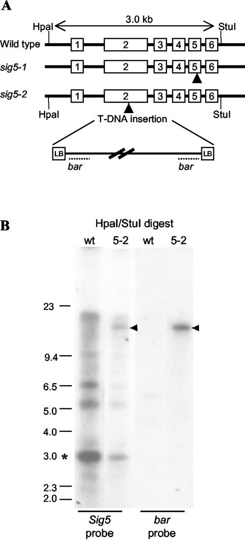
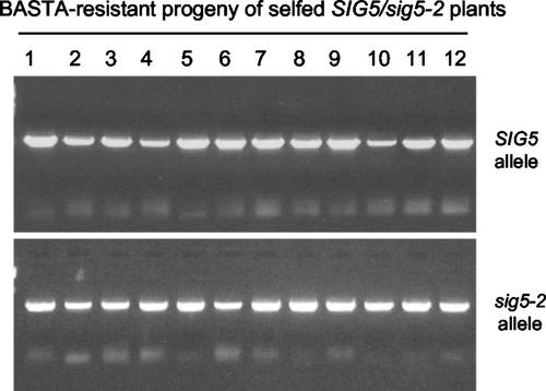
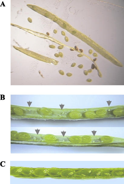
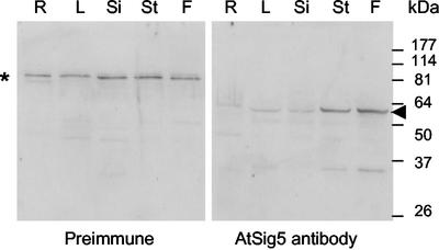
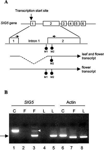
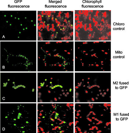
Transcription of chloroplast genes is subject to control by nucleus-encoded proteins. The chloroplast-encoded RNA polymerase (PEP) is a eubacterial-type RNA polymerase that is presumed to assemble with nucleus-encoded sigma-factors mediating promoter recognition. Recently, families of sigma-factor genes have been identified in several plants including Arabidopsis. One of these genes, Arabidopsis SIG5, encodes a sigma-factor, AtSig5, which is phylogenetically distinct from the other family members. To investigate the role of this plant sigma-factor, two different insertional alleles of the SIG5 gene were identified and characterized. Heterozygous mutant plants showed no visible leaf phenotype, but exhibited siliques containing aborted embryos and unfertilized ovules. Our inability to recover plants homozygous for a SIG5 gene disruption indicates that SIG5 is an essential gene. SIG5 transcripts accumulate in flower tissues, consistent with a role for AtSig5 protein in reproduction. Therefore, SIG5 encodes an essential member of the Arabidopsis sigma-factor family that plays a role in plant reproduction in addition to its previously proposed role in leaf chloroplast gene expression.


Figures

Figure 1.
Figure 1.
Characterization of T-DNA insertions in two SIG5 knockout lines, sig5-1 and sig5-2. A, Diagram illustrating the exon-intron structure of the SIG5 gene (exons are depicted as numbered boxes) and the position of T-DNA insertions (arrowheads) in sig5-1 and sig5-2. Recognition sites for restriction enzymes HpaI and StuI and the sizes of fragments generated by digestions of wild-type DNA are indicated. B, DNA gel blot of wild-type and heterozygous sig5-2 plant DNA digested with HpaI and StuI and probed with either a SIG5 (left) or T-DNA (bar, right) probe. The asterisk marks the wild-type SIG5 band, whereas the arrowhead indicates the hybridizing band corresponding to the sig5-2 insertional allele. Additional bands hybridizing to the SIG5 probe are most likely derived from related σ-factor gene family members.
Figure 2.
Figure 2.
Homozygous sig5-2 lines could not be recovered from the selfed progeny of heterozygous sig5-2 plants. DNA prepared from 12 BASTA-resistant progeny was subjected to PCR analysis using primers flanking the T-DNA insertion site (top panel). The presence of a wild-type allele in progeny plants was indicated by the amplification of a 2.5-kb PCR product (SIG5). The large PCR product that would have resulted from the mutant insertional allele was not readily amplified. The same DNA samples were subjected to PCR with a SIG5 primer adjacent to the T-DNA insertion site and a T-DNA left border primer (bottom panel). The amplification of a product of the predicted size for the insertional allele (sig5-2) indicated the presence of the mutant sig5-2 allele in every BASTA-resistant progeny line recovered.
Figure 3.
Figure 3.
Heterozygous sig5-2 lines display a seed phenotype. A, Seeds from a mature silique on a self-fertilized heterozygous sig5-2 plant include brown wrinkled defective seeds and normal round seeds. B, Immature siliques from selfed heterozygous sig5-2 plants contain gaps in the seed rows, indicative of unfertilized ovules (arrowheads). C, A typical silique from a wild-type plant shows round green seeds with no gaps in the seed row.
Figure 4.
Figure 4.
SIG5 is expressed in reproductive and vegetative tissues. Immunoblot of total soluble protein prepared from roots (R), rosette leaves (L), siliques (Si), stems (St), and flowers (F) of 4-week-old wild-type Arabidopsis plants. Immunoblots were probed with preimmune serum (left panel) or anti-AtSig5 antibodies (right panel). A nonspecific cross-reacting band in the preimmune blot (asterisk) serves as a loading control. The AtSig5 cross-reacting band is indicated by an arrow. Migration of Mr markers is shown.
Figure 5.
Figure 5.
Partially unspliced SIG5 transcripts accumulate in flower tissue but not in leaf tissue. A, Diagram of the SIG5 gene indicating exons (numbered boxes). The exon1-intron1-exon2 region is enlarged to indicate the two in-frame Met codons (M1 and M2; •). Transcripts with or without intron 1 are indicated below. The annealing positions of the intron-spanning primers used in RT-PCR are indicated by horizontal arrows. B, Total RNA prepared from leaf (L, lane 5) or flower (F, lane 3) was subjected to RT-PCR analysis using SIG5 primers that span intron 1. Both leaf and flower RNA contain spliced transcripts (black arrow). To control for DNA contamination, the reactions were repeated with no reverse transcription step (lane 2 for flower RNA and lane 4 for leaf RNA). Lane 1 (C) contains SIG5 product amplified from genomic DNA to demonstrate the size of the unspliced SIG5 PCR product. A product of this size is amplified from flower but not leaf RNA (lane 3, white arrowhead). Lanes 6 through 8 show RT-PCR reactions with actin gene primers to control for the amount of RNA in each sample. Lane 6 is a control PCR using intron-spanning actin gene primers on genomic DNA. The lack of this larger product in lanes 7 and 8 reconfirms the absence of genomic DNA in the flower and leaf RNA preparations.
Figure 6.
Figure 6.
Differential targeting of AtSig5-GFP fusion proteins translated from the M1 or M2 Met. NH2-terminal AtSig5 sequences initiated from the M1 (row D) or M2 (row C) Met were fused to GFP and were transiently expressed in Arabidopsis leaf cells. Expression of control constructs with GFP targeted to chloroplasts (row A) or mitochondria (row B) is shown. In each part of the figure, the left panel represents the GFP fluorescent signal, the right panel represents the chlorophyll autofluorescent signal of chloroplasts, and the center panel is the GFP signal merged with the chlorophyll autofluorescence.

References

    1. Allison LA (2000) The role of sigma factors in plastid transcription. Biochimie 82: 537–548 - PubMed
    1. Allison LA, Maliga P (1996) Deletion of rpoB reveals a second distinct transcription system in plastids of higher plants. EMBO J 15: 2802–2809 - PMC - PubMed
    1. Beardslee T, Chowdhury SR, Chang CC, Jaiswal P, Lerbs-Mache S, Stern D, Allison LA (2002) A nucleus-encoded maize protein with sigma factor activity accumulates in both chloroplasts and mitochondria. Plant J 31: 199–209 - PubMed
    1. Burgess RR, Anthony L (2001) How sigma docks to RNA polymerase and what sigma does. Curr Opin Microbiol 4: 126–131 - PubMed
    1. Campbell EA, Muzzin O, Chlenov M, Sun JL, Olson CA, Weinman O, Trester-Zedlitz ML, Darst SA (2002) Structure of the bacterial RNA polymerase promoter specificity sigma subunit. Mol Cell 9: 527–539 - PubMed

Publication types