

The monosaccharide transporter gene, AtSTP4, and the cell-wall invertase, Atbetafruct1, are induced in Arabidopsis during infection with the fungal biotroph Erysiphe cichoracearum
- PMID: 12805612
- PMCID: PMC167022
- DOI: 10.1104/pp.103.021428
The monosaccharide transporter gene, AtSTP4, and the cell-wall invertase, Atbetafruct1, are induced in Arabidopsis during infection with the fungal biotroph Erysiphe cichoracearum
Abstract
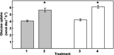
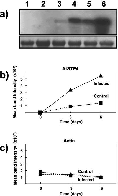
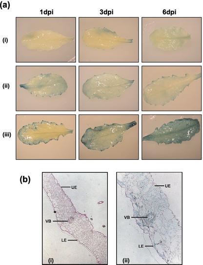
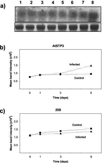
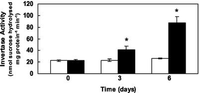
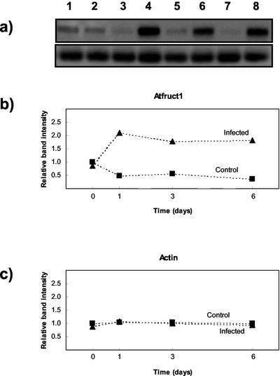
Powdery mildew fungi are biotrophic pathogens that form a complex interface, the haustorium, between the host plant and the parasite. The pathogen acts as an additional sink, competing with host sinks, resulting in considerable modification of photoassimilate production and partitioning within the host tissue. Here, we examine the factors that may contribute to these changes. We show for the first time in one biotrophic interaction (Arabidopsis/Erysiphe cichoracearum) all of the following responses: Glc uptake in host tissues is enhanced after fungal infection; this coincides with the induction of expression of the monosaccharide transporter gene, Arabidopsis sugar transport protein 4 (AtSTP4), in infected leaves; invertase activity and transcript levels for a cell wall invertase, Atbetafruct1, increase substantially in Arabidopsis during attack by this pathogen. Before infection, Arabidopsis plants transformed with an AtSTP4 promoter-beta-glucuronidase construct show expression mainly in sink tissues such as roots; after infection, AtSTP4 expression is induced in the mature leaves and increases over the 6-d time period. Sections of infected leaves stained for beta-glucuronidase show that AtSTP4 expression is not confined to infected epidermal cells but is also evident in a wider range of cells, including those of the vascular tissue. The results are discussed in relation to the possible coordinated expression of hexose transporters and cell wall invertase in the host response to powdery mildew infection.
Figures
Similar articles
-
Involvement of abscisic acid in the coordinated regulation of a stress-inducible hexose transporter (VvHT5) and a cell wall invertase in grapevine in response to biotrophic fungal infection.Plant Physiol. 2010 May;153(1):211-21. doi: 10.1104/pp.110.154765. Epub 2010 Mar 26. Plant Physiol. 2010. PMID: 20348211 Free PMC article.
-
AtSTP8, an endoplasmic reticulum-localised monosaccharide transporter from Arabidopsis, is recruited to the extrahaustorial membrane during powdery mildew infection.New Phytol. 2021 Jun;230(6):2404-2419. doi: 10.1111/nph.17347. Epub 2021 Apr 3. New Phytol. 2021. PMID: 33728642
-
PMR5, an acetylation protein at the intersection of pectin biosynthesis and defense against fungal pathogens.Plant J. 2019 Dec;100(5):1022-1035. doi: 10.1111/tpj.14497. Epub 2019 Oct 12. Plant J. 2019. PMID: 31411777
-
Roles of cell-wall invertases and monosaccharide transporters in the growth and development of Arabidopsis.J Exp Bot. 2003 Jan;54(382):525-31. doi: 10.1093/jxb/erg055. J Exp Bot. 2003. PMID: 12508063 Review.
-
Grapevine powdery mildew (Erysiphe necator): a fascinating system for the study of the biology, ecology and epidemiology of an obligate biotroph.Mol Plant Pathol. 2012 Jan;13(1):1-16. doi: 10.1111/j.1364-3703.2011.00728.x. Epub 2011 Jun 20. Mol Plant Pathol. 2012. PMID: 21726395 Free PMC article. Review.
Cited by
-
Sugar Transporter HmSWEET8 Cooperates with HmSTP1 to Enhance Powdery Mildew Susceptibility in Heracleum moellendorffii Hance.Plants (Basel). 2024 Aug 19;13(16):2302. doi: 10.3390/plants13162302. Plants (Basel). 2024. PMID: 39204738 Free PMC article.
-
Genome-Wide Identification and Expression Profiling of Sugar Transporter Protein (STP) Family Genes in Cabbage (Brassica oleracea var. capitata L.) Reveals their Involvement in Clubroot Disease Responses.Genes (Basel). 2019 Jan 21;10(1):71. doi: 10.3390/genes10010071. Genes (Basel). 2019. PMID: 30669698 Free PMC article.
-
Alcohol dehydrogenase 1 of barley modulates susceptibility to the parasitic fungus Blumeria graminis f.sp. hordei.J Exp Bot. 2011 Jun;62(10):3449-57. doi: 10.1093/jxb/err017. Epub 2011 Feb 21. J Exp Bot. 2011. PMID: 21339386 Free PMC article.
-
BABA and Phytophthora nicotianae Induce Resistance to Phytophthora capsici in Chile Pepper (Capsicum annuum).PLoS One. 2015 May 28;10(5):e0128327. doi: 10.1371/journal.pone.0128327. eCollection 2015. PLoS One. 2015. PMID: 26020237 Free PMC article.
-
Systemic control of plant regeneration and wound repair.New Phytol. 2023 Jan;237(2):408-413. doi: 10.1111/nph.18487. Epub 2022 Oct 2. New Phytol. 2023. PMID: 36101501 Free PMC article. Review.
References
-
- Adam L, Somerville SC (1996) Genetic characterization of five powdery mildew disease resistance loci in Arabidopsis thaliana. Plant J 9: 341–356 - PubMed
-
- Alexopoulos CJ, Mims CW, Blackwell M (1996) Introductory Mycology, Ed 4. Wiley, New York
-
- Ayres PG, Press MC, Spencer-Phillips PTN (1996) Effects of pathogens and parasitic plants on source-sink relationships. In E Zamski, AA Schaffer, eds, Photoassimilate Distribution in Plants and Crops. Marcel Dekker, New York, pp 479–499
-
- Buell CR (1998) Arabidopsis: A weed leading the field of plant-pathogen interactions. Plant Physiol Biochem 36: 177–186
Publication types
MeSH terms
Substances
LinkOut - more resources
Full Text Sources
Molecular Biology Databases
