. 2003 Aug 1;550(Pt 3):731-8.
doi: 10.1113/jphysiol.2003.043778. Epub 2003 Jun 13.

C terminus L-type Ca2+ channel calmodulin-binding domains are 'auto-agonist' ligands in rabbit ventricular myocytes

Affiliations

C terminus L-type Ca2+ channel calmodulin-binding domains are 'auto-agonist' ligands in rabbit ventricular myocytes

Igor Dzhura et al. J Physiol. .

Retraction in

Abstract

L-type Ca2+ channel C terminus calmodulin (CaM)-binding domains are molecular determinants for Ca(2+)-CaM-dependent increases in L-type Ca2+ current (ICa), and a CaM-binding IQ domain mimetic peptide (IQmp) increases L-type Ca2+ channel current by promoting a gating mode with prolonged openings (mode 2), suggesting the intriguing possibility that CaM-binding domains are 'auto-agonist' signalling molecules. In order to test the breadth of this concept, we studied the effect of a second C terminus CaM-binding domain (CB) mp (CBmp), in conjunction with IQmp, on single L-type Ca2+ channel currents in excised cell membrane patches from rabbit ventricular myocytes. Here we show that both CBmp and IQmp are agonist ligands that non-additively increase L-type Ca2+ channel opening probability (Po) by inducing mode 2 gating. CBmp and IQmp agonist effects were lost under conditions favouring calcification of CaM (Ca(2+)-CaM, 150 nM free Ca2+ and 10-20 microM CaM), but persisted in the presence of CaM (0-20 microM) under conditions adverse to Ca(2+)-CaM (20 mM BAPTA), indicating that CaM-binding domains increase L-type Ca2+ channel Po by a low Ca(2+)-CaM activity mechanism. Increasing Ca(2+)-CaM in the bath (cytosol) reduced the efficacy of CBmp and IQmp signals with Ba2+ as charge carrier, suggesting that CaM binding motifs target a site outside of the pore region. We measured the combined effects of CBmp and Ca(2+)-CaM-dependent protein kinase II (CaMKII) on L-type Ca2+ channels by using an engineered Ca(2+)-CaM-independent form of CaMKII that remains active under low Ca(2+)-CaM conditions, permissive for CBmp signalling. CBmp and CaMKII increased L-type Ca2+ channel Po in a non-additive manner, suggesting that low and high Ca(2+)-CaM-dependent L-type Ca2+ channel facilitation pathways converge upon a common signalling mechanism.


Figures
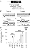
Figure 1
Figure 1. L-type Ca2+ channel C terminus CaM-binding domains increase L-type Ca2+ channel opening probability (Po)
A, schematic representation of the L-type Ca2+ channel with the amino acid sequences and relative positions of the IQ and CB CaM-binding motifs. Numerals I– IV indicate homolgous domains in α1C. B, representative L-type Ca2+ channel current recordings under control conditions and after addition of CB or IQ mimetic peptides (mp) to the bath (cytoplasmic) solution. The scale bars indicate 20 ms (horizontal) and 2 pA (vertical). C, mean L-type Ca2+ channel Po under basal conditions (control) or after stepwise increases in CBmp or IQmp (indicated by horizontal bars, with concentrations on the abscissa). The rightmost bar represents L-type Ca2+ channel Po after 10 μm CBmp and 10 μm IQmp added to the bath solution. * P < 0.001 versus control; † P < 0.05 versus CBmp. Differences between 10 and 20 μm CBmp and IQmp were not significant.
Figure 2
Figure 2. The CaM-binding domain mimetic peptide (CBmp) induces mode 2 gating
A and B, representative L-type Ca2+ channel recordings (top), ensemble-averaged currents (200–300 tracings, middle), and opening probability (Po) diary plots (bottom). C and D, histograms plot logarithmically binned L-type Ca2+ channel open times (abscissa) against the number of events (ordinate). Open-time distributions were best fitted with the sum of two exponentials, and the long (τl) and short (τs) time constants for these fits are shown. The percentage of total openings described by τl and τs are shown in brackets. E and F, quantification of gating modes was performed by plotting Po (top) against the maximum open time (Tomax, bottom) for each non-blank sweep. A vertical line is placed at the minimum of two Gaussian distributions between long and short L-type Ca2+ channel openings, while the horizontal line marks Po = 2 %. Numerals indicate gating modes 0–2 (Yue et al. 1990). Representative gating mode analysis is shown for E, the inactive control peptide, and F, CBmp (10 μm). Data from panels A, C and E are taken from the same cell membrane excised patch treated with inactive control peptide (10 μm) while panels B, D and F are results from a different excised cell membrane patch treated with CBmp (10 μm).
Figure 3
Figure 3. Elevated Ca2+–CaM prevents CBmp actions at L-type Ca2+ channels
A-C, L-type Ca2+ channel open time histograms with two exponential fits, and long (τl) and short (τs) time constants describing opening times are shown, as in Fig. 2C and D. While CBmp (10 μm) with CaM (2 μm) increased the percentage of long L-type Ca2+ channel openings (B), this effect was prevented by increased CaM (20 μm, C). Data in panels A and B are the same as panels C and D in Fig. 2), and are shown for comparison with panel C. Summary data show the percentage of long L-type Ca2+ channel openings (D) and τl and τs (E) with control peptide + CaM (2 μm) (Control, open bar), CBmp + CaM (2 μm) (hatched bar), and CBmp + CaM (20 μm) (black bar), where the number of L-type Ca2+ channels studied is indicated in parentheses. F, summary data show the distribution of gating modes from excised cell membrane patches (n = 3 for each group) in response to control + CaM (2 μm) (open bars), CBmp + CaM (2 μm) (hatched bars) and CBmp + CaM (20 μm) (black bars). The numbers of sweeps (ordinate) with L-type Ca2+ channel openings representative of each gating mode are shown. * P = 0.01 for mode 2 sweeps after CBmp + CaM (2 μm) versus control + CaM (2 μm) and CBmp + CaM (20 μm).
Figure 4
Figure 4. CBmp increases L-type Ca2+ channel Po only under low Ca2+–CaM activity conditions
A, mean L-type Ca2+ channel Po in the presence of a control peptide (Control) + CaM, CBmp + CaM, or CBmp + AC3-I + CaM. CBmp, Control, and AC3-I were all used at 10 μm, and CaM concentrations are indicated (abscissa) in B. The bath free (cytosolic) Ca2+ = 150 nm before CaM addition in A, while the Ca2+ was buffered with BAPTA (20 mm) in B. The number of excised cell membrane patches studied in each group is indicated by numerals (abscissa). * P < 0.001 versus control (A) and * P = 0.02 versus control (B).
Figure 5
Figure 5. CBmp and CaMKII increase L-type Ca2+ channel Po by distinct low- and high-Ca2+–CaM activity mechanisms
Mean L-type Ca2+ channel Po increases significantly from control (0 μm CaM) after CBmp (10 μm, P = 0.008), but this increase is reversed in step-wise fashion by increasing concentrations of CaM up to 20 μm, in the continued presence of CBmp. This trend reverses at CaM = 40 μm (but does not reach significance versus control) in the presence of CBmp. Mean L-type Ca2+ channel Po significantly increases (P = 0.002) with increasing CaM (from 10–20 μm) in the absence of CBmp, and this increase is occluded by the CaMKII inhibitory peptide AC3-I (10 μm), indicating endogenous CaMKII is localized near the L-type Ca2+ channel in excised cell membrane patches (Dzhura et al. 2002). Labelled horizontal bars indicate the presence of CBmp and AC3-I. CaM concentrations and the number of excised cell membrane patches studied are indicated (abscissa). * P < 0.05 compared to the 0 μm CaM-0 μm CBmp condition, and ** P < 0.05 compared to the 2 μm CaM-0 μm CBmp condition.
Figure 6
Figure 6. A model for increasing L-type Ca2+ channel Po in low- and high-Ca2+–CaM conditions
Under basal conditions (centre panel) CaMKII is largely inactive (dark folded, membrane-associated bar) and CaM is bound to the L-type Ca2+ channel C terminus by a low-Ca2+ activity mechanism (Pitt et al. 2001) that does not permit Ca2+–CaM-dependent binding with IQ or CB motifs, or significant interaction of these motifs with a facilitation site (fs). Increased Ca2+–CaM (left panel) results in Ca2+–CaM binding to CaMKII, activation and autophosphorylation of endogenous CaMKII (dark, extended bar), which binds the L-type Ca2+ channel C terminus (Hudmon & Schulman, 2002). CaMKII binding to the L-type Ca2+ channel C terminus disrupts CaM binding to the C terminus CaM-binding domains (Hudmon et al. 2002) and improves the interaction of IQ and CB with the facilitation site (fs). CaMKII activation and C terminus binding results in increased L-type Ca2+ channel Po (Dzhura et al. 2000; Wu et al. 2001a; Dzhura et al. 2002). During low-Ca2+–CaM conditions (right panel), C terminus CaM-binding domain mimetic peptides (CBmp and IQmp) are ‘enhanced’ auto-agonist ligands, by virtue of reduced steric constraint compared to the endogenous counterparts, for interacting with the facilitation site (fs) to induce mode 2 gating. CBmp (Figs 3 and 4) and IQmp (Wu et al. 2001a) cannot interact with the facilitation site while bound to Ca2+–CaM, and so are ineffective during increased Ca2+–CaM activity conditions.

Comment in

  • Findings of research misconduct.
    [No authors listed] [No authors listed] NIH Guide Grants Contracts (Bethesda). 2014 Dec 5:NOT-OD-15-031. NIH Guide Grants Contracts (Bethesda). 2014. PMID: 25528784 Free PMC article. No abstract available.
  • Findings of Research Misconduct.
    [No authors listed] [No authors listed] Fed Regist. 2014 Nov 25;79(227):70187-70188. Fed Regist. 2014. PMID: 27737250 Free PMC article. No abstract available.

References

    1. Adachi-Akahane S, Cleemann L, Morad M. Cross-signaling between L-type Ca2+ channels and ryanodine receptors in rat ventricular myocytes. J Gen Physiol. 1996;108:435–454. - PMC - PubMed
    1. Anderson ME, Braun AP, Schulman H, Premack BA. Multifunctional Ca2+/calmodulin-dependent protein kinase mediates Ca(2+)-induced enhancement of the L-type Ca2+ current in rabbit ventricular myocytes. Circ Res. 1994;75:854–861. - PubMed
    1. Balshaw DM, Xu L, Yamaguchi N, Pasek DA, Meissner G. Calmodulin binding and inhibition of cardiac muscle calcium release channel (ryanodine receptor) J Biol Chem. 2001;276:20144–20153. - PubMed
    1. Bers DM, Patton CW, Nuccitelli R. A practical guide to the preparation of Ca2+ buffers. Methods Cell Biol. 1994;40:3–29. - PubMed
    1. Braun AP, Schulman H. A non-selective cation current activated via the multifunctional Ca(2+)-calmodulin-dependent protein kinase in human epithelial cells. J Physiol. 1995a;488:37–55. - PMC - PubMed

Publication types

MeSH terms

LinkOut - more resources