. 2003 Jun;14(6):2216-25.
doi: 10.1091/mbc.e02-09-0598. Epub 2003 Feb 6.

Tumor necrosis factor-alpha causes accumulation of a ubiquitinated form of hypoxia inducible factor-1alpha through a nuclear factor-kappaB-dependent pathway

Affiliations

Tumor necrosis factor-alpha causes accumulation of a ubiquitinated form of hypoxia inducible factor-1alpha through a nuclear factor-kappaB-dependent pathway

Jie Zhou et al. Mol Biol Cell. 2003 Jun.

Abstract

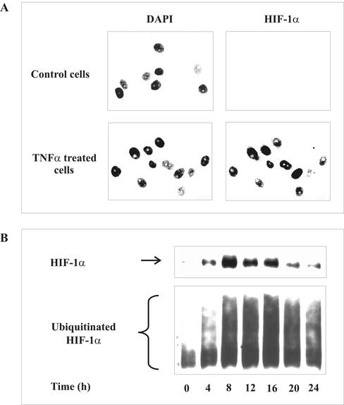
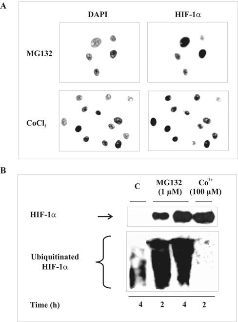
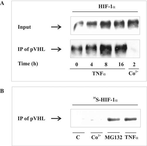
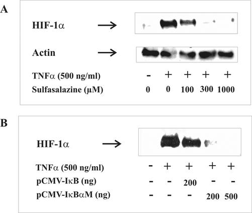
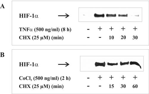
Hypoxia-inducible factor-1 (HIF-1) is a regulator of metabolic adaptation to hypoxia. It is now appreciated that HIF-1alpha accumulation is achieved under normoxic conditions by various factors, such as TNF-alpha. Here, it was our intention to gain insight into the signaling mechanisms used by TNF-alpha to stimulate HIF-1alpha. In tubular LLC-PK1 or human embryonic kidney cells, TNF-alpha induced accumulation of HIF-1alpha protein but not HIF-1alpha mRNA. Blocking nuclear factor (NF)-kappaB with sulfasalazine or expression of an IkappaB superrepressor attenuated HIF-1alpha accumulation, whereas transfection of active p50/p65-NF-kappaB subunits mimicked a TNF-alpha response. Experiments with actinomycin D and cycloheximide also pointed to a transcriptional and translational process in facilitating the TNF-alpha response. Interestingly, and in contrast to established hypoxic signaling concepts, TNF-alpha elicited HIF-1alpha accumulation in a ubiquitinated form that still bound the von Hippel-Lindau (pVHL) protein. These data indicate that HIF-1alpha accumulation by TNF-alpha demands the NF-kappaB pathway, preserves ubiquitination of HIF-1alpha, and allows the HIF-1alpha-pVHL interaction.


Figures

Figure 1.
Figure 1.
HIF-1α accumulation and ubiquitination by TNF-α. (A) LLC-PK1 cells were stimulated with 500 ng/ml TNF-α for 16 h or remained untreated. Nuclei were visualized by DAPI staining. HIF-1α was detected by an anti-HIF-1α mAb and FITC-labeled anti-mouse secondary antibody. (B) HEK293 cells were cotransfected with 3 μg pCMV-HIS6-ubiquitin and 0.3 μg pHIF-1α expression plasmids for 16 h. Medium was changed and incubations continued for another 8-h period before stimulation with 500 ng/ml TNF-α for times indicated. HIF-1α and ubiquitinated HIF-1α were determined by Western analysis as described in MATERIALS AND METHODS. Each experiment was performed at least three times, and representative data are shown.
Figure 2.
Figure 2.
HIF-1α stabilization and ubiquitination by MG132 and CoCl2. (A) LLC-PK1 cells were stimulated with 1 μM MG132 or 100 μM CoCl2 for 4 h. HIF-1α and DAPI staining were performed as described in Figure 1A. (B) HEK293 cells were cotransfected with 3 μg pCMV-HIS6-ubiquitin and 0.3 μg pHIF-1α to follow HIF-1α and ubiquitinated HIF-1α accumulation as described in Figure 1B. HEK293 cells were stimulated with 1 μM of the proteasome inhibitor MG132 or 100 μM CoCl2 for times indicated. Each experiment was performed at least three times, and representative data are shown.
Figure 3.
Figure 3.
Interaction between HIF-1α and pVHL. (A) HEK293 cells were cotransfected with 1 μg pCMV-HA-pVHL and 3 μg pHIF-1α expression plasmids. After medium was changed at 16 h, incubations continued for 8 h before stimulation with 500 ng/ml TNF-α or 100 μM CoCl2 for times indicated. Coimmunoprecipitation was performed with an anti-HA mAb that recognized pVHL- and anti-mouse antibody-coated magnetic beads. Accumulation of HIF-1α in cell lysates (input) and immunoprecipitates (IP) were determined by Western analysis using an anti-HIF-1α mAb as described in MATERIALS AND METHODS. (B) Autoradiography of endogenous [35S]methionine-labeled HIF-1α that coimmunoprecipitated with pVHL. LLC-PK1 cells were treated for 8 h with 500 ng/ml TNF-α, 1 μM MG132, or 100 μM CoCl2 or remained unstimulated (C). For details, see MATERIALS AND METHODS. Each experiment was performed at least three times, and representative data are shown.
Figure 4.
Figure 4.
Actinomycin D attenuated TNF-α-stimulated HIF-1α accumulation. LLC-PK1 cells were preincubated for 30 min with the transcription inhibitor actinomycin D. Thereafter, cells were stimulated with (A) 500 ng/ml TNF-α or (B) 3 μM MG132 for 8 h. HIF-1α was determined by Western analysis. For details, see MATERIALS AND METHODS. Each experiment was performed at least three times, and representative data are shown.
Figure 5.
Figure 5.
Inhibition of NF-κB attenuated TNF-α-stimulated HIF-1α accumulation. (A) LLC-PK1 cells were preincubated for 30 min with the NF-κB inhibitor sulfasalazine at the indicated concentrations. (B) Cells were transfected with pCMV-IκBα and pCMVIκBαM expression plasmids for 24 h. Thereafter, cells were stimulated with 500 ng/ml TNF-α for 16 h. HIF-1α was determined by Western analysis. For details, see MATERIALS AND METHODS. Each experiment was performed at least three times, and representative data are shown.
Figure 6.
Figure 6.
Cotransfection of p50/p65 NF-κB subunits promoted HIF-1α accumulation. LLC-PK1 cells were cotransfected with indicated amount of pRSV-NF-κB1/pRSV-RelA expression plasmids. After transfection for 16 h, medium was changed, and incubations continued for 8 h. As the positive control, cells were treated with 500 ng/ml TNF-α for 8 h. HIF-1α was detected by Western analysis as described in MATERIALS AND METHODS. Each experiment was performed at least three times, and representative data are shown.
Figure 7.
Figure 7.
HIF-1α mRNA under the impact of TNF-α. LLC-PK1 cells were transfected with 500 ng pIκBαM (IκB-mutant) or with 500 ng pRSV-NF-κB1/pRSV-RelA expression plasmids as indicated in Figures 4 and 5. Sulfasalazine (500 μM) was preincubated for 30 min. Cells were then stimulated with 500 ng/ml TNF-α for 8 h. Total RNA was isolated, and 1 μg total RNA was used for reverse transcription. PCR products were visualized with ethidium bromide on 2% agarose gels as described in MATERIALS AND METHODS. Each experiment was performed at least three times, and representative data are shown.
Figure 8.
Figure 8.
CHX affects TNF-α-induced HIF-1α accumulation. LLCPK1 cells were stimulated with (A) 500 ng/ml TNF-α for 8 h or (B) 100 μM CoCl2 for 2 h. Thereafter, the protein translation inhibitor CHX was added (25 μM), and incubations continued for 10-60 min. HIF-1α was detected by Western analysis using the anti-HIF-1α mouse mAb as described in MATERIALS AND METHODS. Each experiment was performed at least three times, and representative data are shown.

References

    1. Albina, J.E., Mastrofrancesco, B., Vessella, J.A., Louis, C.A., Henry, W.L., Jr., and Reichner, J.S. (2001). HIF-1 expression in healing wounds: HIF-1alpha induction in primary inflammatory cells by TNF-alpha. Am. J. Physiol. Cell Physiol. 281, C1971-C1977. - PubMed
    1. Alvarez-Tejado, M., Alfranca, A., Aragones, J., Vara, A., Landazuri, M.O., and del Peso, L. (2002). Lack of evidence for the involvement of the phosphoinositide 3-kinase/akt pathway in the activation of hypoxia-inducible factors by low oxygen tension. J. Biol. Chem. 277, 13508-13517. - PubMed
    1. Arsham, A.M., Plas, D.R., Thompson, C.B., and Simon, M.C. (2002). Phosphatidylinositol 3-kinase/Akt signaling is neither required for hypoxic stabilization of HIF-1alpha nor sufficient for HIF-1-dependent target gene transcription. J. Biol. Chem. 277, 15162-15170. - PubMed
    1. Baud, V., and Karin, M. (2001). Signal transduction by tumor necrosis factor and its relatives. Trends Cell Biol. 11, 372-377. - PubMed
    1. Chan, D.A., Sutphin, P.D., Denko, N.C., and Giaccia, A.J. (2002). Role of prolyl hydroxylation in oncogenically stabilized hypoxiainducible factor-1alpha. J. Biol. Chem. 277, 40112-40117. - PubMed

Publication types

MeSH terms