. 2003 Jun;14(6):2292-302.
doi: 10.1091/mbc.e02-10-0636. Epub 2003 Apr 4.

MTG1 codes for a conserved protein required for mitochondrial translation

Affiliations

MTG1 codes for a conserved protein required for mitochondrial translation

Antoni Barrientos et al. Mol Biol Cell. 2003 Jun.

Abstract

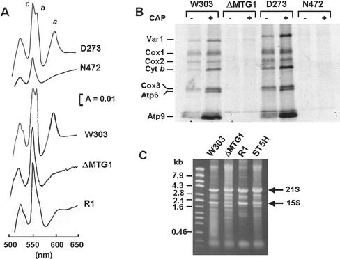
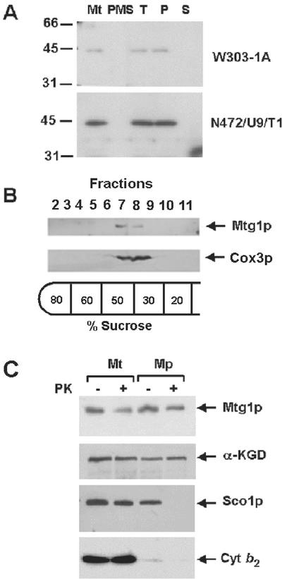
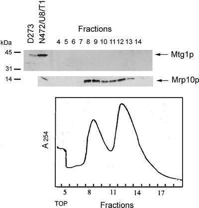
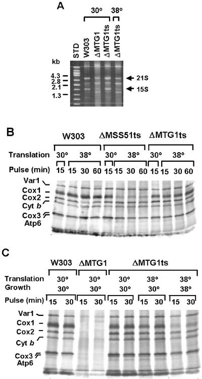
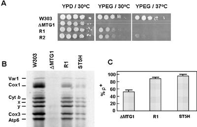
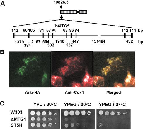
The MTG1 gene of Saccharomyces cerevisiae, corresponding to ORF YMR097c on chromosome XIII, codes for a mitochondrial protein essential for respiratory competence. A human homologue of Mtg1p capable of partially rescuing the respiratory deficiency of a yeast mtg1 mutant has also been localized in mitochondria. Mtg1p is a member of a family of GTPases with largely unknown functions. The respiratory deficiency of mtg1 mutants stems from a defect in mitochondrial protein synthesis. Mutations in the 21S rRNA locus are able to suppress the translation defect of mtg1 null mutants. This points to the 21S rRNA or the large ribosomal subunit as the most likely target of Mtg1p action. The presence of mature size 15S and 21S mitochondrial rRNAs in mtg1 mutants excludes Mtg1p from being involved in transcription or processing of these RNAs. More likely, Mtg1p functions in assembly of the large ribosomal subunit. This is consistent with the peripheral localization of Mtg1p on the matrix side of the inner membrane and the results of in vivo mitochondrial translation assays in a temperature-sensitive mtg1 mutant.


Figures

Figure 2.
Figure 2.
Cloning of MTG1. Restriction maps of pG132/T1 and of subclones. The locations of the restriction sites for EcoRI (E), HindIII (H), SacI (Sa), and SphI (Sp) are shown above the nuclear DNA insert in pG132/T1. The regions of this insert subcloned in YEp24 (Botstein and Davis, 1982) or YEp352 (Hill et al., 1986) are represented by the solid bars in the top part of the figure. The plus and minus signs indicate complementation or lack thereof, respectively, of the mtg1 mutant N472/U9. The MTG1 reading frame and the direction of the transcription of the gene are indicated by the solid arrow in the pG132/T1 insert.
Figure 1.
Figure 1.
Characterization of mtg1 mutants. (A) Cytochrome spectra. Mitochondria were prepared from the wild-type strains D273-10B/A1 (D273) and W303-1A (W303), from the mtg1 mutants N472 and W303ΔMTG1 (ΔMTG1), and from the revertant aW303ΔMTG1/R1 (ΔMTG1/R1). They were extracted at a protein concentration of 5 mg/ml with potassium deoxycholate under conditions that quantitatively solubilize all the cytochromes (Tzagoloff et al., 1975). Difference spectra of the reduced (sodium dithionite) vs. oxidized (potassium ferricyanide) extracts were recorded at room temperature. The α absorption bands corresponding to cytochromes a and a3 have maxima at 603 nm (a). The maxima for cytochrome b (b) and for cytochrome c and c1 (c) are 560 nm and 550 nm, respectively. (B) In vivo labeling of mitochondrial gene products. The wild-type strains W303-1A and D273-10B/A1, and the mtg1 mutants N472 and W303ΔMTG1 (ΔMTG1) were grown in minimal 2% galactose medium supplemented with the appropriate prototrophic requirements. One half of the culture was incubated in the presence (+) of 2 mg/ml chloramphenicol during the last 2 h of growth. Cells were harvested from both media and washed two times with a solution containing 40 mM potassium phosphate plus 2% galactose before labeling with [35S]methionine at 30°C for 15 min in the presence of cycloheximide as described (Barrientos et al., 2002). Equivalent amounts of total cellular proteins were separated by SDS-PAGE on a 17.5% polyacrylamide gel, transferred to a nitrocellulose membrane, and exposed to an x-ray film. The mitochondrially translated ribosomal protein Var1, subunits 1 (Cox1), subunit 2 (Cox2), subunit 3 (Cox3) of cytochrome oxidase, cytochrome b (Cyt b), and subunit 6 (Atp6) and subunit 9 (Atp9) of the oligomycin-sensitive ATPase are identified in the margin. (C) Mitochondrial rRNAs. The wild-type strain W303-1A, the mutant W303ΔMTG1 (ΔMTG1), the revertant aW303ΔMTG1/R1 (R1), and the transformant W303ΔMTG1/ST5H (ST5H) harboring the human hMTG1 gene were grown to early stationary phase in YPGal (2% galactose, 1% yeast extract, 2% peptone). Mitochondria were prepared by the method of Glick and Pon (1995) and total RNA was extracted (Myers et al., 1985) from equivalent amounts of mitochondria. The RNA extracts were separated on a 1% agarose gel and stained with ethidium bromide. The migration of the mitochondrial 21S and 15S rRNAs is indicated in the margin.
Figure 3.
Figure 3.
Mitochondrial localization of Mtg1p. (A) Mitochondria and the postmitochondrial supernatant fractions were prepared from the wild-type W303-1A and from N472/U8/T1, the mtg1 point mutant transformed with MTG1 on a high-copy plasmid. A sample of mitochondria was sonically irradiated and centrifuged at 100,000 × gav for 30 min. The pellet, consisting of submitochondrial particles, was suspended in the starting volume of buffer. Equivalent volumes of mitochondria (Mt), sonicated mitochondria (T), submitochondrial particles (P), and the supernatant obtained after centrifugation of the sonicated mitochondria (S) were separated on a 12% polyacrylamide gel. The amount of mitochondrial and postmitochondrial supernatant proteins (PMS) loaded on the gel was 40 μg. Proteins were transferred to nitrocellulose paper, and the Western blot was treated with antiserum against Mtg1p followed by a second incubation with 125I-labeled protein A (Schmidt et al., 1984). The antibody-antigen complexes were visualized by exposure of the blot to Kodak x-ray film. (B) Mitochondria of wild-type yeast, at a protein concentration of 10 mg/ml, were converted to submitochondrial particles by sonic disruption as in A. After centrifugation, the particles were resuspended in the starting volume of 10 mM Tris-Cl, pH 7.5, and 0.5 ml was layered on 5 ml of a discontinuous sucrose gradient prepared in 10 mM Tris-Cl, pH 7.5. The gradient was centrifuged at 260,000 × gav for 2 h. The gradient was fractionated into 12 equal fractions. Fractions 2 through 11 were separated on a 12% polyacrylamide gel, transfer to nitrocellulose, and treated with antibodies against cytochrome oxidase subunit 3 (Cox3p) and Mtg1p. Proteins were visualized with anti-rabbit IgG peroxidase-conjugated secondary antibody (Sigma, St. Louis, MO) using the Super Signal chemiluminescent substrate kit (Pierce, Rockford, IL). (C) Mitochondria were prepared by the method of Glick and Pon (1995) from W303-1A and the overexpressor N472/U8/T1. The mitochondria were suspended at a protein concentration of 8 mg/ml in 0.6 M sorbitol, 20 mM HEPES, pH 7.5. To prepare mitoplasts (Mp) the mitochondrial suspension was diluted with 8 volumes of 20 mM HEPES, pH 7.5. For controls, mitochondria (Mt) were diluted with 8 volumes of 0.6 M sorbitol, 20 mM HEPES, pH 7.5. Proteinase K (prot K) was added to one half of each sample at a final concentration of 100 μg/ml and incubated for 60 min on ice. The reaction was stopped by addition of phenylmethylsulfonyl fluoride to a final concentration of 2 mM and the mitochondria and mitoplasts were recovered by centrifugation at 100,000 × gav. The pellets were suspended in, 0.6 M sorbitol, 20 mM HEPES, pH 7.5, and proteins were precipitated by addition of 0.1 volume of 50% trichloroacetic acid and heated for 10 min at 65°C. Mitochondrial and mitoplast proteins from wild-type (80 μg) and the transformant (40 μg) were separated by SDS-PAGE on a 12% polyacrylamide gel, transferred to nitrocellulose, and probed with antibody against Mtg1p, Sco1p, α-ketoglutarate dehydrogenase (αKGD), and cytochrome b2 (Cyt b2). Antibody-antigen complexes were visualized as in B.
Figure 4.
Figure 4.
Analysis of yeast mitochondrial ribosomes. Mitochondria of the wild-type strain W303-1A were lysed with 1% potassium deoxycholate and clarified by centrifugation at 14,000 × gav for 15 min. Mitochondrial ribosomes were enriched by centrifugation of the extract through a 50% sucrose cushion (Myers et al., 1987). The ribosomal pellet was suspended in AMT (500 mM ammonium chloride, 10 mM MgCl2, 10 mM Tris-Cl, pH 7.5, 6 mM β-mercaptoethanol) and was layered on a 5-ml column of a 10–30% linear sucrose gradient in AMT buffer. After centrifugation at 65,000 rpm in a Beckman SW65Ti rotor for 100 min, the gradient was fractionated into 18 fractions. Only fractions containing the large and small subunits were analyzed as in Figure 3 with antibody against Mtg1p and Mrp10p (Jin et al., 1997).
Figure 5.
Figure 5.
Characterization of the mtg1 ts mutant. (A) The respiratory competent strain W303-1A (W303) and the mtg1 null mutant aW303ΔMTG1 (ΔMTG1) were grown at 30°C, and the mtg1 ts mutant aW303ΔMTG1ts (ΔMTG1ts) was grown at 30 and 38°C in YPGal to early stationary phase. Mitochondria were prepared (Hell et al., 1998) and total mitochondrial RNAs, extracted (Myers et al., 1985) from equivalent amounts of mitochondria were separated on a 1% agarose gel and stained with ethidium bromide. The migration of DNA molecular size standards (STD) and the bands corresponding to the 15S and 21S rRNAs are identified in the margins. (B) The wild-type W303-1A (W303), the mtg1 and mss51 ts mutants aW303ΔMTG1ts (ΔMTG1ts), and aW303ΔMSS51ts (ΔMSS51ts) were grown at 30°C in minimal 2% galactose medium. Mitochondrial protein synthesis was assayed as in Figure 1C either at 30 or 38°C for the indicated period of time. The incorporation of [35S]methionine into the mitochondrial translation products was essentially completed after 15 min of incubation at either temperature. (C) The wild-type W303-1A (W303) and the mtg1 null mutant aW303ΔMTG1 (ΔMTG1) were grown in 2% minimal galactose medium at 30°C, and the mtg1 ts mutant aW303ΔMSS51ts (ΔMSS51ts) was grown in the same medium at 30 and 38°C. The different strains were incubated for 15 min at 30 or 38°C before translation was uninitiated at the same temperature. Samples were taken 15 and 30 min after addition of [35S]methionine. The radioactively labeled products are identified in the margin as in Figure 1C.
Figure 6.
Figure 6.
Properties of mtg1 revertants. (A) Serial dilutions of the respiratory competent strain W303-1A (W303), the mtg1 null mutant aW303ΔMTG1 (ΔMTG1), and two revertants, aW303ΔMTG1/R1 (R1) and R2, were spotted on YPD and YPEG plates and incubated at 30 and 37°C for 2.5 d. (B) In vivo labeling of mitochondrial products with [35S]methionine in the presence of cycloheximide. The wild-type strain W303-1A (W303), the mtg1 null mutant aW303ΔMTG1 (ΔMTG1), the revertant aW303ΔMTG1/R1 (R1), and W303ΔMTG1/ST5H, the null mutant transformed with hMTG1 on a high-copy plasmid, were grown in 20 ml YPGal overnight. The cells were collected and transferred to 10 ml of fresh YPGal medium containing 2 mg/ml chloramphenicol. After a 2-h incubation at 30°C the cells were collected, washed two times in 50 ml of sterile water and transferred to 10 ml of minimal 2% galactose medium (Difco, nitrogen base without amino acids) supplemented with the prototrophic requirements and cycloheximide at a concentration of 0.5 mM. After 2 min, 0.2 mCi of [35S]methionine was added and incorporation allowed to proceed for 30 min. Protein synthesis was quenched by addition of 1.2 M sorbitol containing 1 mM cycloheximide. The cells were collected, washed several times in the same buffer, and digested with zymolyase for 5 min in 2 ml of 1.2 M sorbitol, 20 mM potassium phosphate, pH 7.5, 1 mM EDTA, 140 mM β-mercaptoethanol, and 20 mg/ml zymolyase. The spheroplasts were lysed in 0.5 M sorbitol and 0.2 mg/ml phenylmethylsulfonylfluoride, and mitochondria were isolated by differential centrifugation of the lysate. Total mitochondrial proteins (80 μg) were separated on a 12.5% SDS-PAGE gel containing 6 M urea and 6% glycerol (Claisse et al., 1980). The gel was dried before autoradiography. The mitochondrial translation products are identified in the left-hand margin as in Figure 1C. The two bands below Cyt b (labeled X and Y) have not been identified. They are consistently seen under these conditions of labeling and sample preparation. (C) Stability of mtDNA. The mtg1 null mutant (ΔMTG1), the revertant (R1), and the mutant transformed with the human hMTG1 gene (ST5H) were purified and 5 ρ+ colonies were inoculated into YPGal and grown to early stationary phase. The cultures were spread for single colonies on YPD and after several days of growth at 30°C they were replicated on a lawn of a ρo tester spread on minimal glucose medium. The diploid colonies formed on minimal glucose were replicated on YPEG and growth scored after overnight incubation at 30°C. The bars represent the average ± SD of the results corresponding to five independent colonies.
Figure 7.
Figure 7.
Locations of the suppressor mutations in the 21S rRNA. Schematic representation of a portion of domain V of the yeast mitochondrial 21S RNA. The mutations of W303ΔMTG1/R1 and R2 (ΔMTG1/R1,R2), W303ΔMTG1/R3 (ΔMTG1/R3), and W303ΔMTG1/R8 (ΔMTG1/R8) are indicated. The nucleotide numbering is based on the DNA sequence of the gene reported in GenBank (NC 001224). The corresponding E. coli (Ec) nucleotides numbers are indicated in parenthesis. Two mutations in the stem structure, previously reported to produce erythromycin resistance (eryR); (Sor and Fukuhara, 1982; Cui and Mason, 1989), are also shown.
Figure 8.
Figure 8.
Properties and expression of the human gene homologue of yeast MTG1. (A) Localization and genomic structure of human hMTG1 gene based on the result of a BLAST search of the human genome for homology to the hMTG1 cDNA sequence. The closed and open bars depict the exon and intron regions, respectively. Their lengths in nucleotides are indicated above and below the gene. (B) Subcellular localization of hMtg1p in human osteosarcoma 143B cells. Cells were transiently transfected with a cDNA coding for a hemagluttin-tagged hMtg1p (HA). The protein was visualized by indirect immunofluorescence using antibodies to human Cox1p and to the hemagluttin epitope (HA). A merged image is shown on the right. (C) Serial dilutions of the respiratory competent strain W303-1A (W303), the mtg1 null mutant aW303ΔMTG1 (ΔMTG1), and aW303ΔMTG1/ST5H (ST5H), the null mutant transformed with the hMTG1 cDNA in the high-copy shuttle plasmid YEp351. The plates were incubated at 30°C in YPD for 2.5 d and at 30 or 37°C for 4 d in YPEG (compare with the growth of revertants after 2.5 d shown in Figure 5A).

References

    1. Ansmant, I., Massenet, S., Grosjean, H., Motorin, Y., and Branlant, C. (2000). Identification of the Saccharomyces cerevisiae RNA: pseudouridine synthase responsible for formation of psi(2819) in 21S mitochondrial ribosomal RNA. Nucleic Acids Res. 28, 1941-1946. - PMC - PubMed
    1. Ban, N., Nisson, P., Hansen, J., Moore, P.B., and Steitz, T.S. (2000). The complete atomic structure of the large ribosomal subunit at 2.4Å resolution. Science 289, 905-920. - PubMed
    1. Barrientos, A., Korr, D., and Tzagoloff, A. (2002). Shy1p is necessary for full expression of mitochondrial COX1 in the yeast model of Leigh's syndrome. EMBO J. 21, 43-52. - PMC - PubMed
    1. Bassler, J., Grandi, P., Gadal, O., Lessman, T., Petfalski, E., Tollerway, D., Lechner, J., and Hurt, E. (2001). Identification of a 60S preribosomal particle that is closely linked to nuclear export. Mol. Cell 8, 517-529. - PubMed
    1. Beers, J., Glerum, D.M., and Tzagoloff, A. (1997). Purification, characterization, and localization of yeast Cox17p, a mitochondrial copper shuttle. J. Biol. Chem. 272, 33191-33196. - PubMed

Publication types

MeSH terms

Substances