. 2003 Jun;14(6):2357-71.
doi: 10.1091/mbc.e02-10-0693. Epub 2003 Mar 7.

A novel Golgi membrane protein is a partner of the ARF exchange factors Gea1p and Gea2p

Affiliations

A novel Golgi membrane protein is a partner of the ARF exchange factors Gea1p and Gea2p

Sophie Chantalat et al. Mol Biol Cell. 2003 Jun.

Abstract

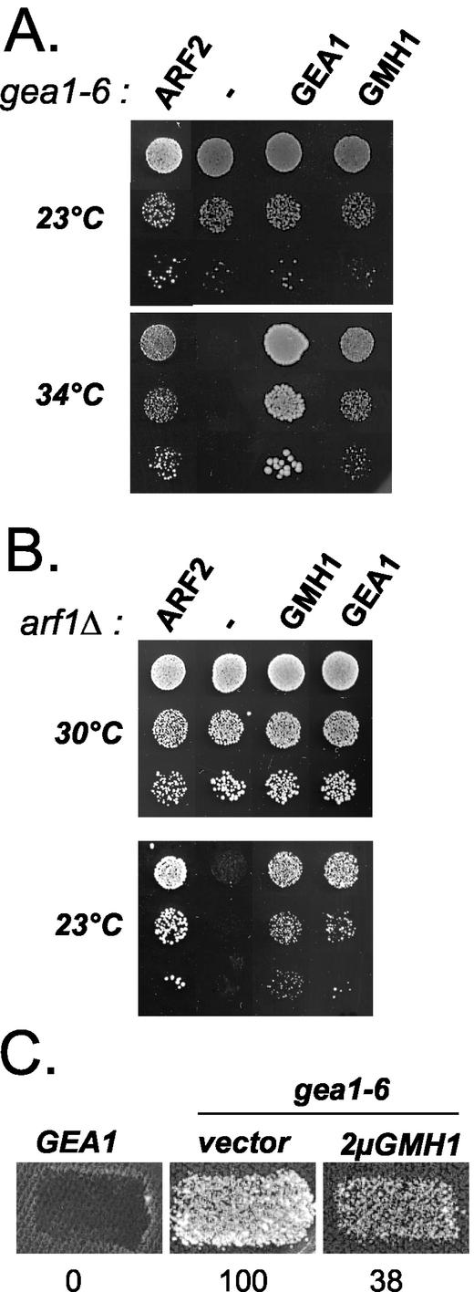
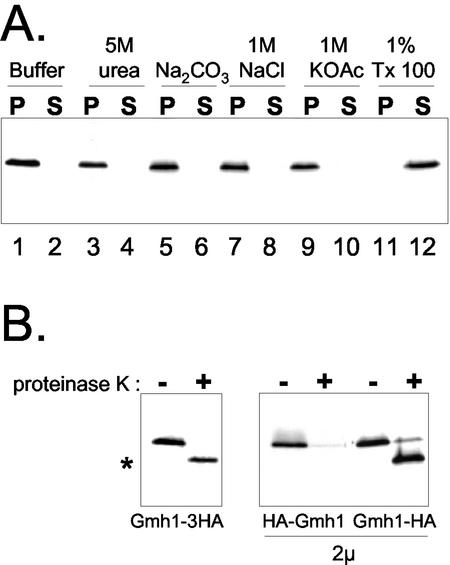
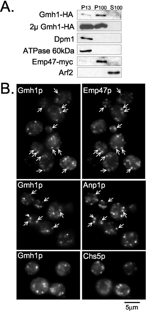
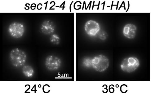
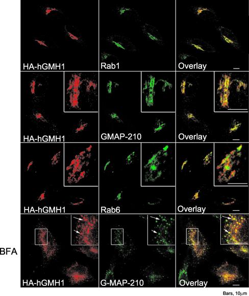
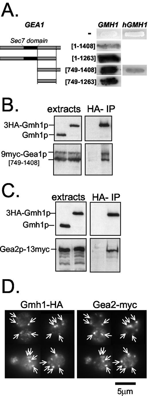
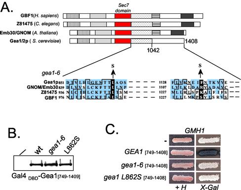
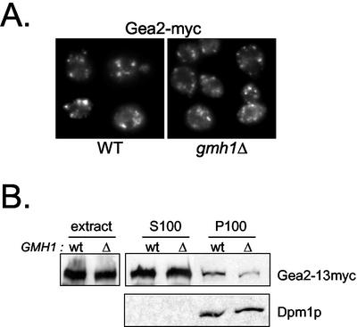
The Sec7 domain guanine nucleotide exchange factors (GEFs) for the GTPase ARF are highly conserved regulators of membrane dynamics and protein trafficking. The interactions of large ARF GEFs with cellular membranes for localization and/or activation are likely to participate in regulated recruitment of ARF and effectors. However, these interactions remain largely unknown. Here we characterize Gmh1p, the first Golgi transmembrane-domain partner of any of the high-molecular-weight ARF-GEFs. Gmh1p is an evolutionarily conserved protein. We demonstrate molecular interaction between the yeast Gmh1p and the large ARF-GEFs Gea1p and Gea2p. This interaction involves a domain of Gea1p and Gea2p that is conserved in the eukaryotic orthologues of the Gea proteins. A single mutation in a conserved amino acid residue of this domain is sufficient to abrogate the interaction, whereas the overexpression of Gmh1p can compensate in vivo defects caused by mutations in this domain. We show that Gmh1p is an integral membrane protein that localizes to the early Golgi in yeast and in human HeLa cells and cycles through the ER. Hence, we propose that Gmh1p acts as a positive Golgi-membrane partner for Gea function. These results are of general interest given the evolutionary conservation of both ARF-GEFs and the Gmh proteins.


Figures

Figure 1.
Figure 1.
Overexpression of GMH1 rescues gea1-6 and arf1Δ mutant cells. APY022 gea1-6 (A) or CJY045-5-3 arf1Δ (B) mutant cells were transformed either with multicopy vector alone or with the same vector containing ARF2, GEA1 or GMH1(YKR030w). Serial dilutions of the transformants were then spotted on selective medium and grown at indicated temperatures for 3 d. (C) Mat a wild-type (ALY014-2) or gea1-6 (ALY013-1) transformed either with multicopy vector alone or with the same vector containing GMH1(YKR030w) were grown on nonselective medium and replica plated to a lawn of wild-type cells Mat α SEY6210 and incubated at 33°C for 6 h. Mating was then analyzed by growth on medium selective for diploids. Numbers indicate the efficiency of mating in arbitrary units (average of three independent experiments).
Figure 2.
Figure 2.
Structure of Gmh1p and its putative orthologues. (A) Sequence comparison of Gmh1p and its putative orthologues. The amino acid sequence of Gmh1p from S. cerevisiae (Sc; accession number Z28255) is aligned with those of related proteins present in H. sapiens (H.s; accession number AF077038 for hGMH1a and AL080115 for hGMH1b), in R. norgevicus (R.n; accession number U96638), in C. elegans (C.e; accession number Z48055), in A. thaliana (A.th; accession number AF378872), and in D. melanogaster (D.m; accession number AE003680). Identical amino acid residues in the analogous positions of at least four proteins are in gray boxes. Lines indicate the predicted hydrophobic transmembrane domains (TDM1 to TDM5). Boxes indicate highly conserved motifs I, II, and III. (B) Hydropathy profiles of Gmh1p and hGMH1. Predicted transmembrane domains are numbered 1–5.
Figure 3.
Figure 3.
Gmh1p is an integral membrane protein. (A) Membrane association of HA-tagged Gmh1p expressed in yeast. Gently lysed RCY006 cells were treated as indicated and then submitted to 100,000 × g centrifugation. Pellet (P) and supernatant (S) fractions were then analyzed by immunoblotting with anti-HA antibodies. (B) Topology of Gmh1p. Gently lysed extracts were prepared from RCY006, YPH499/pAP100 or YPH499/pAP101 strains. Membrane pellets containing the proteins tagged at either Nor C terminus, as indicated, were digested (+) or not (-) with proteinase K and then analyzed by immunoblotting with anti-HA antibodies. Asterisk indicates the shorter polypeptide corresponding to a N-terminus truncated form of Gmh1p.
Figure 4.
Figure 4.
Intracellular localization of Gmh1p. (A) Analysis by sequential fractionation. RCY006 cells containing pRS-Emp47m were spheroplasted and gently lysed as indicated in MATERIALS AND METHODS. Lysates were submitted to sequential centrifugation at 13,000 and 100,000 × g. Aliquots of pellet (P13 and P100) and supernatant (S100) fractions were analyzed by western-blot with antibodies against the different proteins as indicated. The same experiment was performed with YPH499 cells containing HA-tagged Gmh1p on a 2-μm multicopy plasmid (from pAP100). (B) Analysis by immunofluorescence. HA-tagged Gmh1p (expressed from pRC7) was compared with a coexpressed Emp47-myc protein (from pRS-Emp47m) by double-immunofluorescence (top panel) or with endogenous Anp1p (middle panel). On the lower panel, double-immunofluorescence was performed from CHSY004-2 cells transformed with pRC7 to reveal Gmh1p and Chs5p with HA and Myc antibodies, respectively. Arrows indicate identical position in each pair of image.
Figure 5.
Figure 5.
Gmh1p cycles from the Golgi to the ER. The temperature-sensitive sec12-4 strain (RSY253) expressing HA3-GMH1 from pRC8 was incubated at 25°C (permissive) or 36°C (nonpermissive) for 1 h after addition of cycloheximide (20 mg/ml). Cells were then processed for immunofluorescence using anti-HA antibodies.
Figure 6.
Figure 6.
immunofluorescence localization of human hGMH1b in HeLa cells. HeLa cells transfected with HA-hGMH1b plasmid (pEM2) were fixed 20 h after transfection and processed for immunofluorescence. BFA (5 mg/ml) was added 2 h before fixation when indicated. Cells were double-labeled with a mAb against HA epitope and a polyclonal antibody against either Rab1, Rab6 or GMAP-210 as indicated. All pictures shown here represent stacks of four medial optical slices obtained by confocal microscopy. Arrows indicate identical position in each pair of image.
Figure 7.
Figure 7.
Physical interaction between Gmh1p and Gea1/2p proteins. (A) Two-hybrid interactions of Gmh1 proteins with full-length and truncated forms of Gea1p. Y190 strain containing pAPDH15 (pACTII-GMH1) was transformed with plasmids containing different fragments of GEA1 fused to Gal4 DNA binding domain (corresponding respectively to pSC59, pSC58, pSC6b, and pSC4b). Alternatively, human Gmh1p (hGMH1) fused to Gal4 activation domain (pSC37b) was used as a bait. Numbers indicate first and last amino acid residues of Gea1p encompassed within the fusion protein. Coloration in presence of X-Gal assays for β-galactosidase activity. (B) Coimmunoprecipitation of Gmh1p and a C-terminal part of Gea1p. Coimmunoprecipitation experiments were performed from strain c13-ABYS86 expressing a myc-tagged version of Gea1p[749–1408] (from pSC64) and a wild-type or HA-tagged version of Gmh1p (from pAP68 or pRC11 respectively). One percent of total extracts and 10% or 35% of eluates were analyzed by immunoblotting either with anti-Gmh1p antibodies (top panel) or with A14 antimyc antibodies (lower panel). (C) Coimmunoprecipitation of Gmh1p and full-length Gea2p. Coimmunoprecipitation experiments were carried out with detergent-lysed cells extracts prepared from GEA2-myc SCY002 strain expressing Gmh1p or HA-Gmh1p (from pAP52 or pRC8, respectively). Eluates were analyzed as in B. (D) Gea2p and Gmh1p colocalize in yeast cells. Yeast cells expressing a chromosomal modified myc-tagged Gea2 protein (SCY002) were transformed with HA3-GMH1 expressing plasmid (pRC11). Double immunofluorescence experiments were then performed with anti-Myc (A14) and anti-HA (HA-11) antibodies. Arrows indicate identical positions in each pair of image.
Figure 8.
Figure 8.
Physical interaction between Gmh1p and Gea1p is compromised in gea1-6. (A) The Gea/GBF/GNOM family. Representative members of this subfamily of Sec7-domain proteins are shown. The Sec7 domains are represented by red boxes. Regions showing significant level of sequence similarity are represented by drawn boxes. The position of the amino acid substitutions in the gea1-6 allele is indicated on the sequence alignments with three other ARF GEFs of the Gea/GBF/GNOM family. GenBank accession numbers for the sequences shown are Gea1p (Z49531); Gnom/Emb30 (U36433); C. elegans ARF GEF (Z81475); and GBF1 (AF068755). (B) Western-blot analysis of Gea1[749–1408] wild-type, gea1-6 or gea1 L862S expressed from pASΔ. Crude extracts of Y190 cells expressing the different fusions were loaded on a polyacrylamide gel and Gal4 (DBD) fusion proteins were revealed by immunoblot with anti-Gal4 (DBD) antibodies. (C) Gea1p-Gmh1p interaction is impaired with gea1 mutant forms. Y190 strain containing pAPDH15 (pACTII-GMH1) was transformed with Gea1[749–1408] wild-type, gea1-6 or gea1 L862S fused to Gal4 (DBD) in pASΔ. Transformants were streaked on selective medium (+H) and tested for expression of β-galactosidase on X-Gal containing plates.
Figure 9.
Figure 9.
Membrane association of Gea2p in gmh1Δ cells. (A) Immunofluorescence of Gea2p in gmh1Δ yeast cells. A chromosomal modified myc-tagged Gea2 protein was introduced in wild-type cells (SCY002) or in gmh1Δ cells (APY130-2). Immunofluorescence experiments were then performed with anti-Myc (9E10). (B) Effect of GMH1 expression on membrane association of myc-tagged Gea2p by fractionation. Wild-type cells (SCY002) or gmh1Δ cells (APY130-2) were gently lysed (extracts) and then submitted to 100,000 × g centrifugation. Pellet (P100) and supernatant (S100) fractions were then analyzed by immunoblotting with antimyc antibodies (9E10) or anti-Dpm1p for control of loading.

References

    1. Alcalde, J., Egea, G., and Sandoval, I.V. (1994). gp74 a membrane glycoprotein of the cis-Golgi network that cycles through the endoplasmic reticulum and intermediate compartment. J. Cell Biol. 124, 649-665. - PMC - PubMed
    1. Antony, C., Cibert, C., Geraud, G., Santa Maria, A., Maro, B., Mayau, V., and Goud, B. (1992). The small GTP-binding protein rab6p is distributed from medial Golgi to the trans-Golgi network as determined by a confocal microscopic approach. J. Cell Sci. 103, 785-796. - PubMed
    1. Benli, M., Doring, F., Robinson, D.G., Yang, X., and Gallwitz, D. (1996). Two GTPase isoforms, Ypt31p and Ypt32p, are essential for Golgi function in yeast. EMBO J. 15, 6460-6475. - PMC - PubMed
    1. Boehm, M., and Bonifacino, J.S. (2001). Adaptins: the final recount. Mol. Biol. Cell. 12, 2907-2920. - PMC - PubMed
    1. Brenner, S. (1974). The genetics of Caenorhabditis elegans. Genetics 77, 71-94. - PMC - PubMed

Publication types

MeSH terms

Substances