. 2003 Jun;14(6):2410-24.
doi: 10.1091/mbc.e02-07-0446. Epub 2003 Mar 7.

Golgi localization of Syne-1

Affiliations

Golgi localization of Syne-1

Lisa Lucio Gough et al. Mol Biol Cell. 2003 Jun.

Abstract

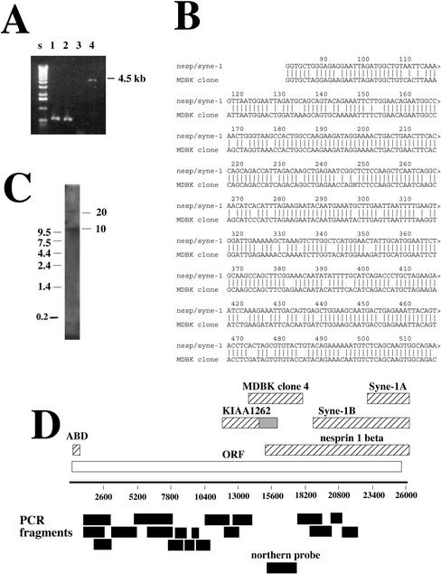
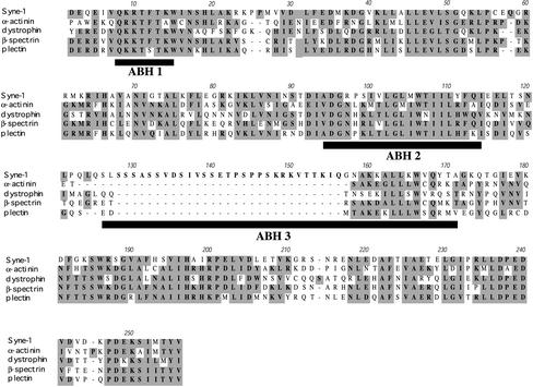
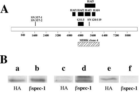
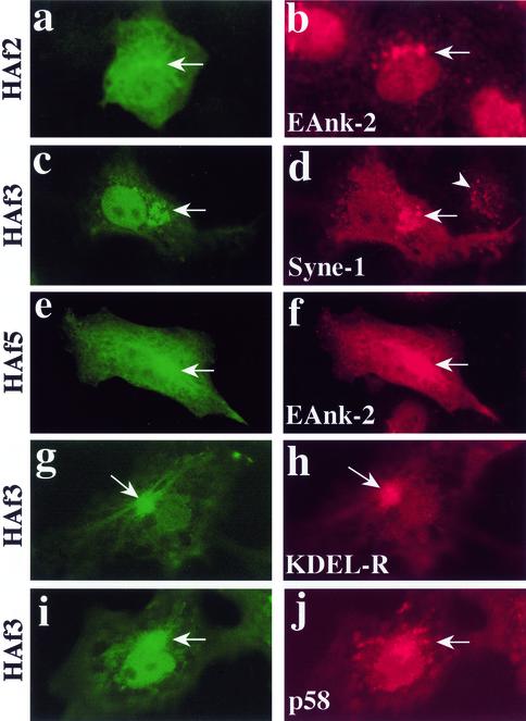
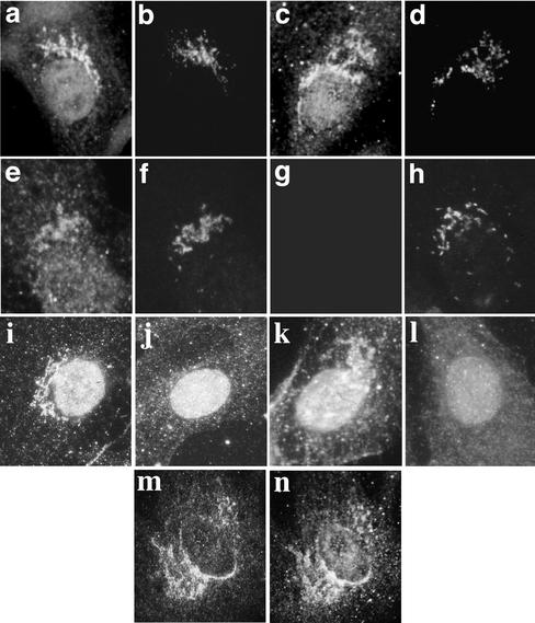
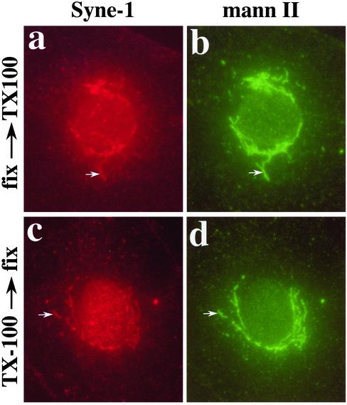
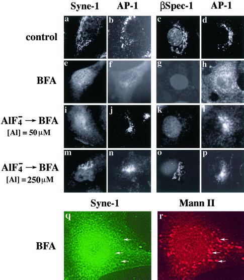
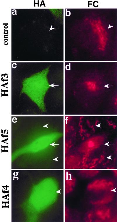
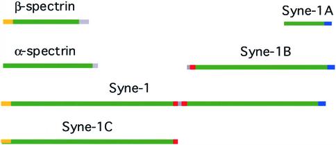
We have previously identified a Golgi-localized spectrin isoform by using an antibody to the beta-subunit of erythrocyte spectrin. In this study, we show that a screen of a lambdagt11 expression library resulted in the isolation of an approximately 5-kb partial cDNA from a Madin-Darby bovine kidney (MDBK) cell line, which encoded a polypeptide of 1697 amino acids with low, but detectable, sequence homology to spectrin (37%). A blast search revealed that this clone overlaps with the 5' end of a recently identified spectrin family member Syne-1B/Nesprin-1beta, an alternately transcribed gene with muscle-specific forms that bind acetylcholine receptor and associate with the nuclear envelope. By comparing the sequence of the MDBK clone with sequence data from the human genome database, we have determined that this cDNA represents a central portion of a very large gene ( approximately 500 kb), encoding an approximately 25-kb transcript that we refer to as Syne-1. Syne-1 encodes a large polypeptide (8406 amino acids) with multiple spectrin repeats and a region at its amino terminus with high homology to the actin binding domains of conventional spectrins. Golgi localization for this spectrin-like protein was demonstrated by expression of epitope-tagged fragments in MDBK and COS cells, identifying two distinct Golgi binding sites, and by immunofluorescence microscopy by using several different antibody preparations. One of the Golgi binding domains on Syne-1 acts as a dominant negative inhibitor that alters the structure of the Golgi complex, which collapses into a condensed structure near the centrosome in transfected epithelial cells. We conclude that the Syne-1 gene is expressed in a variety of forms that are multifunctional and are capable of functioning at both the Golgi and the nuclear envelope, perhaps linking the two organelles during muscle differentiation.


Figures

Figure 1.
Figure 1.
Cloning of a Golgi-specific spectrin family member. A λgt11 expression library constructed from MDBK cDNA was screened with the antiserum βSpec-1. (A) λ DNA was isolated from four positive clones and PCR amplification of the insert (MDBK cDNA) was performed with primers complementary to λ genome sequences flanking the insert site. Only one clone was large enough to be considered a spectrin (clone 4, 4.5 kb, lane 4). (B) Sequence alignment of a portion of MDBK clone 4 with the 5′ end of Syne-1B/Nesprin-1β. The homology begins at nucleotide 81 of Syne-1B/Nesprin-1β. (C) A Northern blot of poly-A+ MDBK RNA with a probe derived from clone 4 (see D for the region used to construct the probe) revealed two transcripts of ∼10 and ∼25 kb. (D) A schematic of the predicted full-length cDNA for Syne-1, including regions homologous to the MDBK clone isolated by antibody screening. KIAA1262, including its untranslated region (gray bar), is shown. Also shown are the full-length Nesprin-1β, Syne-1A, the N-terminal actin binding domain (ABD), and the partial sequence initially reported for Syne-1B. To determine whether MDBK cells do indeed express this full-length sequence, RT-PCR was performed on HEK-293 cell extracts by using primer pairs that defined several overlapping segments of the clone (black bars). All RT-PCR reactions generated single reaction products of correct sizes (our unpublished data).
Figure 2.
Figure 2.
The Syne-1 actin binding domain. A 257 amino acid region of the sequence of hSyne-1 near its amino terminus is aligned with the actin binding domains of human erythroid β-spectrin and three other spectrin-related proteins: α-actinin, dystrophin, and plectrin. Conserved amino acids are highlighted. Note how this region of hSyne-1 possesses high homology with the actin binding domains of the other spectrin family members. Also shown are the positions of the three α-helical segments of the utrophin actin-binding domain, which have been shown to interact directly with actin (black bars, ABH 1, ABH 2, and ABH 3). The putative Syne-1 actin binding domain differs from other actin binding domains in that it contains a unique 28 amino acid proline and serine-rich insert at the start of the third actin binding helix (ABH 3)
Figure 3.
Figure 3.
Erythroid spectrin-specific antibodies react with ectopically expressed Syne-1. (A) A schematic map of the amino acid sequence of the predicted Syne-1 gene illustrating the location of MDBK clone 4 (hatched bar). Also shown are the positions of the various epitope-tagged fragments of Syne-1 (HAf 1–5, black bars) used in these studies, as well as the positions of the peptides (SN357-1, SN357-2, SN-120, and SN119) and fusion protein (GS1.5) used to raise antibodies to Syne-1. (B) Epitope tagged (influenza HA tags) constructs encoding various regions of Syne-1 were expressed in HEK-293 cells. Cell extracts were prepared and analyzed by SDS-PAGE and immunoblotting with either the epitope tag specific antibody (HA, lanes a, c, and e) or the erythroid β-spectrin antibody (βspec-1, lanes b, d, and f) originally used to clone MDBK clone 4: lanes a and b, cells expressing HAf3; lanes c and d, cells expressing HAf5; lanes e and f, cells expressing HAf4.
Figure 4.
Figure 4.
Ectopically expressed fragments of Syne-1 identify Golgi binding determinants. To demonstrate Golgi localization for Syne-1, we expressed epitope-tagged fragments HAf2 (a and b), HAf3 (c, d, g, h, i, and j) and HAf5 (e–f) in MDCK (a–f) or COS-1 (g–j) cells and localized them by indirect immunofluorescence microscopy. Twenty-four to 48 h after transfection, cells where double stained with the HA epitope tag antibody (a, c, e, g, and i) and with antibodies to a Golgi-specific ankyrin (Eank-2, b and f), Syne-1 (d), the cis-Golgi marker KDEL receptor (KDEL-R, h) or the Golgi intermediate compartment marker p58 (j). HAf2, HAf3, and HAf5 were prominently localized to perinuclear Golgi structures (arrows) which were stained with Golgi marker antibodies. When the anti-Syne-1 antibody SN120 was used to stain cells transfected with HAf3, transfected cells (d, arrow) showed a higher reactivity to the antibody than untransfected cells (d, arrowhead), confirming that the epitope for this antibody is present in HAf3.
Figure 5.
Figure 5.
Antibodies specific for Syne-1 stain the Golgi complex. To confirm that Syne-1 is a Golgi localized protein, peptide antibodies raised against two peptides derived from Syne-1 (EESGEEGTNSEIS for antibodies SN120 and SN119; i and k respectively; and EAKASSPEMDISAD for antibody SN357-2, m), as well as an antibody to a bacterially expressed fragment of Syne-1 (GS1.5; a, c, e, and g) were prepared and used to localize Syne-1 by indirect immunofluorescence microscopy. MDBK (a–l) or LB10 (m and n) cells were fixed and double stained with antibodies specific for Syne-1 (a, c, e, i, k, and m) and the Golgi markers p58 (b, f, h, and n) or β-COP (d). The Syne-1 antibodies prominently stained reticular, perinuclear structures that costained with Golgi markers. The specificity of the GS1.5 antiserum was evaluated by affinity purification on either immobilized erythrocyte spectrin (spectrin-Sepharose; g) or immobilized Syne-1 fragment (GS1.5-Sepharose; e). Antibodies eluted from these affinity resins were tested for reactivity with a Golgi antigen by indirect immunofluorescence (e and g). Double staining with the Golgi marker p58 (f and h) is also shown. Although a Golgi-reactive antibody was eluted from the GS1.5 resin (e), specific Golgi staining was not detected when the GS1.5 antibody was affinity purified on spectrin-Sepharose (g). To test the specificity of the peptide antibodies SN120 and 119, these antibodies were preadsorbed with peptide before staining (j and l). Preadsorbtion specifically blocked the ability of these antibodies to stain perinuclear Golgi structures. Note that nuclear staining persists after preadsorbtion (j and l). Nuclear staining was not observed with peptide antibody SN357-2, which reacted predominantly with perinuclear Golgi structures (m) that costained with p58 (n)
Figure 6.
Figure 6.
Localization of Syne-1 to a detergent-insoluble Golgi ghost. We have shown previously that Golgi spectrin and Golgi ankyrin localize to a detergent-insoluble structure that maintains the basic morphology of the Golgi complex. We refer to these structures as detergent-insoluble Golgi ghosts. To determine whether Syne-1 also localizes to Golgi ghosts, normal rat kidney (NRK) cells were extracted with Triton X-100 (0.5%, 10 min, 20°C) either before (c and d) or after fixation (a and b). Cells were then double stained with GS1.5 antibody (a and c) and antimannosidase II, which we have previously shown to act as a detergent-resistant Golgi marker (Beck et al., 1997). The extraction conditions used have been previously shown to be sufficient to extract ceramide-labeled lipid in the trans-Golgi, unassembled cytoplasmic tubulin, as well as the integral membrane protein TGN 38 (Beck et al., 1997). The GS1.5 antiserum was able to stain Golgi ghosts in extracted cells (c) that colocalized with mannosidase II and was virtually identical in morphology to the Golgi of control cells (a and b) extracted after fixation.
Figure 7.
Figure 7.
Syne-1 associates with the Golgi in a BFA and aluminum fluoride sensitive manner. MDBK cells were untreated (control, a–d) or treated with 5 μM BFA for 10 min (e–h, q, and r); aluminum fluoride (50 μM aluminum) for 20 min before treatment with BFA (i–l); or aluminum fluoride (250 μM aluminum) for 20 min before treatment with BFA (m–p). Fixed cells were double stained with antibodies to either Syne-1 (GS1.5; a, e, i, and m) and AP-1 (b, f, j, and n); Golgi spectrin (βspec-1; c, g, k, and o) and AP-1 (d, h, l, and p); or Syne-1 (SN120; q) and mannosidase II (r). When MDBK cells are treated with BFA for 10 min, a uniform cytoplasmic distribution of Syne-1, distinct from the Golgi pattern seen in control cells (a) was observed. Identical results were observed by staining with antibodies to erythroid β-spectrin and the Golgi-associated clathrin coat protein AP-1 (f and h). Under these conditions, cells double stained with antibodies to syne-1 and the Golgi marker mannosidase II revealed that Syne-1 no longer localized to Golgi membranes (q and r, arrows), as we have shown previously for the antigen recognized by the βspec-1 antiserun (Beck et al., 1997). Aluminum fluoride pretreatment blocked the effect of BFA on the distribution of Syne-1 and AP-1, but the effects of aluminum fluoride were concentration dependent. At 50 μM aluminum, BFA insensitivity was observed for the clathrin coat protein AP-1 (j and l) but not for Syne-1 (i) or the βspec-1–reactive epitope (k). However, at 250 μM aluminum fluoride both Syne-1 (m) and spectrin (o) were rendered insensitive to BFA.
Figure 8.
Figure 8.
Ectopic expression of a Golgi binding fragments of Syne-1 alter the structure of the Golgi. MDBK cells were transiently transfected with epitope tagged Golgi binding fragments HAf3 (c and d), HAf5 (e and f), as well as the control fragment HAf4 (g and h). Transfected cells were stained with antibodies to HA (a, c, e, and g) and the TGN marker furin convertase (b, d, f, and h). In untransfected cells (a, b, e, and f, arrowheads) and cells transfected with the control fragment (g and h, arrowhead), the Golgi complex has a normal morphology comprised of reticular elements that surround portions of the nucleus. In cells transfected with the Golgi binding fragments HAf3 and HAf5 (c, d, e, and f, arrows) the Golgi complex collapsed into a compacted structure located near the center of the cell.
Figure 9.
Figure 9.
The major Syne-1 isoforms. A schematic representation of the domain structure of the major Syne-1 forms is compared with erythrocyte spectrin; spectrin repeat regions (green), actin binding domain (yellow), Golgi binding sites (red), and nuclear envelope binding (blue). Assuming that Syne-1 folds similarly to spectrin, this schematic illustrates the relative sizes of spectrin and the various Syne-1 isoforms.

References

    1. Apel, E.D., Lewis, R.M., Grady, M., and Sanes, J.R. (2000). Syne-1, a dystrophin- and klarsicht-related protein associated with synaptic nuclei at the neuromuscular junction. J. Biol. Chem. 275, 31986-31995. - PubMed
    1. Babiá, T., Ayala, I., Valderrama, F., Mato, E., Bosch, M., Santarén, J.F., Renau-Piqueras, J., Kok, J.W., Thomson, T.M., and Egea, G. (1999). N-Ras induces alterations in Golgi complex architecture and in constitutive protein transport. J. Cell Sci. 112, 477-489. - PubMed
    1. Beck, K., Buchanan, J., Malhotra, V., and Nelson, W.J. (1994). Golgi spectrin: identification of an erythroid beta-spectrin homolog associated with the Golgi complex. J. Cell Biol. 127, 707-723. - PMC - PubMed
    1. Beck, K., Buchanan, J.A., and Nelson, W.J. (1997). Golgi membrane skeleton: identification, localization and oligomerization of a 195 kD ankyrin isoform associated with the Golgi complex. J. Cell Sci. 110, 1239-1249. - PubMed
    1. Beck, K., and Nelson, W.J. (1998). A spectrin membrane skeleton of the Golgi complex. Biochim. Biophys. Acta 1404, 153-160. - PubMed

Publication types