. 2003 Jun;14(6):2508-19.
doi: 10.1091/mbc.e02-11-0729. Epub 2003 Feb 21.

Alpha-smooth muscle actin is crucial for focal adhesion maturation in myofibroblasts

Affiliations

Alpha-smooth muscle actin is crucial for focal adhesion maturation in myofibroblasts

Boris Hinz et al. Mol Biol Cell. 2003 Jun.

Abstract

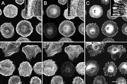
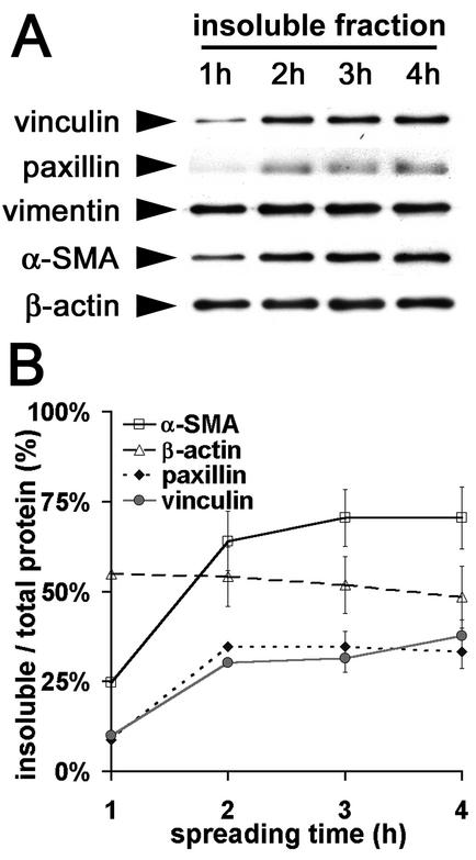
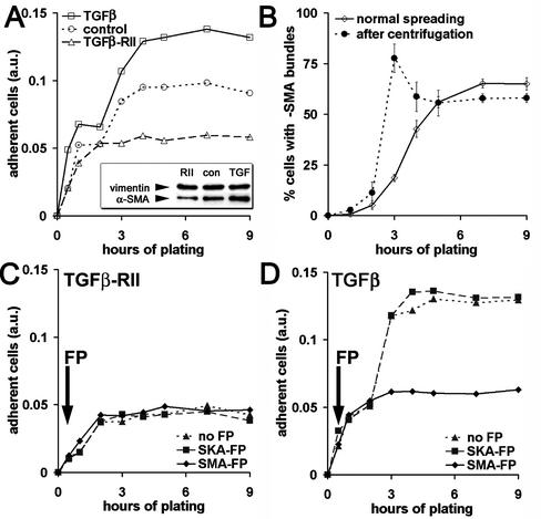
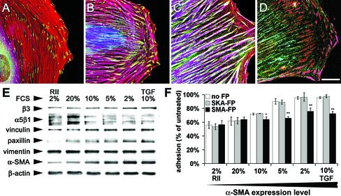
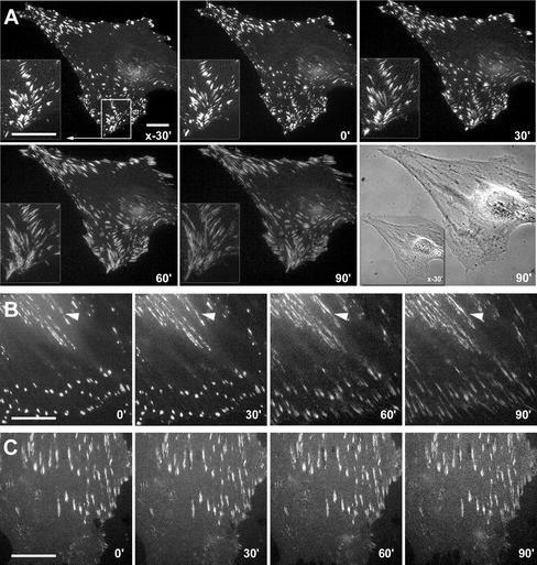
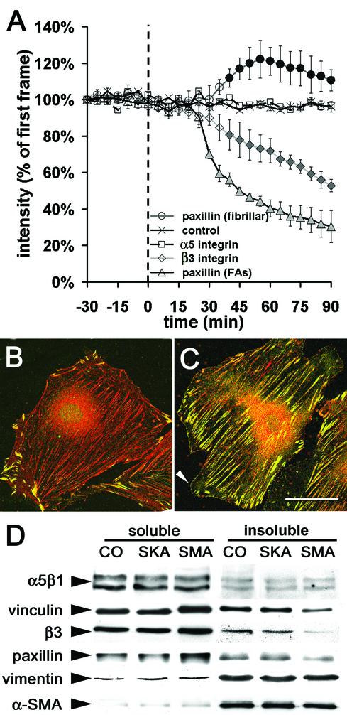
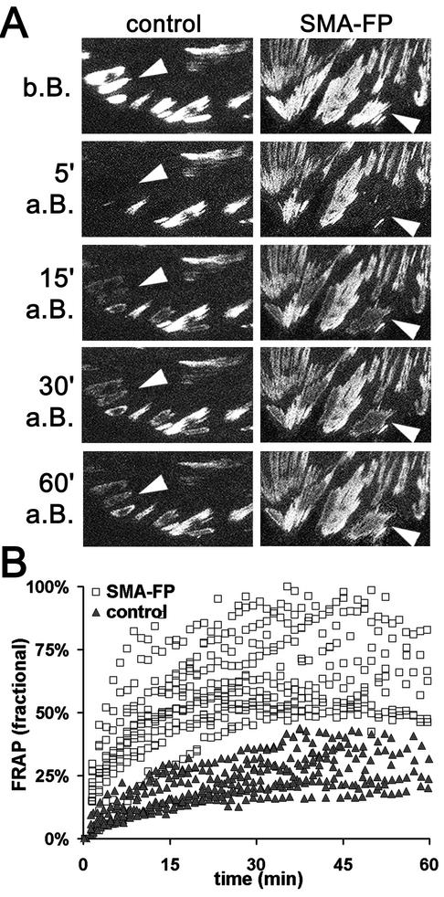
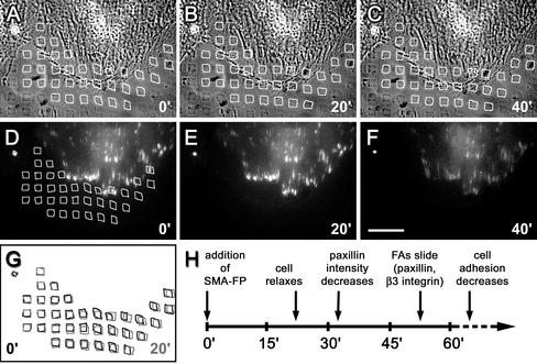
Cultured myofibroblasts are characterized by stress fibers, containing alpha-smooth muscle actin (alpha-SMA) and by supermature focal adhesions (FAs), which are larger than FAs of alpha-SMA-negative fibroblasts. We have investigated the role of alpha-SMA for myofibroblast adhesion and FA maturation. Inverted centrifugation reveals two phases of initial myofibroblast attachment: during the first 2 h of plating microfilament bundles contain essentially cytoplasmic actin and myofibroblast adhesion is similar to that of alpha-SMA-negative fibroblasts. Then, myofibroblasts incorporate alpha-SMA in stress fibers, develop mature FAs and their adhesion capacity is significantly increased. When alpha-SMA expression is induced in 5 d culture by TGFbeta or low serum levels, fibroblast adhesion is further increased correlating with a "supermaturation" of FAs. Treatment of myofibroblasts with alpha-SMA fusion peptide (SMA-FP), which inhibits alpha-SMA-mediated contractile activity, reduces their adhesion to the level of alpha-SMA negative fibroblasts. With the use of flexible micropatterned substrates and EGFP-constructs we show that SMA-FP application leads to a decrease of myofibroblast contraction, shortly followed by disassembly of paxillin- and beta3 integrin-containing FAs; alpha5 integrin distribution is not affected. FRAP of beta3 integrin-EGFP demonstrates an increase of FA protein turnover following SMA-FP treatment. We conclude that the formation and stability of supermature FAs depends on a high alpha-SMA-mediated contractile activity of myofibroblast stress fibers.


Figures

Figure 1.
Figure 1.
Filament bundles of early spreading myofibroblasts contain only β-cytoplasmic actin. REF-52 fibroblasts were trypsinized and plated for 2 h (A–C) and 3 h (D–F) and then fixed and triple-immunostained against β-cytoplasmic actin (A and D), α-SMA (B and E), and vinculin (C and F). Insets show at high magnification diffuse organization of α-SMA in the presence of β-cytoplasmic actin filament bundles. Incorporation of α-SMA into stress fibers starts 3 h after plating, correlating with FA maturation and cell polarization. Bar, 50 μm.
Figure 2.
Figure 2.
FA proteins and α-SMA codistribute in the TX-100–insoluble cytoskeletal fraction of spreading myofibroblasts. (A) TX-100 cytoskeletons were prepared of REF-52 fibroblasts after 1–4 h plating and investigated by means of Western blot. (B) Protein content in both, TX-100–soluble and –insoluble fractions were quantified by densitometry and the ratio of TX-100–insoluble protein/total protein was averaged from three independent experiments. Increase of FA proteins in the TX-100–insoluble fraction correlates with the increase of insoluble α-SMA.
Figure 3.
Figure 3.
Appearance of α-SMA in filament bundles during spreading correlates with increased cell adhesion. (A) REF-52 with low (TGFβ-RII), medium (control), and high α-SMA expression (TGFβ) were plated for 0.5–9 h in serum-containing medium. Highly adhesive cells were selected by inverted centrifugation, stained with crystal violet, and quantified by photometry. After a first phase of low adhesion (phase 1), α-SMA–positive fibroblasts exhibit a second phase of strong adhesion (phase 2), which is absent in α-SMA–negative cells. (B) After plating, REF-52 were either centrifuged (♦) or directly fixed (⋄) and stained for α-SMA and actin-phalloidin to determine the percentages of fibroblasts containing α-SMA filament bundles. After 3–4 h plating, centrifugation selects essentially cells with α-SMA filament bundles. (C and D) REF-52 were plated in serum-free conditions, optionally treated with FPs after 30 min (arrows), and centrifuged and adhesion was evaluated by crystal violet. SMA-FP has no effect on α-SMA–negative REF-52 (C), but inhibits phase 2 of α-SMA–positive fibroblast populations (D); the SKA-FP has no effect. One representative result out of five independent experiments is shown in A, C, and D; mean values from three independent experiments ±SD are presented in B.
Figure 4.
Figure 4.
The adhesion capacity of fibroblasts in long-term culture correlates with their level of α-SMA expression and the degree of FA supermaturation. REF-52 were grown for 5 d in culture medium containing 20% FCS (A), 10% FCS (B and D), and 2% FCS (C). (A–C) Cells were immunostained for α-SMA (blue), vinculin (green), and β-cytoplasmic actin (red) and observed by confocal microscopy; shift from red to purple indicates increase in α-SMA expression. (D) Cells were stained for α5β1 integrin (green), tensin (blue), and β3 integrin (red); tensin colocalizes with β3 integrin in supermature FAs (pink) and with α5β1 integrin in fibrillar adhesions (turquoise). Bar, 25 μm. (E) REF-52 were plated for 5 d in different serum conditions, and protein expression was assessed by Western blotting. The expression levels of α-SMA, paxillin, vinculin, and β3 integrin increase with decreasing serum concentration. To test their adhesion capacity, REF-52 with different levels of α-SMA expression were trypsinized and plated for 2 d. After treatment with FPs and 0.02% EDTA for 60 min, weakly adherent cells were removed by centrifugation. Cell adhesion increases with increasing α-SMA expression. SMA-FP reduces the adhesion of α-SMA-expressing fibroblasts to the level of α-SMA–negative fibroblasts; SKA-FP has no effect.
Figure 5.
Figure 5.
Application of the SMA-FP leads to dissociation of FAs. REF-52 myofibroblasts, transfected with EGFP-constructs of β3 integrin (A), paxillin (B), and α5 integrin (C) were treated with SMA-FP and observed by videomicroscopy; times of recording are indicated in the lower right corner. Application of SMA-FP leads to centripetal sliding and disassembly of β3 integrin- and paxillin-containing FAs. However, fibrillar adhesions that exhibit traces of paxillin (B, arrowheads) and strong expression of α5 integrin (C) remain stable. Bar, 20 μm.
Figure 6.
Figure 6.
Application of SMA-FP leads to destabilization of FA components. (A) Sequences of fluorescence images were recorded from REF-52, transfected with β3 integrin-, paxillin-, and α5 integrin-EGFP. The fluorescence intensities of FAs and fibrillar adhesions were measured every 5 min, starting 30 min before and ending 90 min after application of SMA-FP and SKA-FP (control, shown for β3 integrin-EGFP). Each data point represents the average of 5–10 cells and 20 FAs per cell ±SD; filled symbols indicate p ≤ 0.01 compared with control. REF-52 were treated for 90 min with SKA-FP (B) or SMA-FP (C) and immunostained against paxillin (green) and tensin (red). Note the shift of paxillin from supermature FAs to fibrillar adhesions and de novo formation of focal complexes (arrowhead). Bar, 50 μm. (D) TX 100–insoluble cytoskeletons were prepared 90 min after SMA-FP application and processed for Western blotting. Compared with controls, treatment with SMA-FP leads to a decrease of FA proteins in the TX 100–insoluble cytoskeletal fraction; fibrillar adhesion protein α5β1 integrin was not affected.
Figure 7.
Figure 7.
Turnover of β3 integrin increases in FAs after application of SMA-FP. (A) FAs of β3 integrin-EGFP-transfected REF-52 were photo-bleached (arrowheads) and FRAP in control conditions was compared with SMA-FP-treated cells (b.B., a.B.: before and after bleaching). Box width, 50 μm. (B) Fluorescence intensities were measured every minute and related to the intensity measured in the first video frame. Each data point represents the mean fractional FRAP of 12 FAs per cell; 12 cells were analyzed per condition. Note the increase in β3 integrin turnover after SMA-FP application.
Figure 8.
Figure 8.
On SMA-FP application, myofibroblasts first loose contractile activity followed by FA destabilization. Paxillin-EGFP transfected REF-52 were grown on micropatterned flexible polyacrylamide substrates and observed by video microscopy. (A–C) Substrate deformation is visualized by distortions in the 5 × 5-μm pattern (highlighted by white squares) in phase contrast simultaneously to the observation of paxillin-EGFP–positive FAs in immunofluorescence (D–F). Video frames are shown immediately before (0 min, A and D), 20 min (B and E), and 40 min (C and F) after SMA-FP treatment. Bar, 50 μm. (G) Square pattern positions before (black) and 20 min after SMA-FP (gray) are superposed to highlight cell relaxation. The time course of cell relaxation and FA changes is schematized in H.

References

    1. Adams, J.C. (2001). Cell-matrix contact structures. Cell Mol. Life Sci. 58, 371-392. - PMC - PubMed
    1. Arora, P.D., and McCulloch, C.A. (1994). Dependence of collagen remodelling on alpha-smooth muscle actin expression by fibroblasts. J. Cell. Physiol. 159, 161-175. - PubMed
    1. Arora, P.D., Narani, N., and McCulloch, C.A. (1999). The compliance of collagen gels regulates transforming growth factor-beta induction of alpha-smooth muscle actin in fibroblasts. Am. J. Pathol. 154, 871-882. - PMC - PubMed
    1. Balaban, N.Q. et al. (2001). Force and focal adhesion assembly: a close relationship studied using elastic micropatterned substrates. Nat. Cell Biol. 3, 466-472. - PubMed
    1. Ballestrem, C., Hinz, B., Imhof, B.A., and Wehrle-Haller, B. (2001). Marchingatthefrontanddraggingbehind:differentialalphaVbeta3-integrin turnover regulates focal adhesion behavior. J. Cell Biol. 155, 1319-1332. - PMC - PubMed

Publication types