. 2003 Jul;23(13):4449-60.
doi: 10.1128/MCB.23.13.4449-4460.2003.

Sequential protein association with nascent 60S ribosomal particles

Affiliations

Sequential protein association with nascent 60S ribosomal particles

Cosmin Saveanu et al. Mol Cell Biol. 2003 Jul.

Abstract

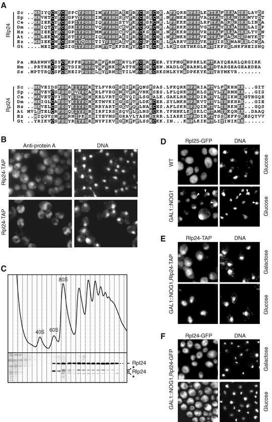
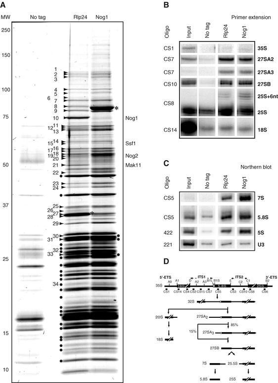
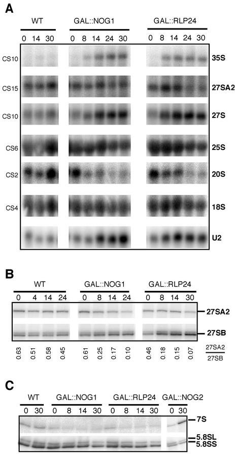
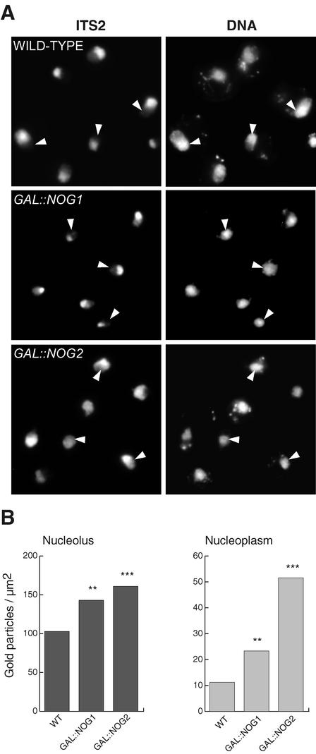
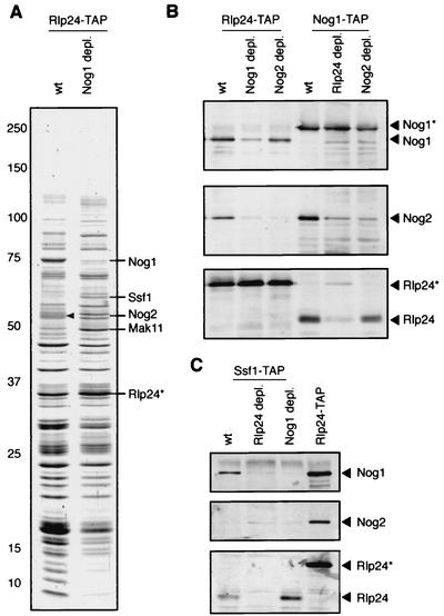
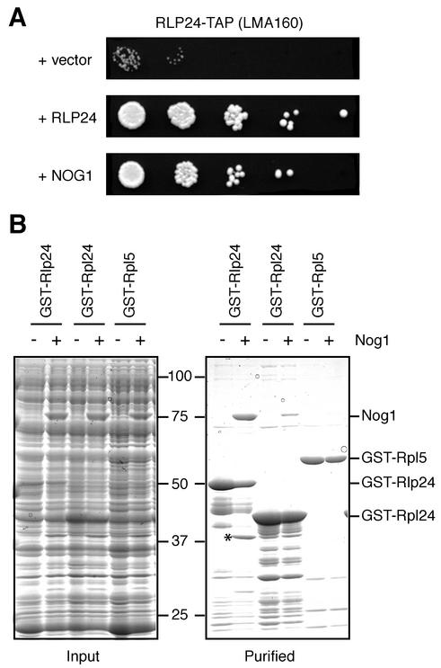
Ribosome biogenesis in eukaryotes depends on the coordinated action of ribosomal and nonribosomal proteins that guide the assembly of preribosomal particles. These intermediate particles follow a maturation pathway in which important changes in their protein composition occur. The mechanisms involved in the coordinated assembly of the ribosomal particles are poorly understood. We show here that the association of preribosomal factors with pre-60S complexes depends on the presence of earlier factors, a phenomenon essential for ribosome biogenesis. The analysis of the composition of purified preribosomal complexes blocked in maturation at specific steps allowed us to propose a model of sequential protein association with, and dissociation from, early pre-60S complexes for several preribosomal factors such as Mak11, Ssf1, Rlp24, Nog1, and Nog2. The presence of either Ssf1 or Nog2 in complexes that contain the 27SB pre-rRNA defines novel, distinct pre-60S particles that contain the same pre-rRNA intermediates and that differ only by the presence or absence of specific proteins. Physical and functional interactions between Rlp24 and Nog1 revealed that the assembly steps are, at least in part, mediated by direct protein-protein interactions.


Figures

FIG. 1.
FIG. 1.
Rlp24 and Rpl24 are homologous proteins of different cellular localizations. (A) N-terminal sequences of Rlp24-like and Rpl24-like proteins encoded by genes present in eukaryotes or Archaea were aligned with ClustalX. Gray, amino acid residues identical in several sequences; black, conserved cysteines of the putative zinc finger present in Rlp24 sequences. Accession numbers (TrEMBL, SwissProt, or Genpept) for theRlp24-like sequences are as follows: Saccharomyces cerevisiae (Sc), Q07915; Schizosaccharomyces pombe (Sp), Q10353; Caenorhabditis elegans (Ce), Q17606; Drosophila melanogaster (Dm), Q9VGN9; Homo sapiens (Hs), Q9UHA3; Arabidopsis thaliana (At), O22165; Encephalitozoon cuniculi (Ez), gi-19074000; G. theta nucleomorph (Gt), gi-13812425; Pyrococcus abyssi (Pa), Q9V0W3; H. marismortui (Hm), P14116; Sulfolobus sulfataricus (Ss), Q980Q6. Accession numbers for the Rpl24-like sequences are as follows: S. cerevisiae, P04449; S. pombe, O74884; C. elegans, O01868; D. melanogaster, Q9VJY6; H. sapiens, P38663; A. thaliana, Q9LF73; E. cuniculi, gi-19074515 (starting residue 14). For the G. theta nucleomorph pseudo-Rpl24, see Materials and Methods. (B) Unusual localization of Rlp24 from the nucleolus to the cytoplasm. Cells expressing Rlp24-TAP and Rpl24-TAP under the control of their own promoters were fixed with formaldehyde, washed, and incubated with anti-protein A antibodies (Sigma) followed by Cy3-conjugated secondary antibodies (Jackson Immunoresearch). (C) Rlp24 is neither free nor associated with polysomes. The sedimentation behavior of Rpl24-TAP was assessed by sucrose gradient ultracentrifugation. Twenty-four fractions were collected, and tagged proteins were detected by immunoblotting using peroxidase-antiperoxidase conjugates (Sigma) and Rlp24-specific antibodies. Asterisks, cross-reactive bands. The same profile of sedimentation was obtained with an Rlp24-TAP strain. (D) Cytoplasmic Rlp24 localization depends on active ribosome biogenesis. Preribosomes are blocked in the nucleus in absence of Nog1. Wild-type (WT) and GAL1::NOG1 cells expressing Rpl25-GFP were grown for 16 h in glucose-containing medium. The fluorescence signals of GFP and Hoechst 33342-stained DNA were observed by microscopy. Arrowheads, position of the nucleoplasm. The localizations of Rlp24-TAP (E) and Rpl24-GFP (F) expressed under the control of their own promoters were observed in GAL1::NOG1 cells either grown exclusively on galactose or shifted to glucose-containing medium for 16 h. Rlp24-TAP was detected as described for panel B. Similar results (data not shown) were obtained when we blocked pre-60S export by overexpressing the Nmd3Δ100 dominant-negative allele (16).
FIG. 2.
FIG. 2.
Similar compositions of Rlp24- and Nog1-associated preribosomal complexes. Tandem affinity purification of Nog1- and Rlp24-associated complexes was followed by separation of proteins by denaturing electrophoresis on a 5 to 20% polyacrylamide gel and colloidal Coomassie blue staining. Asterisks, positions of the tagged proteins used as baits. Numbers correspond to excised gel bands in which the preribosomal factors listed in Table 2 were identified, with the exception of the band marked 34, identified as ribosomal protein Rpl24A/B. Dots correspond to bands in which ribosomal proteins of the large subunit were identified. The result for an identically treated extract from wild-type cells is shown in the left lane. (B and C) Rlp24 and Nog1 are associated with pre-60S RNAs. RNAs were isolated by one-step purification of extracts of Nog1-TAP, Rlp24-TAP, or wild-type cells and detected by primer extension (B) (35S, 27SA2, 27SA3, 27SB, 25S, and 18S) or Northern blotting (C) (7S, 5.8S, 5S, U3 and U2 snRNA) with radiolabeled oligonucleotides. (D) Schematic representation of the steps involved in pre-rRNA processing in S. cerevisiae. The largest 35S intermediate pre-rRNA undergoes extensive modifications and processing to generate the mature 18S, 5.8S, 25S rRNAs (19). The relative positions of the oligonucleotides used in this study are indicated.
FIG. 3.
FIG. 3.
Rlp24 and Nog1 are involved in 27SB pre-rRNA processing. (A) Steady-state levels of mature and precursor rRNA in strains depleted of Rlp24 or Nog1. Cells grown on galactose-containing medium were shifted to glucose for the indicated number of hours, and total RNAs were analyzed by Northern blotting after denaturing 1.2% formaldehyde agarose electrophoresis. The positions of the probes are indicated in the Fig. 2D. WT, wild type. (B) Accumulation of 27SB over 27SA2 intermediates. Following primer extension with oligonucleotide CS10 (specific for the ITS2 sequence), the changes in the ratio of 27SA2 to 27SB in cells depleted of Rlp24 and Nog1 were quantified with a PhosphorImager and the ImageQuant software (Molecular Dynamics) and are shown below the gel image. (C) Small RNAs separated on denaturing urea-5% polyacrylamide gels were analyzed by Northern blotting using a probe specific for 7S and 5.8S intermediates (CS5).
FIG. 4.
FIG. 4.
In the absence of Nog1, pre-rRNAs accumulate in the nucleolus. (A) Intranuclear distribution of the preribosomal particles in Nog1- or Nog2-depleted cells was determined by FISH with a probe specific for the ITS2 sequence. DNA was stained with the DAPI (4′,6′-diamidino-2-phenylindole) fluorescent dye. Arrowheads, position of the nucleoplasm. (B) ITS2 containing pre-RNAs were detected by electron microscopy in situ hybridization in cells depleted of Nog1 or Nog2 by growth on glucose for 14 h, and the labeling densities of the nucleolus and the nucleoplasm were determined. One-to-one comparisons with the wild-type (WT) strain showed significant differences (∗∗, P < 0.01; ∗∗∗, P < 0.001). The nucleoplasmic labeling in Nog2-depleted cells is also significantly higher (P < 0.001) than that in cells lacking Nog1.
FIG. 5.
FIG. 5.
Nog1 association with pre-60S complexes depends on Rlp24 and is a prerequisite for Nog2 assembly. (A) Early pre-60S complexes accumulate in the absence of Nog1. Rlp24-associated complexes were purified from wild-type (wt) or Nog1-depleted cells (GAL1::NOG1 cells shifted to glucose for 14 h), and the proteins were separated on a 5 to 20% polyacrylamide gel and stained with colloidal Coomassie blue. Asterisks (A to C), positions of the tagged proteins used as baits. The proteins are the same as those in Fig. 2A for comparison. Arrowhead, the Nog2 band identified by mass spectrometry. (B) The role of Nog1 and Rlp24 in Nog2 assembly on pre-60S particles was confirmed by immunoblots with tandem affinity-purified complexes and specific antibodies. Cells were shifted to glucose for 14 h to deplete Nog1 or Rlp24 and for 18 h to deplete Nog2. (C) The requirement of Nog1 for Rlp24 assembly on pre-60S particles was tested by using Ssf1-TAP purified complexes and immunoblots in wild-type cells or after Nog1 or Rlp24 depletion for 14 h.
FIG. 6.
FIG. 6.
Rlp24 and Nog1 genetically and physically interact. (A) The Rlp24-TAP strain was transformed with a high-copy-number LEU2 vector (pFL46S) carrying no insert or inserts corresponding to Nog1 or Rlp24. The rescue of the slow-growth phenotype was tested by spotting transformants on minimal-medium plates in 10−1-dilution steps. The plates were incubated at 25°C for 4 days. (B) Total protein extracts from bacteria expressing GST-Rlp24, GST-Rpl24B, and GST-Rpl5 were incubated with extracts containing Nog1 and then with glutathione-Sepharose beads (Pharmacia). After extensive washing, the bound proteins were eluted under denaturing conditions and separated by electrophoresis. The Coomassie blue-stained gels representing the input mixture of bacterial protein extracts (left) and the purified proteins (right) are shown side by side for comparison. A band of about 38 kDa that copurifies with GST-Rlp24 only in the presence of Nog1 (∗) is a C-terminal Nog1 fragment (matching peptides between amino acids 361 and 599 as determined by MALDI-TOF mass spectrometry and assignment of peptide masses to the Nog1 sequence).
FIG. 7.
FIG. 7.
A model for sequential protein assembly and intracellular trafficking of pre-60S particles. Early pre-60S particles formed in the nucleolus contain Mak11, Ssf1, and Rlp24. The arrival of Nog1 is probably concomitant with Mak11 dissociation. After 27SA2-to-27SB processing, Nog2 associates with pre-60S concomitantly or after Ssf1 has left the particles. Late complexes containing Nog1, Nog2, and Rlp24 are transported to the nucleoplasm, where Nog1 and Nog2 dissociate, apparently before export to the cytoplasm. Finally, Rlp24 dissociates from cytoplasmic pre-60S particles and is likely to be exchanged for its ribosomal homologue, Rpl24.

References

    1. Bachellerie, J. P., B. Michot, M. Nicoloso, A. Balakin, J. Ni, and M. J. Fournier. 1995. Antisense snoRNAs: a family of nucleolar RNAs with long complementarities to rRNA. Trends Biochem. Sci. 20:261-264. - PubMed
    1. Ban, N., P. Nissen, J. Hansen, P. B. Moore, and T. A. Steitz. 2000. The complete atomic structure of the large ribosomal subunit at 2.4 Å resolution. Science 289:905-920. - PubMed
    1. Bassler, J., P. Grandi, O. Gadal, T. Lessmann, E. Petfalski, D. Tollervey, J. Lechner, and E. Hurt. 2001. Identification of a 60S preribosomal particle that is closely linked to nuclear export. Mol. Cell 8:517-529. - PubMed
    1. Baudin, A., O. Ozier-Kalogeropoulos, A. Denouel, F. Lacroute, and C. Cullin. 1993. A simple and efficient method for direct gene deletion in Saccharomyces cerevisiae. Nucleic Acids Res. 21:3329-3330. - PMC - PubMed
    1. Bertrand, E., F. Houser-Scott, A. Kendall, R. H. Singer, and D. R. Engelke. 1998. Nucleolar localization of early tRNA processing. Genes Dev. 12:2463-2468. - PMC - PubMed

Publication types

MeSH terms