. 2003 Jul;23(13):4532-41.
doi: 10.1128/MCB.23.13.4532-4541.2003.

Transcriptional program of apoptosis induction following interleukin 2 deprivation: identification of RC3, a calcium/calmodulin binding protein, as a novel proapoptotic factor

Affiliations

Transcriptional program of apoptosis induction following interleukin 2 deprivation: identification of RC3, a calcium/calmodulin binding protein, as a novel proapoptotic factor

Laxminarayana R Devireddy et al. Mol Cell Biol. 2003 Jul.

Abstract

Apoptosis of mature T lymphocytes preserves immune system homeostasis by counteracting transient increases in T-cell number. This process is regulated, at least in part, by the cytokine interleukin 2 (IL-2): T cells deprived of IL-2 undergo apoptosis. The mechanism of apoptosis induction by IL-2 deprivation remains to be determined but is known to require RNA synthesis, implying the existence of transcriptionally activated genes whose products induce cell death. To identify such genes, we have performed expression profiling in IL-2-dependent T cells following cytokine deprivation. Our results reveal an intricate transcriptional program entailing the induction of known proapoptotic factors and the simultaneous repression of known antiapoptotic factors. Surprisingly, one gene whose transcription substantially increased was RC3 (also called neurogranin), which encodes a calmodulin binding protein thought to be a neural-specific factor involved in learning and memory. We show that ectopic expression of RC3 in IL-2-dependent T cells increases the intracellular Ca(2+) concentration and induces apoptosis even in the presence of cytokine. Buffering the Ca(2+) increase with the cytoplasmic Ca(2+) chelator BAPTA-AM [1,2-bis(2-aminophenoxy)ethane-N,N,N1,N-tetraacetic acid] blocks RC3-induced apoptosis, indicating that the rise in intracellular Ca(2+) is required for apoptotic death. RC3 mutants unable to bind calmodulin fail to increase intracellular Ca(2+) levels and to induce apoptosis. Based upon these results, we propose that IL-2 deprivation raises the level of RC3 and other apoptotic factors, which induce apoptosis by increasing the intracellular Ca(2+) concentration.


Figures
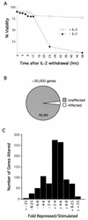
FIG. 1.
FIG. 1.
A transcriptional program of apoptosis induction following IL-2 deprivation. (A) Time course of apoptosis following IL-2 deprivation. The percentage of viable HT-2 cells was determined by annexin V-FITC staining. (B) Summary of microarray analysis. Affected is defined as a twofold or greater change following IL-2 deprivation. (C) Graphical representation of the fold changes of genes stimulated or repressed.
FIG. 2.
FIG. 2.
Induction of RC3 transcription following IL-2 deprivation. (A) Transcriptional activation of RC3 upon IL-2 deprivation. Expression of RC3, ATFx, and GAPDH (as a loading control) in HT-2 cells was assessed by Northern blot analysis with 1 μg of poly(A)+ mRNA or 10 μg of total RNA 8 h after IL-2 deprivation. (B) Time course of RC3 induction. RC3 mRNA levels in HT-2 cells were analyzed at the indicated times following IL-2 deprivation. The Northern blot was stripped and reprobed with a 28S rRNA probe as a loading control. (C) Immunoblot analysis of whole-cell extracts (Lysate) or culture supernatants (Supt) from HT-2 cells grown in the presence or absence of IL-2. The same blot was stripped and reprobed with an α-tubulin antibody (Sigma) as a loading control. The exposure time was 10 s.
FIG. 3.
FIG. 3.
Cell specificity of RC3 transcription activation. (A) RC3 is transcriptionally activated in CTLL-2 cells upon IL-2 deprivation. Expression of RC3 and GAPDH (as a loading control) was assessed by Northern blot analysis with 1 μg of poly(A)+ mRNA or 10 μg of total RNA 8 h after IL-2 removal. (B) Northern blot analysis of poly(A)+ mRNA prepared from primary human T lymphocytes 12 to 14 h after IL-2 removal. (C) Northern blot analysis of poly(A)+ mRNA prepared from FL5.12 cells 8 h after IL-3 removal. The same blot was sequentially stripped and reprobed with 24p3 (as an induction control), ATFx (as a repression control), and GAPDH probes. (D) Northern analysis of poly(A)+ mRNA from 32D cells. The blot was probed sequentially with RC3 and GAPDH probes. (E) Immunoblot analysis of whole-cell extracts from Jurkat cells after staurosporine (STS) treatment, which induces apoptosis. The same blot was stripped and reprobed with a caspase-3 antibody (Santa Cruz Biotechnology) as a control; STS treatment results in proteolytic activation of caspase 3.
FIG. 4.
FIG. 4.
Expression of RC3 induces apoptosis in HT-2 and CTLL-2 cells. (A) HT-2 and CTLL-2 cells were transiently transfected with 5 μg of either pcDNA3.1 (empty vector) or pcDNA3.1/RC3 or left untransfected (control). One day after transfection, cells were collected and stained with annexin V-FITC and analyzed by FACS. PI, propidium iodide. (B) Extracts were prepared from a portion of the transfected cells and analyzed by immunoblotting with an antihemagglutinin (anti-HA) monoclonal antibody or an α-tubulin antibody. (C) HT-2 cells were cotransfected with 1 μg of GFP plasmid and 4 μg of either pcDNA3.1 (empty vector), pcDNA3.1+/RC3 (sense orientation), or pcDNA3.1-/RC3 (antisense orientation). Cotransfected cells were stained with annexin V-PE and analyzed by FACS. The percentage of GFP-positive cells that were also positive for annexin V-PE was quantitated and plotted. Error bars indicate standard deviations. (D) HT-2 and CTLL-2 cells transfected with empty vector plasmid (pcDNA3.1) or an RC3 expression plasmid were subjected to DNA fragmentation analysis.
FIG. 5.
FIG. 5.
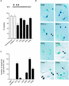
A functional RC3 CaM binding domain is required for induction of apoptosis. (A) HT-2 cells grown in the presence of IL-2 were transfected with 4 μg of empty vector plasmid (pcDNA3.1), wild-type RC3 (WT), or RC3 mutants. Cells were collected 24 h after transfection, stained with annexin V-FITC, and subjected to FACS analysis to monitor cell viability. The protein sequence of the 20-amino-acid CaM binding domain (IQ motif) of RC3 is shown; mutated residues are indicated by arrows. Error bars indicate standard deviations. (B) HEK-293 cells were transiently cotransfected with a β-galactosidase (β-gal) reporter plasmid (0.5 μg) and the experimental plasmid (4 μg), as indicated. The cells were washed 24 h after transfection, fixed, and stained with 0.2% X-Gal. (C) Quantitative analysis of the experiment for panel B. Four hundred β-galactosidase-positive cells were analyzed, and the number of blue cells displaying an apoptotic morphology was assessed.
FIG. 6.
FIG. 6.
Both IL-2 deprivation and RC3 expression increase intracellular Ca2+ concentration. (A) HT-2 cells grown in the presence or absence of IL-2 for 12 h were stained with the fluorescent Ca2+ indicator fura-2 AM, and the intracellular Ca2+ concentration was measured (see Materials and Methods for details). As a control, HT-2 cells were treated with 2 μM thapsigargin (TSG), a Ca2+ store depletor, for 1 min before staining with fura-2 AM. Error bars indicate standard deviations. (B) HT-2 cells were transfected with a plasmid expressing RC3 or an RC3 mutant and analyzed as described for panel A. WT, wild type. (C) HT-2 cells were transfected with either pcDNA3.1 or an RC3 expression plasmid; transfected cells were isolated from untransfected cells (see Materials and Methods for details) and, in parallel with controls with or without IL-2, stained with fura-2 AM. The left panels show the pseudocolor images of intracellular Ca2+ within fura-2-loaded cells; high Ca2+ levels are shown in red, and low Ca2+ levels are shown in blue or violet. Cells cultured in the absence of IL-2 or expressing RC3 display an apoptotic morphology. The average intracellular Ca2+ concentration of the cells shown in each field of view was calculated from background corrected images (see Materials and Methods) and is shown in the panel on the right. (D) Primary human T lymphocytes were grown in the presence or absence of IL-2 for 12 to 14 h and stained with fura-2 AM. The left panels show the pseudocolor images, and the right panel shows the average intracellular Ca2+ concentration as described for panel C.
FIG. 7.
FIG. 7.
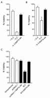
An increase in intracellular Ca2+ is required for induction of apoptosis. (A) HT-2 cells grown in the presence or absence of IL-2 were incubated with BAPTA-AM, and the number of cells undergoing apoptosis was quantitated by annexin V-FITC staining and FACS analysis. Error bars indicate standard deviations. (B) Primary human T lymphocytes grown in the presence or absence of IL-2 were incubated with BAPTA-AM, and the number of cells undergoing apoptosis was quantitated as described for panel A. (C) HT-2 cells transfected with either pcDNA3.1 or an RC3 expression plasmid were treated with BAPTA-AM, and the number of cells undergoing apoptosis was analyzed as described for panel A.

References

    1. Ayllon, V., C. Martinez-A, A. Garcia, X. Cayla, and A. Rebollo. 2000. Protein phosphatase 1 α is a Ras-activated Bad phosphatase that regulates interleukin-2 deprivation-induced apoptosis. EMBO J. 19:2237-2246. - PMC - PubMed
    1. Baffy, G., T. Miyashita, J. R. Williamson, and J. C. Reed. 1993. Apoptosis induced by withdrawal of interleukin-3 (IL-3) from an IL-3-dependent hematopoietic cell line is associated with repartitioning of intracellular calcium and is blocked by enforced Bcl-2 oncoprotein production. J. Biol. Chem. 268:6511-6519. - PubMed
    1. Bahler, M., and A. Rhoads. 2002. Calmodulin signaling via the IQ motif. FEBS Lett. 513:107-113. - PubMed
    1. Baudier, J., J. C. Deloulme, A. Van Dorsselaer, D. Black, and H. W. Matthes. 1991. Purification and characterization of a brain-specific protein kinase C substrate, neurogranin (p17). Identification of a consensus amino acid sequence between neurogranin and neuromodulin (GAP43) that corresponds to the protein kinase C phosphorylation site and the calmodulin-binding domain. J. Biol. Chem. 266:229-237. - PubMed
    1. Broome, H. E., C. M. Dargan, E. F. Bessent, S. Krajewski, and J. C. Reed. 1995. Apoptosis and Bcl-2 expression in cultured murine splenic cells. Immunology 84:375-382. - PMC - PubMed

Publication types

MeSH terms

LinkOut - more resources